
Cell Injury, Cell Death, and Adaptations Cells


cellular_injury_and_cellular_death.ppt
- Размер: 6.4 Mегабайта
- Количество слайдов: 121
Описание презентации Cell Injury, Cell Death, and Adaptations Cells по слайдам
Cell Injury, Cell Death, and Adaptations
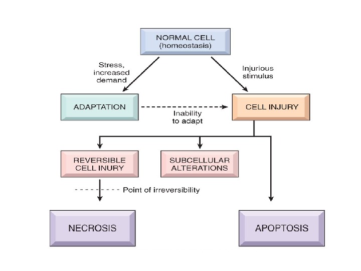
Cells are active participants in their environment, constantly adjusting their structure and function to accommodate changing demands and extracellular stresses. Cells tend to maintain their intracellular milieu within a fairly narrow range of physiologic parameters; that is, they maintain normal homeostasis.
CELLULAR ADAPTATIONS As cells encounter physiologic stresses or pathologic stimuli, they can undergo: • adaptation • achieving a new steady state • preserving viability and function The principal adaptive responses are hypertrophy, hyperplasia, atrophy, metaplasia.
CELLULAR ADAPTATIONS Broadly speaking, such physiologic and pathologic adaptations occur by • Decreasing or increasing their size (atrophy and hypertrophy respectively). • By changing the pathway of phenotypic differentiation of cells (metaplasia and dysplasia). In general, the adaptive responses are reversible on withdrawal of stimulus.
Cells may adapt to a pathological (disease) stimulus by extending the three normal physiological adaptive responses: 1. Increased cellular activity — increased functional demand on a tissue can be met by increase in cell number (hyperplasia), as well as by increase in cell size (hypertrophy). 2. Decreased cellular activity — cell atrophy. 3. Alteration of cell morphology (degeneration, apoptosis, necrosis).
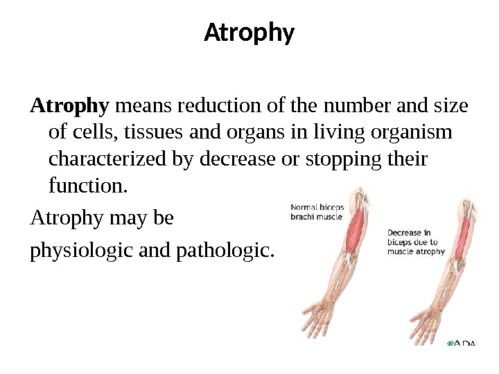
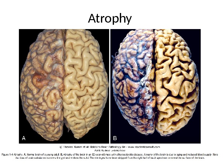
Atrophy means reduction of the number and size of cells, tissues and organs in living organism characterized by decrease or stopping their function. Atrophy may be physiologic and pathologic.
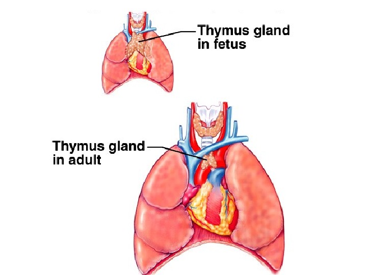
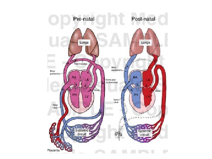
A. Physiologic atrophy. It is a normal process of aging in some tissues: 1. Atrophy of lymphoid tissue in lymph nodes, appendix and thymus. 2. Atrophy of gonads after menopause. 3. Atrophy of brain. 4. Atrophy of bones. It may be obliteration of the umbilical arteries and arterial duct (Botallow’s) after birth.
Atrophy
B. Pathologic atrophy may be general and local. General atrophy is observed in cachexia due to • Oncologic and chronic diseases. • Starvation. • Injury of hypophysis (endocrine cachexia). • Injury of hypothalamus (cerebral cachexia).
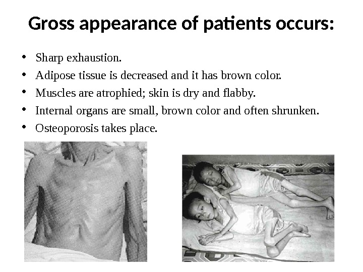
Gross appearance of patients occurs: • Sharp exhaustion. • Adipose tissue is decreased and it has brown color. • Muscles are atrophied; skin is dry and flabby. • Internal organs are small, brown color and often shrunken. • Osteoporosis takes place.
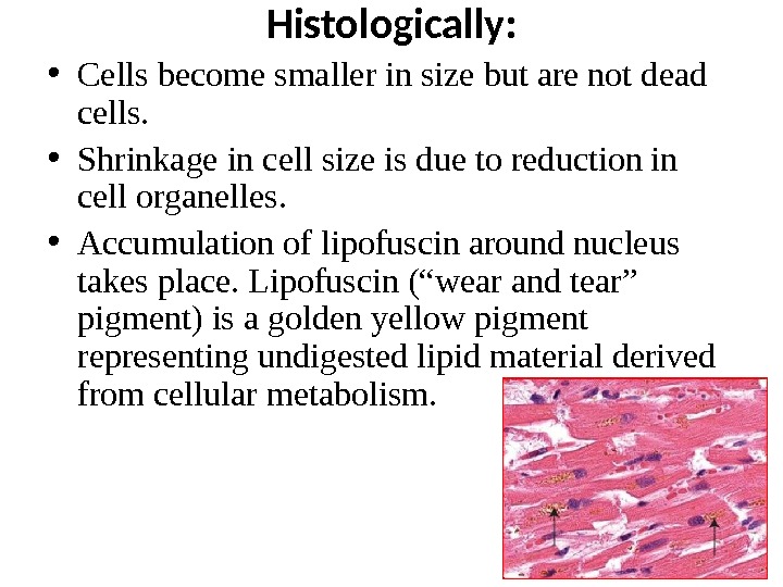
Histologically: • Cells become smaller in size but are not dead cells. • Shrinkage in cell size is due to reduction in cell organelles. • Accumulation of lipofuscin around nucleus takes place. Lipofuscin (“wear and tear” pigment) is a golden yellow pigment representing undigested lipid material derived from cellular metabolism.
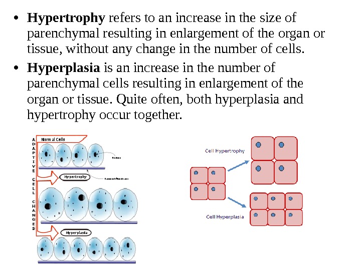
• Hypertrophy refers to an increase in the size of parenchymal resulting in enlargement of the organ or tissue, without any change in the number of cells. • Hyperplasia is an increase in the number of parenchymal cells resulting in enlargement of the organ or tissue. Quite often, both hyperplasia and hypertrophy occur together.
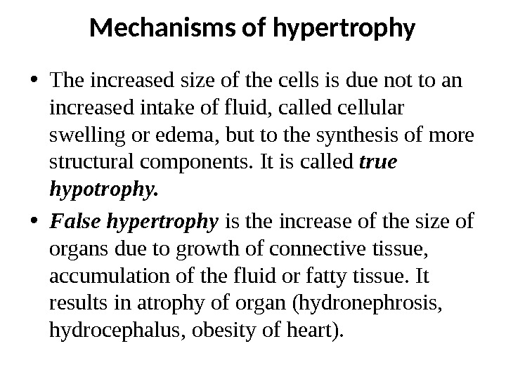
Mechanisms of hypertrophy • T he increased size of the cells is due not to an increased intake of fluid, called cellular swelling or edema, but to the synthesis of more structural components. It is called true hypotrophy. • False hypertrophy is the increase of the size of organs due to growth of connective tissue, accumulation of the fluid or fatty tissue. It results in atrophy of organ (hydronephrosis, hydrocephalus, obesity of heart).
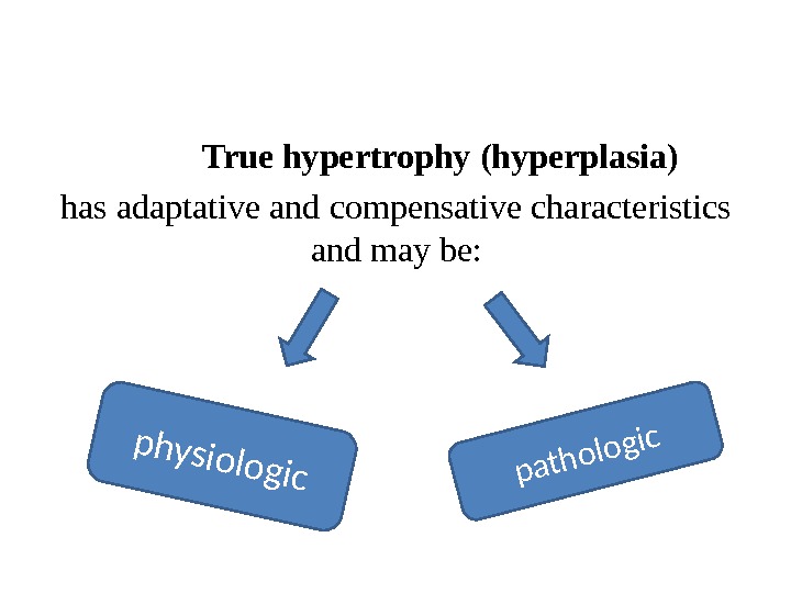
True hypertrophy (hyperplasia) has adaptative and compensative characteristics and may be: physiologicpathologic
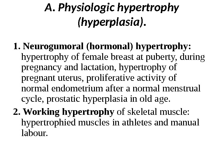
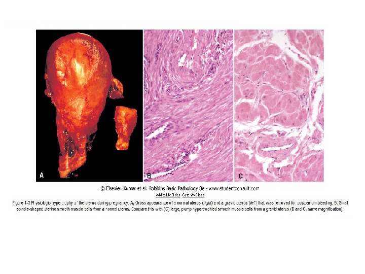
A. Physiologic hypertrophy (hyperplasia). 1. Neurogumoral (hormonal) hypertrophy: hypertrophy of female breast at puberty, during pregnancy and lactation, hypertrophy of pregnant uterus, proliferative activity of normal endometrium after a normal menstrual cycle, prostatic hyperplasia in old age. 2. Working hypertrophy of skeletal muscle: hypertrophied muscles in athletes and manual labour.
B. Pathologic hypertrophy (hyperplasia). Neurogumoral hypertrophy develops due to impairment of endocrine functions. Endometrial glandular hyperplasia following estrogen excess which it occurs by metrorrhagia; atrophy of testis leads to increase of breast (gynecomastia); hyperfunction of anterior lobus hypophisis (adenoma) leads to increase skeleton (acromegaly). Working hypertrophy develops in tissues consisting of stable undivided cells due to increase of size it one. It may be often in cardiac muscle at some cardiac diseases, such as: systemic hypertension, aortic valve disease (stenosis and insufficiency), mitral insufficiency; hypertrophy of smooth muscle: cardiac achalasia (in esophagus), pyloric stenosis (in stomach), and intestinal stricture; hypertrophy of urine bladder in adenoma of prostatic glands.
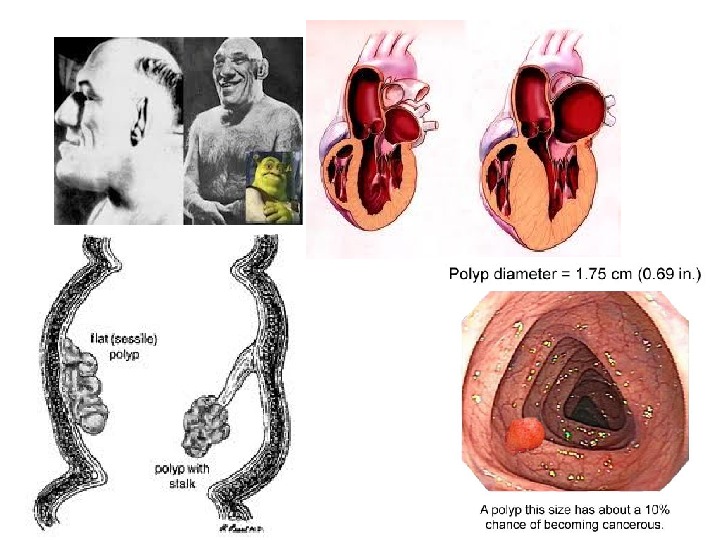
3. Compensatory reparative hypertrophy : regeneration of the liver following partial hepatectomy, regeneration of epidermis after skin abrasion; hypertrophy of myocardium in postinfarctional cardiosclerosis. 4. Vicarious (substitutional) hypertrophy: following nephrectomy on one side in a young patient there is compensatory hypertrophy as well as hyperplasia of the nephrons of the other kidney. 5. Hypertrophic vegetations develop due to chronic inflammation in mucous membranes (polyps and condilomas) ; lymphostasis leads to ingrowth of connective tissue, examples of false hypertrophy. In wound healing, there is formation of granulation tissue.
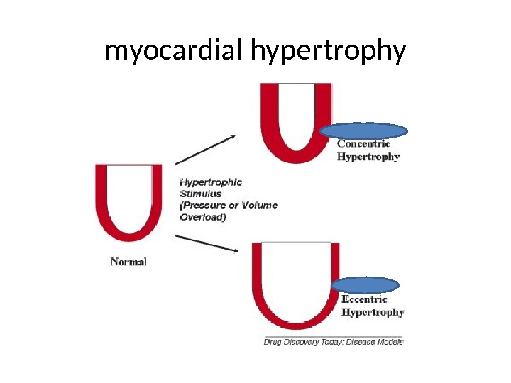
According to stage of adaptation two types of myocardial hypertrophy have been described: • Concentric. In concentric hypertrophy (clinically, no insufficiency) the musculature is clearly enlarged, measuring till 1. 8 cm, but chambers of the heart are not dilated. • Eccentric. In eccentric hypertrophy myocardium is enlarged but chambers of the heart are dilated. This leads to hemodynamic disorder with cardiac insufficiency. It is called myogenic dilatation.
myocardial hypertrophy
Metaplasia is defined as a reversible change of one type to another type of adult epithelial or mesenchymal cells, usually in response to abnormal stimuli, and often reverts back to normal on removal of stimulus. Metaplasia is broadly divided into 2 types: 1. Epithelial metaplasia 2. Mezenhymal metaplasia
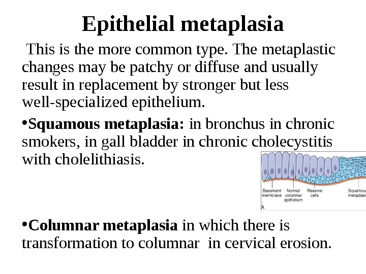
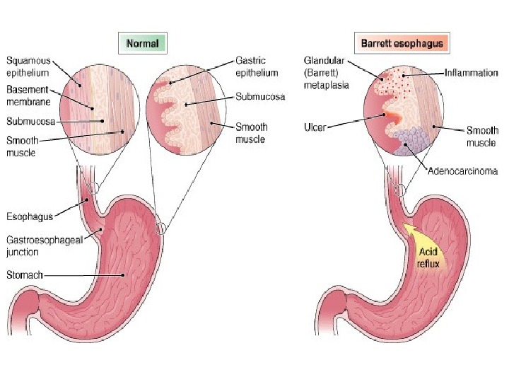
Epithelial metaplasia This is the more common type. The metaplastic changes may be patchy or diffuse and usually result in replacement by stronger but less well-specialized epithelium. • Squamous metaplasia: in bronchus in chronic smokers, in gall bladder in chronic cholecystitis with cholelithiasis. • Columnar metaplasia in which there is transformation to columnar in cervical erosion.
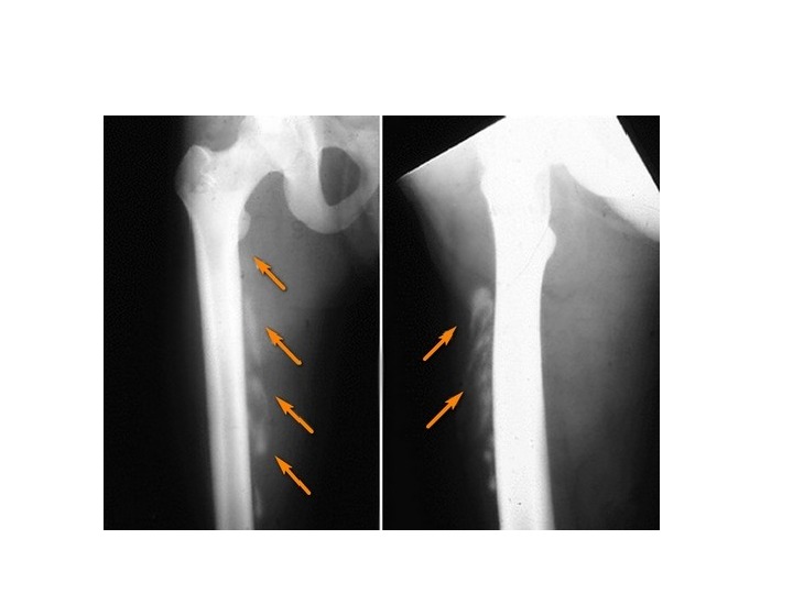
Mezenhymal metaplasia. Transformation of one adult type of mesenchymal tissue to another. • Osseous metaplasia is formation of bone in fibrous tissue, cartilage or Less often, there myxoid tissue: in arterial wall in old age, in soft tissues in myositis ossificans, in cartilage of larynx and bronchi in elderly people, in scar of chronic inflammation of prolonged duration, in the fibrous stroma of tumor. • Cartilaginous metaplasia. In healing of fractures, cartilaginous metaplasia may occur where there is undue mobility.
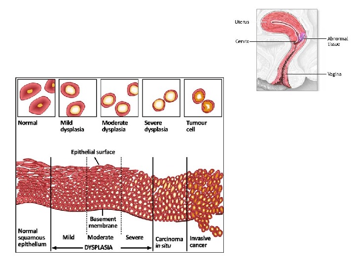
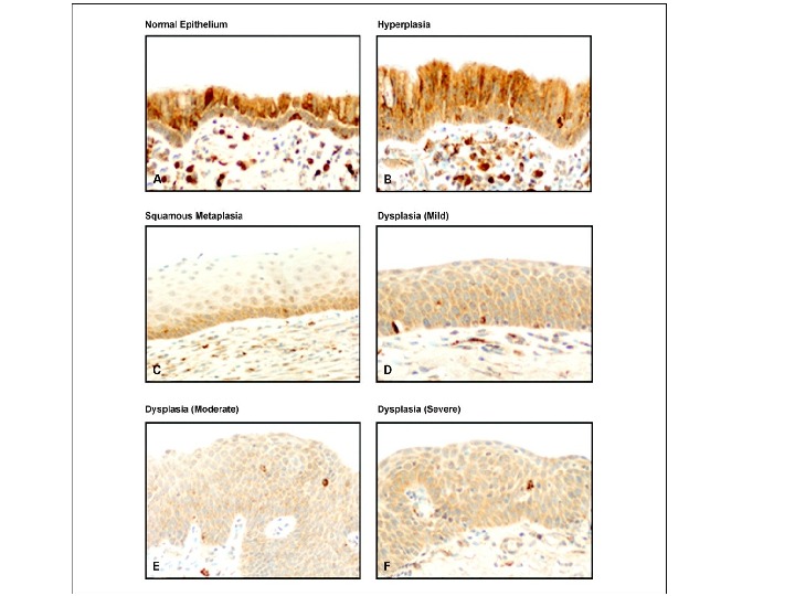
Dysplasia means “disordered cellular development”, often accompanied with metaplasia and hyperplasia, it is therefore also referred to as atypical hyperplasia. Epithelial dysplasia is characterized by cellular proliferation and cytological changes, which include: • Hyperplasia of epithelial layers. • Disorderly arrangement of cells from basal layer to the surface layer. • Cellular and nuclear pleomorphism. • Increased nucleocytoplasmic ratios. • Nuclear hyperchromatism. • Increased mitotic activity. The two most common examples of dysplastic changes are the uterine cervix and respiratory tract.
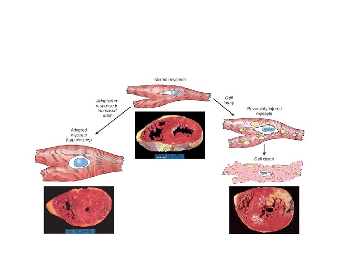
If the adaptive capability is exceeded or if the external stress is inherently harmful, cell injury develops. Within certain limits injury is reversible , and cells return to a stable baseline; however, severe or persistent stress results in irreversible injury and death of the affected cells.
IRREVERSIBLE CELLULAR INJURY: Cell death is a state of irreversible injury. It may occur in the living body as a local change (i. e. autolysis, necrosis and apoptosis ), or result in end of the life (somatic death).
Cell death is One of the most crucial events in the evolution of disease in any tissue or organ. It results from diverse causes, including ischemia (lack of blood flow), infections, toxins, and immune reactions. Cell death is also a normal and essential process in embryogenesis, the development of organs, and the maintenance of homeostasis.
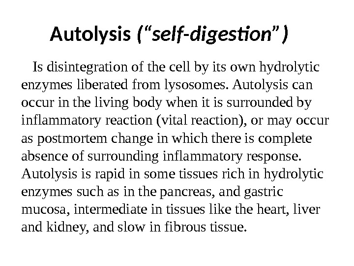
Autolysis (“self-digestion”) I s disintegration of the cell by its own hydrolytic enzymes liberated from lysosomes. Autolysis can occur in the living body when it is surrounded by inflammatory reaction (vital reaction), or may occur as postmortem change in which there is complete absence of surrounding inflammatory response. Autolysis is rapid in some tissues rich in hydrolytic enzymes such as in the pancreas, and gastric mucosa, intermediate in tissues like the heart, liver and kidney, and slow in fibrous tissue.
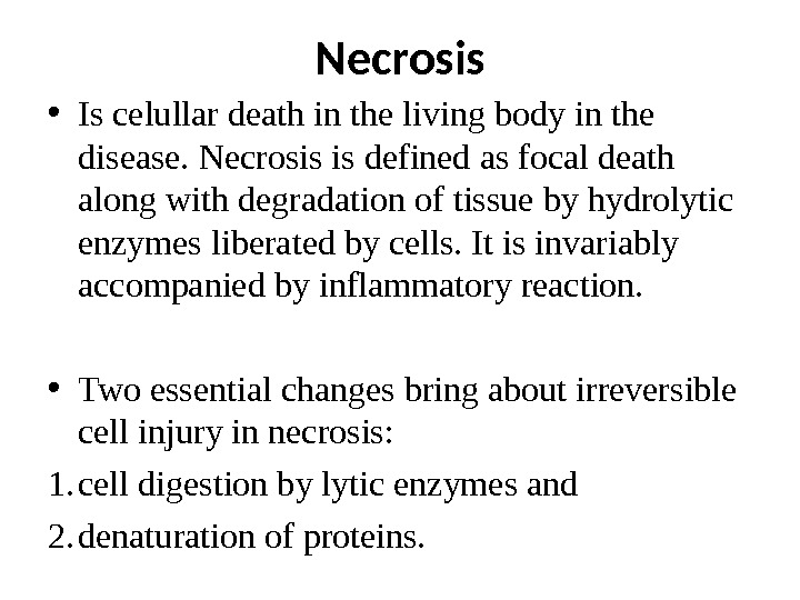
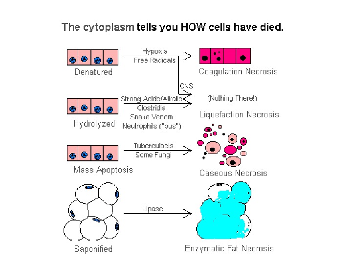
Necrosis • I s celullar death in the living body in the disease. Necrosis is defined as focal death along with degradation of tissue by hydrolytic enzymes liberated by cells. It is invariably accompanied by inflammatory reaction. • Two essential changes bring about irreversible cell injury in necrosis : 1. cell digestion by lytic enzymes and 2. denaturation of proteins.
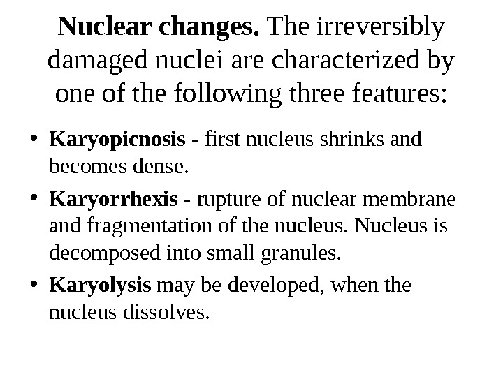
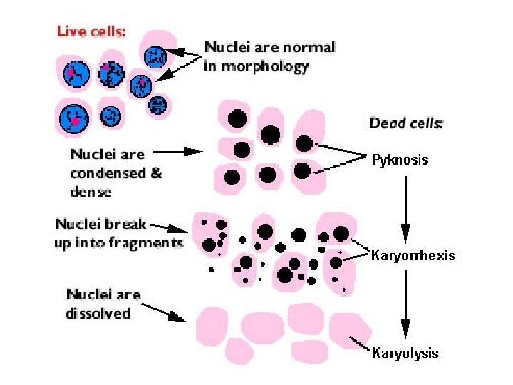
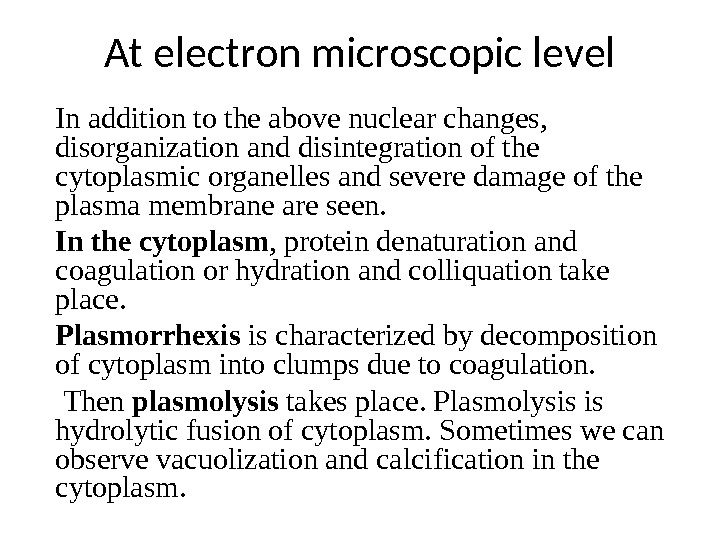
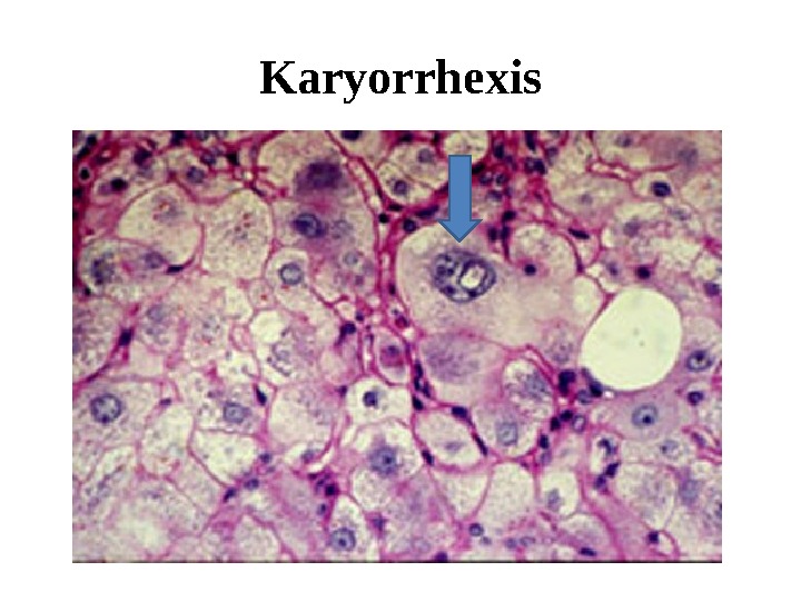
Nuclear changes. The irreversibly damaged nuclei are characterized by one of the following three features: • K aryopicnosis — first nucleus shrinks and becomes dense. • K aryorrhexis — rupture of nuclear membrane and fragmentation of the nucleus. Nucleus is decomposed into small granules. • K aryolysis may be developed, when the nucleus dissolves.
At electron microscopic level I n addition to the above nuclear changes, disorganization and disintegration of the cytoplasmic organelles and severe damage of the plasma membrane are seen. In the cytoplasm , protein denaturation and coagulation or hydration and colliquation take place. Plasmorrhexis is characterized by decomposition of cytoplasm into clumps due to coagulation. Then plasmolysis takes place. Plasmolysis is hydrolytic fusion of cytoplasm. Sometimes we can observe vacuolization and calcification in the cytoplasm.
K aryorrhexis
K aryolysis
The presence of the grains of chromatin in a focus of caseous necrosis is a manifestation of: A. Karyolysis B. Karyopyknosis C. Karyorhexis D. Mitotic activity of nuclei
Stages of necrosis (or morphogenesis): 1. Paranecrosis — reversible changes; as a rule, reversible degeneration. 2. Necrobiosis — irreversible degenerative changes. 3. Death of cells. 4. Autolysis is the enzymic digestion of the dead cell due to effect of catalytic enzymes derived from lysosomes.
Types of necrosis According to the mechanisms of development: 1. Direct (from influence of mechanical, physical, chemical, and toxic factors). 2. Indirect (vascular and neurogenous).
Types of necrosis According to the cause: 1. Traumatic. 2. Toxic. 3. Trophoneurotic. 4. Allergic. 5. Vascular or ischemic.
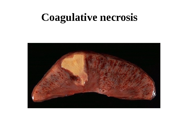
Coagulative necrosis Is associated with inhibition of lytic enzymes. Foci of coagulative necrosis in the early stage are pale, firm, and slightly swollen. With progression they become more yellowish, softer, and shrunken. The cells do not lyse; thus, their outlines are relatively preserved. Nuclei disappear and the acidified cytoplasm becomes eosiniphilic. Waxy (Zenker’s) necrosis of muscle may occur at typhoid fever.
Coagulative necrosis
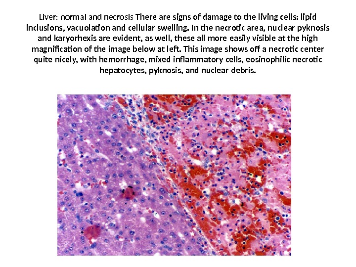
Liver: normal and necrosis There are signs of damage to the living cells: lipid inclusions, vacuolation and cellular swelling. In the necrotic area, nuclear pyknosis and karyorhexis are evident, as well, these all more easily visible at the high magnification of the image below at left. This image shows off a necrotic center quite nicely, with hemorrhage, mixed inflammatory cells, eosinophilic necrotic hepatocytes, pyknosis, and nuclear debris.
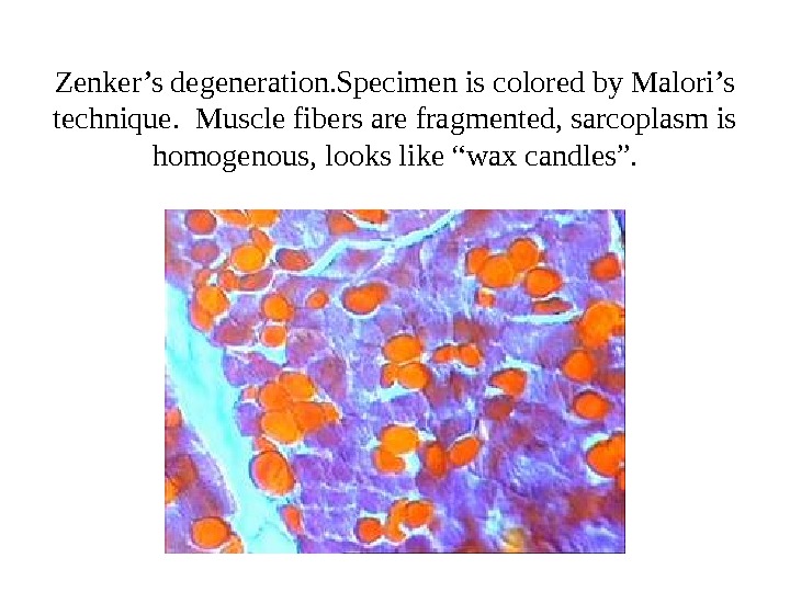
Zenker’s degeneration. Specimen is colored by Malori’s technique. Muscle fibers are fragmented, sarcoplasm is homogenous, looks like “wax candles”.
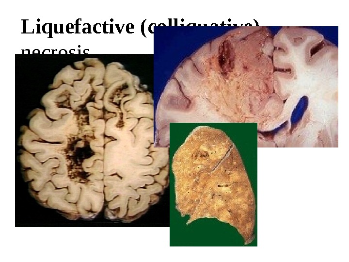
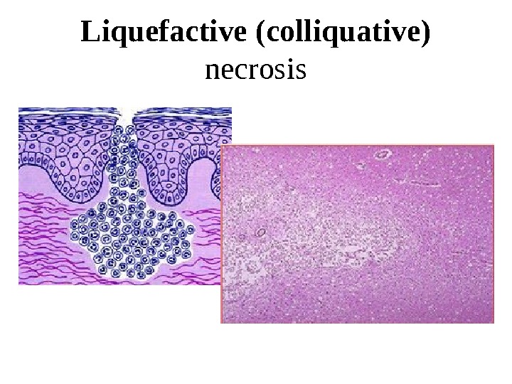
Liquefactive (colliquative) necrosis Is marked by dissolution of tissue due to enzymatic lysis of dead cells. Typically, it takes place in the brain when autocatalytic enzymes are released from dead cells. Liquefactive necrosis occurs also in purulent inflammation due to the heterolytic action of polymorphonuclear leucocytes in pus. Liquefied tissue is soft, diffluent and composed of disintegrated cells and fluid.
Liquefactive (colliquative) necrosis
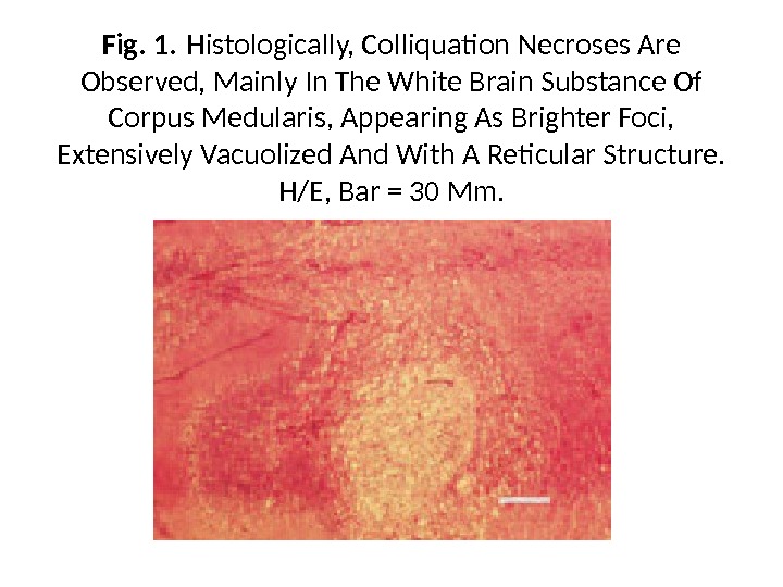
Fig. 1. Histologically, Colliquation Necroses Are Observed, Mainly In The White Brain Substance Of Corpus Medularis, Appearing As Brighter Foci, Extensively Vacuolized And With A Reticular Structure. H/E, Bar = 30 Μm.
Liquefactive (colliquative) necrosis
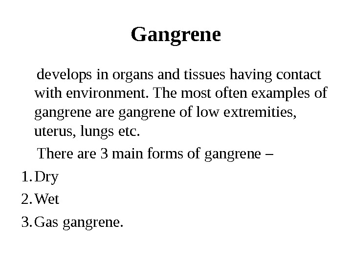
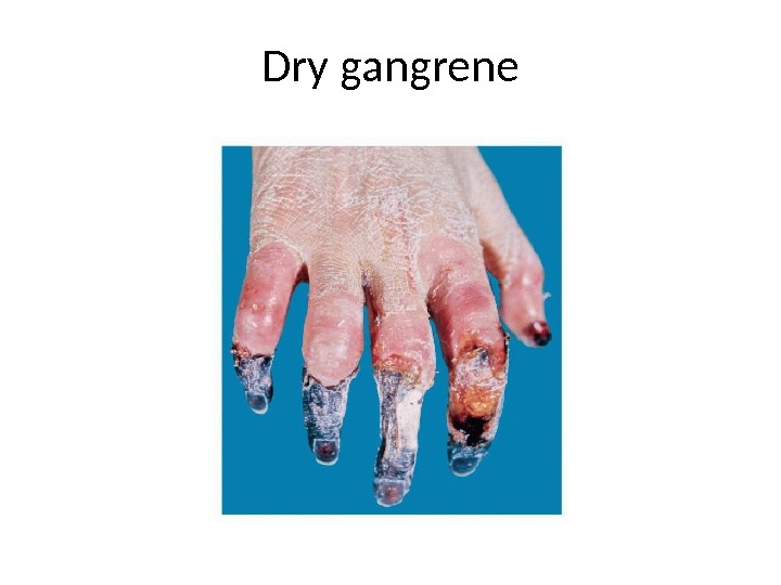
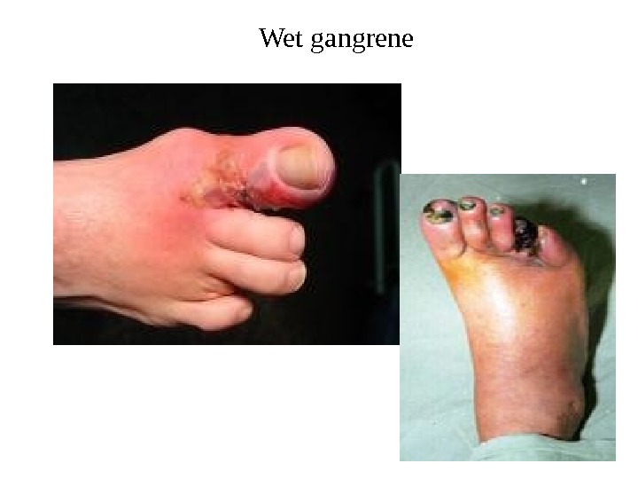
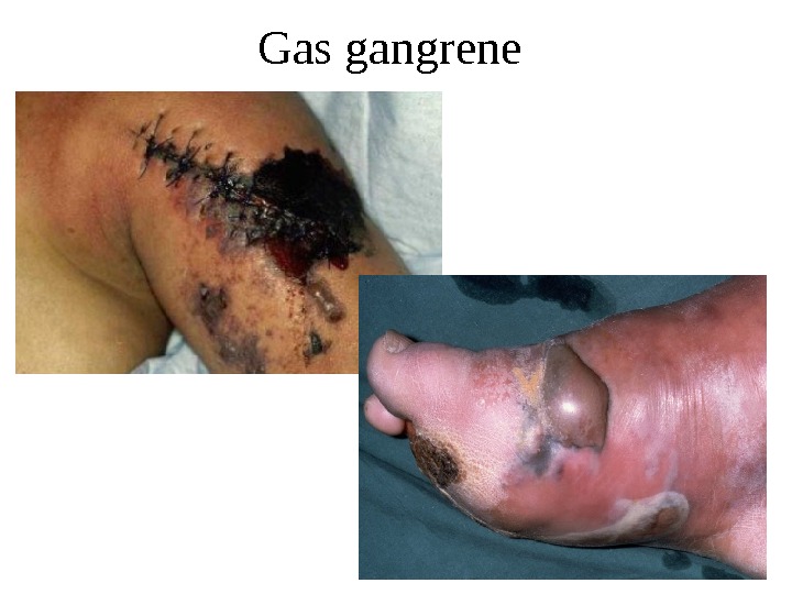
Gangrene develops in organs and tissues having contact with environment. The most often examples of gangrene are gangrene of low extremities, uterus, lungs etc. There are 3 main forms of gangrene – 1. Dry 2. Wet 3. Gas gangrene.
Dry gangrene
Wet gangrene
Gas gangrene
Gangrene does not appear in… • Skin • Kidney • Lungs • Uterus • Intestine (bowel)
On autopsy it is revealed enlarged dense right lung, fibrin layers on the pleura. Lung tissue is light green color on incision with muddy liqued exudates. What lung disease are these symptoms typical for? A. Lung-fever B. Bronchopneumonia, С. Pulmonary gangrene D. Fibrosing alveolitis E. Interstitial pneumonia
A patient with diabetes mellitus suddenly began having sharp pain in his right foot. The examination revealed black hallux, swallen foot tissues, focuses of epidermis detachment, secreta with unpleasant smell. What clinico-morphological form of necrosis developed in a patient? A. Wet gangrene B. Bedsore C. Dry gangrene D. Sequester E. Infarction
In 77 -year-old patient suffered with atherosclerosis the pain has appeared in the right foot. The foot is enlarged in size, its skin has black color and is macerated; the demarcation line is not clear. What pathological process takes place in a patient? A. Coagulative necrosis B. Sequestrum C. Dry gangrene D. Wet gangrene E. Noma
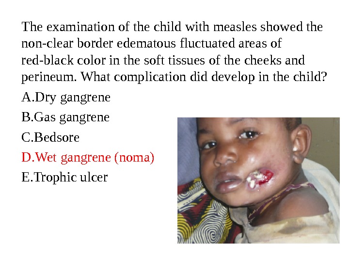
The examination of the child with measles showed the non-clear border edematous fluctuated areas of red-black color in the soft tissues of the cheeks and perineum. What complication did develop in the child? A. Dry gangrene B. Gas gangrene C. Bedsore D. Wet gangrene (noma) E. Trophic ulcer
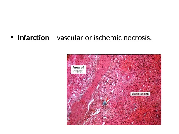
• Infarction – vascular or ischemic necrosis.
A patient died from acute cardiac insufficiency. The histological examination of his heart revealed in myocardium of the left ventricle the necrotized section, which was separated from undamaged tissue by the zone of hyperimic vessels, small hemorrhages and leukocytic infiltration. What is the most likely diagnosis? A. Focal exudate myocarditis B. Diffuse exudate myocarditis C. Productive myocarditis D. Myocardial ischemic dystrophy E. Myocardial infarction
Infarction is… • Hyperemia • Stasis • Ichemical necrosis • Secquestrum • Degeneration
A 65 -year-old patient, who suffered from arteriosclerosis, has been hospitalized in surgical department because he had purulent peritonitis. Thrombosis of mesenteric arteries was found during operation. What is the most probable cause of peritonitis? A. Angiospastic ischemia B. Hemorrhagic infarction C. Angioneurotic edema D. Stasis E. Chronic congestion
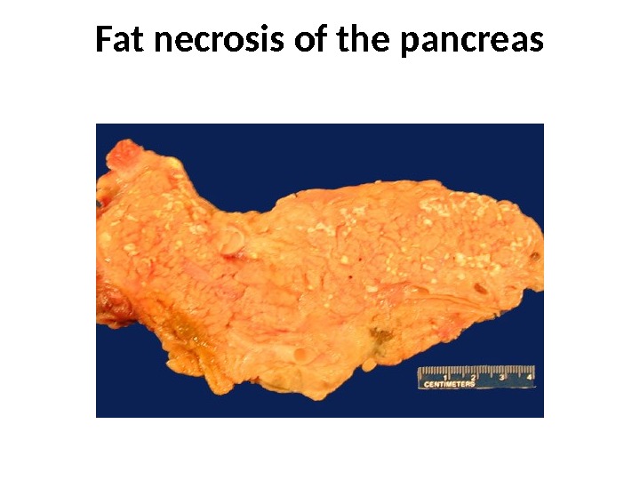
Fat necrosis is encountered in adipose tissue contiguous to the pancreas and more rarely at distant sites, as a result of leakage of lipase after acute injury to pancreatic acinar tissue, most commonly from obstruction of pancreatic ducts. Grossly, fat necrosis appears as firm, yellow-white deposits in peripancreatic and mesenteric adipose tissue. Histologically, necrotic fat cells are distinguishable as pale outlines, and their cytoplasm is filled with an amorphous-appearing, faintly basophilic material (soap).
Fat necrosis of the pancreas
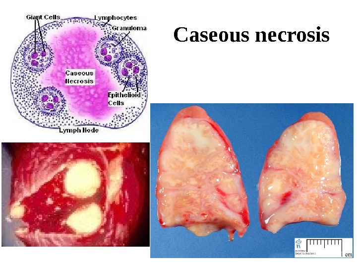
Caseous necrosis Has features of both coagulative and liquefactive necrosis. Typically, it occurs in the center of tuberculous granulomas, which contain a white or yellow “cheesy” material (Latin caseum = cheese) that accounts for the name of this lesion. Histologically, the outlines of necrotic cells are not preserved, but the tissue has not been liquefied either. The remnants of the cells appear as finely granular, amorphous material.
Caseous necrosis
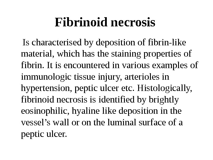
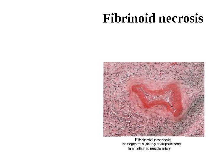
Fibrinoid necrosis Is characterised by deposition of fibrin-like material, which has the staining properties of fibrin. It is encountered in various examples of immunologic tissue injury, arterioles in hypertension, peptic ulcer etc. Histologically, fibrinoid necrosis is identified by brightly eosinophilic, hyaline like deposition in the vessel’s wall or on the luminal surface of a peptic ulcer.
Fibrinoid necrosis
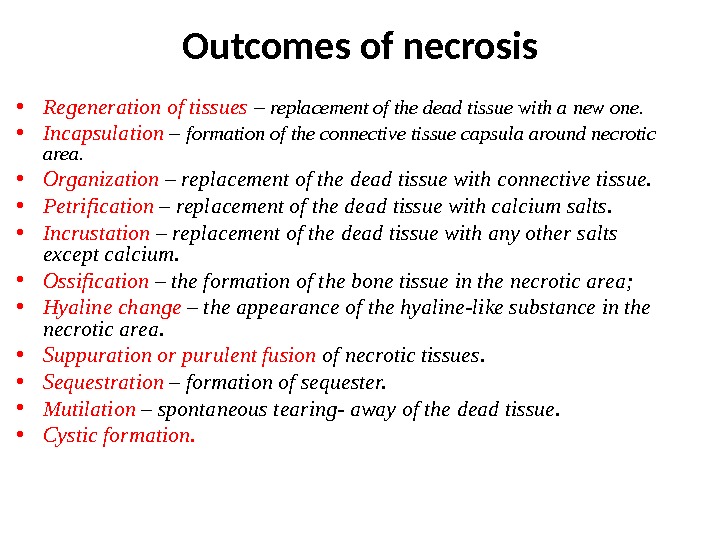
Outcomes of necrosis • Regeneration of tissues – replacement of the dead tissue with a new one. • Incapsulation – formation of the connective tissue capsula around necrotic area. • Organization – replacement of the dead tissue with connective tissue. • Petrification – replacement of the dead tissue with calcium salts. • Incrustation – replacement of the dead tissue with any other salts except calcium. • Ossification – the formation of the bone tissue in the necrotic area; • Hyaline change – the appearance of the hyaline-like substance in the necrotic area. • Suppuration or purulent fusion of necrotic tissues. • Sequestration – formation of sequester. • Mutilation – spontaneous tearing- away of the dead tissue. • Cystic formation.
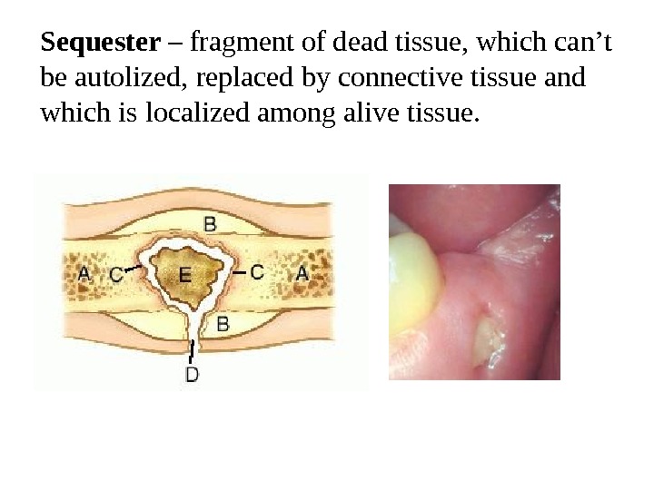
Sequester – fragment of dead tissue, which can’t be autolized, replaced by connective tissue and which is localized among alive tissue.
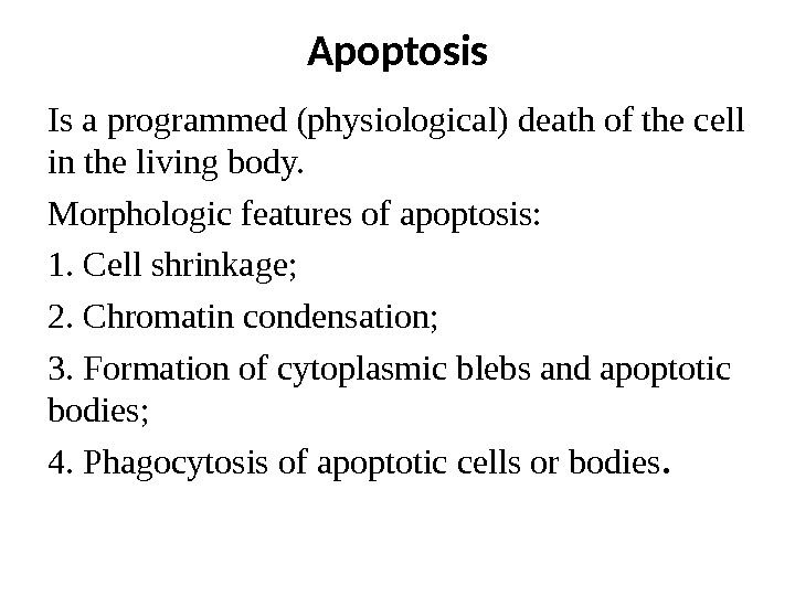
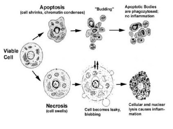
Apoptosis I s a programmed (physiological) death of the cell in the living body. Morphologic features of apoptosis: 1. Cell shrinkage; 2. Chromatin condensation; 3. Formation of cytoplasmic blebs and apoptotic bodies; 4. Phagocytosis of apoptotic cells or bodies.
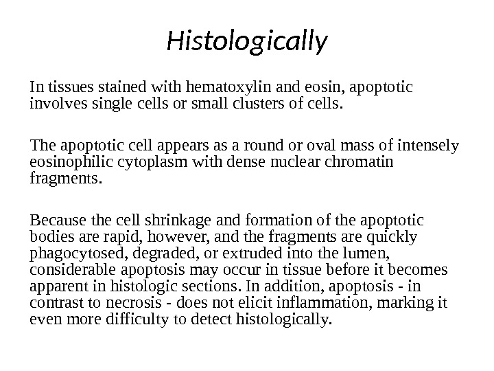
Histologically I n tissues stained with hematoxylin and eosin, apoptotic involves single cells or small clusters of cells. The apoptotic cell appears as a round or oval mass of intensely eosinophilic cytoplasm with dense nuclear chromatin fragments. Because the cell shrinkage and formation of the apoptotic bodies are rapid, however, and the fragments are quickly phagocytosed, degraded, or extruded into the lumen, considerable apoptosis may occur in tissue before it becomes apparent in histologic sections. In addition, apoptosis — in contrast to necrosis — does not elicit inflammation, marking it even more difficulty to detect histologically.
Mechanisms of the development of intra- and extracellular (stromal) degenerations (dystrophies) are the followings: • Infiltration – redundant accumulation (deposition) of metabolites into the cells and extracellular matrix. • Decomposition (phanerosis) – disintegration of membranous structures of the cells and extracellular matrix. • Perverted synthesi s — synthesis of abnormal substances in the cells and tissues. • Transformation – formation of one type of metabolism’s products from common initial substances for proteins, fats and carbohydrates.
Intracellular fatty degenerations For identification of different kind of fats we usually use special reactions (staining): • Sudan III — stains fat red, • Sudan IV — black, • Nile blau sulfat stains fatty acids blue and neutral fats red.
Disturbance of fat metabolism may manifest as: • appearance in the place where it does not appear under normal conditions (e. g. in the myocardium); • appearance of fat of unusual composition; • increase of fat amount in the places where it is present under normal conditions (e. g. in the fat depots). The main cause of fatty change is hypoxia which may be due to: a) disturbances in transportation systems (e. g. in patient with chronic cardiovascular and chronic pulmonary insufficiency); b) chronic intoxications (e. g. alcoholism); c) cachexia, avitaminosis; d) infections (e. g. Diphtheria, tuberculosis).
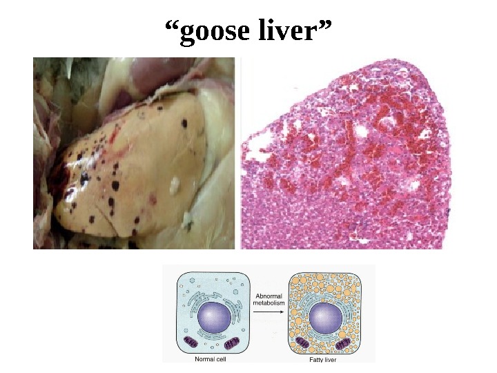
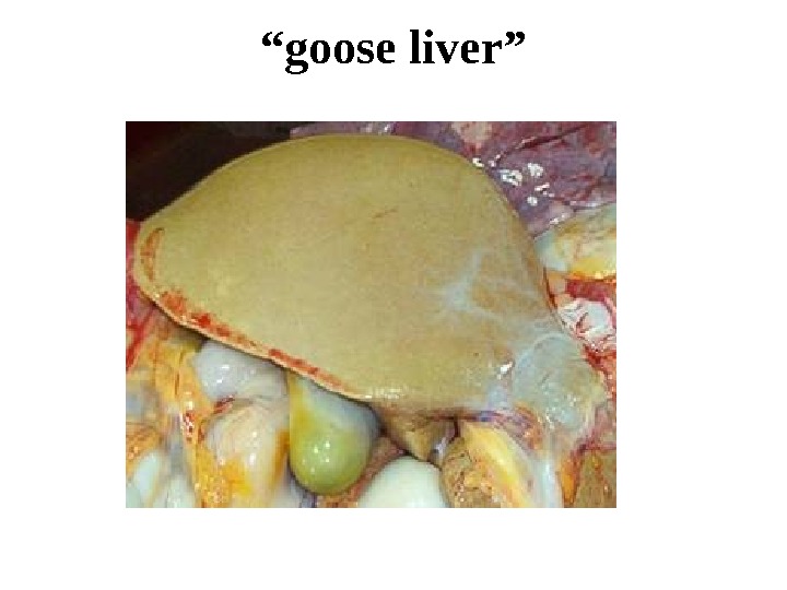
Fatty degeneration of the liver Macroscopically the fatty liver is enlarged with rounded margins. The cut surface bulges slightly and is pale-yellow and is greasy to touch. It is called “goose liver”. Microscopically: there are numerous lipid vacuoles in the cytoplasm of hepatocytes. The vacuoles are initially small (microvesicular), but with progression of the process, the vacuoles become larger pushing the nucleus to the periphery of the cells (macrovesicular). At times, the hepatocytes laden with large lipid vacuoles may rupture and lipid vacuoles coalesce to form fatty cysts. Infrequently, lipogranulomas may appear.
“ goose liver”
“ goose liver”
Autopsy of the patient who had suffered from tuberculosis demonstrated that the liver weighed 1600 g, was flabby, light brown ( goose ). Microscopy showed sudanophilic formations in the hepatocyte cytoplasm on the periphery of the lobules. Which parenchymatous dystrophy was present? A. Protein B. Hyalin drop C. Fatty D. Horny E. Mucus •
Fatty degeneration of the heart It is also called “Tiger’s” heart. Macroscopically the heart is enlarged, the chambers are stretched, flabby. Microscopically we can see dust-like fatty vacuoles in the cardiomyocytes. It is observed in the papillary muscles and trabecules of the ventricles in the form of bands (surrounding the veins).
External examination of a newborn revealed dry dull pale skin Autopsy of the patient who had been ill with leukemia and died of increasing chronic anemia revealed an enlarged heart, dull, flabby, pale gray myocardium. There were yellow plaques and bands under the endocardium. Which pathologic process is observed in the heart? A. Parenchymal fatty degeneration B. Vacuole degeneration C. Hyalin-drop degeneration D. Mesenchymal fatty degeneration E. Functional hypertrophy
Autopsy of the patient who had suffered from hypertension disease revealed considerable enlarged flabby heart with widened cavities. The myocardium was dull, clay-like, with white strips from the side of the myocardium which were more pronounced in the papillary muscles and trabeculas of the heart ventricles ( tiger’s heart ). Which parenchymatous dystrophy was present? A. Protein B. Fat C. Horny D. Mucous E. Carbohydrate
• The kidneys look like “large white kidney”. They are enlarged, flabby. The cortical substance is gray with yellow drops. • Outcomes of fatty degenerations are seldom reversible. Necrosis or sclerosis may develop.
• Stromal fatty infiltration is the deposition of mature adipose cells in the stromal connective tissue. The condition occurs most often in patients with obesity. • As a rule it is a generalized process when the amount of fat in the depots increases. Stromal vascular fatty degenerations
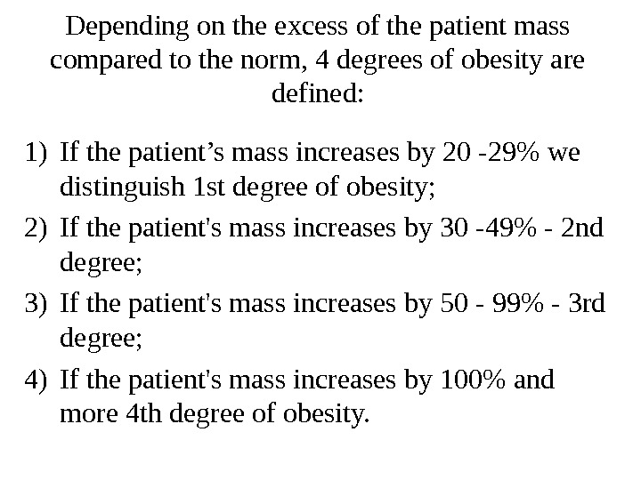
Depending on the excess of the patient mass compared to the norm, 4 degrees of obesity are defined: 1) If the patient’s mass increases by 20 -29% we distinguish 1 st degree of obesity; 2) If the patient’s mass increases by 30 -49% — 2 nd degree; 3) If the patient’s mass increases by 50 — 99% — 3 rd degree; 4) If the patient’s mass increases by 100% and more 4 th degree of obesity.
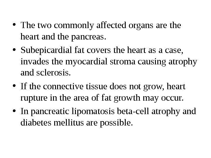
• The two commonly affected organs are the heart and the pancreas. • Subepicardial fat covers the heart as a case, invades the myocardial stroma causing atrophy and sclerosis. • If the connective tissue does not grow, heart rupture in the area of fat growth may occur. • In pancreatic lipomatosis beta-cell atrophy and diabetes mellitus are possible.
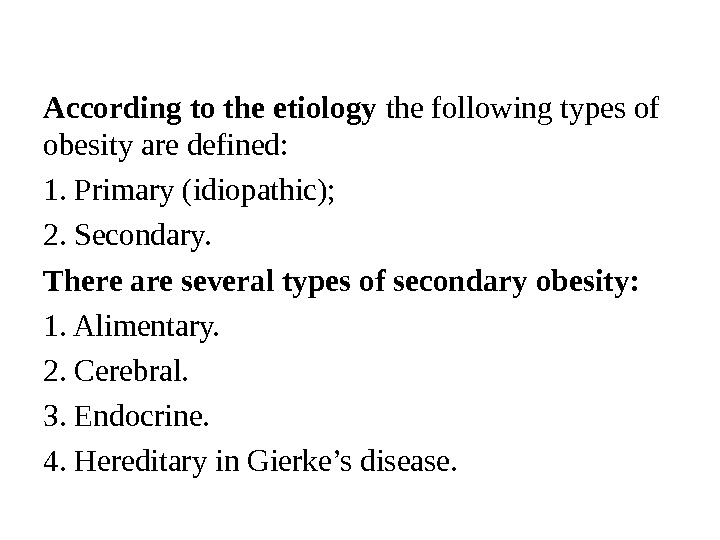
According to the etiology the following types of obesity are defined: 1. Primary (idiopathic); 2. Secondary. There are several types of secondary obesity: 1. Alimentary. 2. Cerebral. 3. Endocrine. 4. Hereditary in Gierke’s disease.
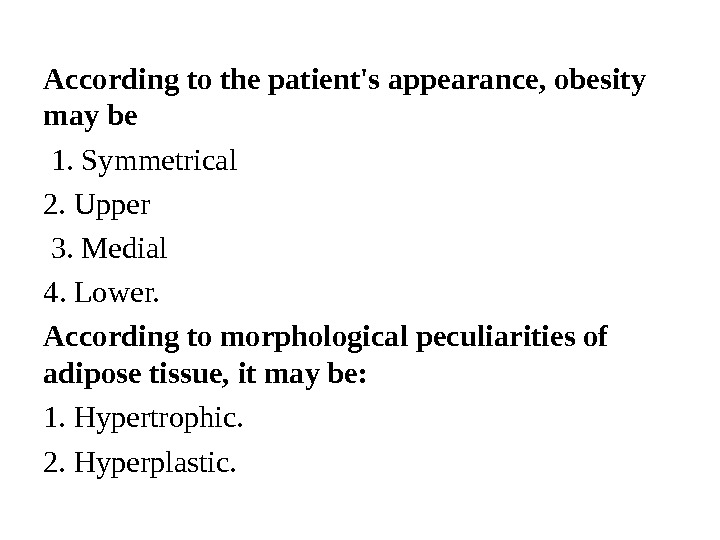
According to the patient’s appearance, obesity may be 1. Symmetrical 2. Upper 3. Medial 4. Lower. According to morphological peculiarities of adipose tissue, it may be: 1. Hypertrophic. 2. Hyperplastic.
• In hypertrophic type adipose tissue enlarges due to increased volume of fatty cells, in hyperplastic due to increase in their number. Obesity is a severe complication of mainly endocrine and nervous diseases. Alimentary obesity is also unfavorable for the organism. As a rule such patients develop ischemic heart disease. • Local enlargement of adipose tissue (lipomatosis) occurs in Dercum’s disease when painful fat nodes appear in the subcutaneous fat of the lower and upper extremities and trunk. • Sharp reduction in the amount of neutral fat in the whole organism is called cachexia.
• A 45 -year old man died of sudden cardiac arrest. Symmetrical stage III obesity, rupture of the wall of the right ventricle with hemopericardium, excessive fat under the epicardium were revealed. Microscopy showed that fatty tissue of the epicardium involved the myocardium causing muscular fiber atrophy. Which pathological process is most probable? A. Fatty degeneration of the myocardium B. Obesity of the heart C. Coronary artery disease D. Acute myocardial infarction E. Hypertension disease
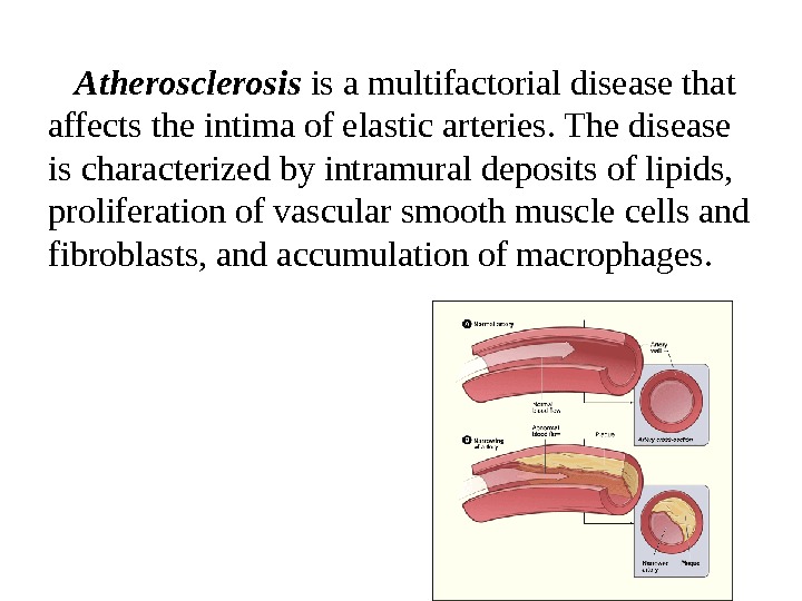
Atherosclerosis is a multifactorial disease that affects the intima of elastic arteries. The disease is characterized by intramural deposits of lipids, proliferation of vascular smooth muscle cells and fibroblasts, and accumulation of macrophages.
A ccumulations of proteins
A granular degeneration (dystrophy). • Macroscopical kind of organs at this dystrophy it is determined as “muddy or dim swelling”. At a section the organs are dim, swollen. Microscopical descriptions of cells on electronics level: presence of electrondense granules in cytoplasm of the cells.
Hyaline-drop degeneration is characterized by the aggregation of small proteins granules into cytoplasm of cells. It is not determined macroscopically. This dystrophy occurs in kidneys, liver and myocardium. The cytoplasm of plasma cells shows pink hyaline inclusions called Russell’s bodies representing synthesised immunoglobulins , the cytoplasm of hepatocytes shows eosinophilic globular deposits of a mutant protein. Mallory’s body or alcoholic hyaline in the hepatocytes is intracellular accumulation of intermediate filaments of cytokeratin. The outcome is negative. The focal or total coagulative necrosis develops.
Hydropic (cloudy, vacuolar, balloon) I s characterized by accumulation of water within the cell due to cytoplasmic vacuolation. The common causes include bacterial toxins, chemicals, poisons, burns, and high fever. The affected organ such as kidney, liver or heart muscle is enlarged. The cut surface bulges outwards and is slightly opaque. Microscopically: the cells are swollen and the microvasculature compressed.
These vacuoles represent distended cisternas of the endoplasmic reticulum. Ultrastructural changes in hydropic swelling include the following: • Dilation of endoplasmic reticulum. • Mitochondrial swelling. • Blebs on the plasma membrane. • Loss of fibrillanty of nucleolus. The outcome is negative, because the focal or total colliquative cellular necrosis develops.
• Microscopy of the kidneys from the dead patient who had suffered from chronic glomerulonephritis showed enlarged epithelial cells of the renal tubules, their cytoplasm was filled with vacuoles with transparent fluid, the nucleus was displaced to the periphery. Which parenchymatous dystrophy was present? A. Protein B. Horny C. Hyalin drop D. Hydropic E. Fatty
Keratoid (horney) degeneration I s characterized by increase production of keratin substance. This process may be local and general. The intracellular keratin may be located in epidermis of skin, keratinic squamous epithelial cells, cervix, and esophagus. Leucoplakia means hyperkeratosis in mucosa. Leucoplakia may lead to malignization. For example: Squamous cell carcinoma with keratinization. The groups of keratinized cells can be found in the center of squamous cell carcinoma’s areas. These cell’s complexes here and there look like rose color homogenous found forms (“canceromatous perls”).
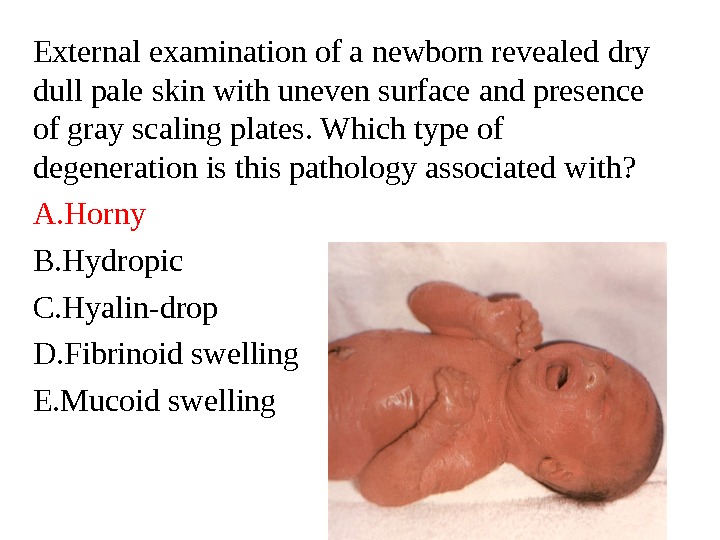
External examination of a newborn revealed dry dull pale skin with uneven surface and presence of gray scaling plates. Which type of degeneration is this pathology associated with? A. Horny B. Hydropic C. Hyalin-drop D. Fibrinoid swelling E. Mucoid swelling
• Mescnchymal (stromal vascular) degenerations develop in the connective tissue as a result of metabolic disturbances in it. 1. Mucoid swelling 2. Fibrinoid swelling 3. Hyaline changes
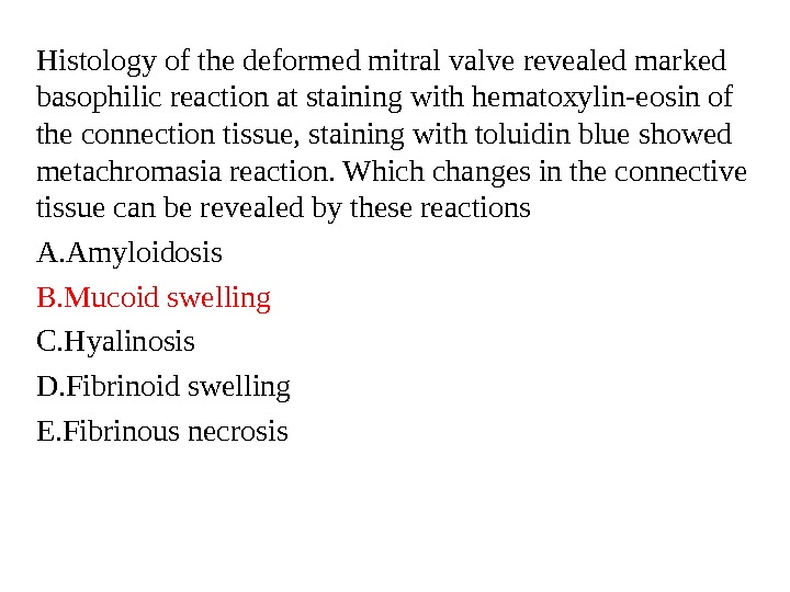
1. Mucoid swelling • Mucoid swelling is superficial reversible disorganization of the connective tissue. • These processes are associated with swelling of collagen fibers, increased vascular permeability (due to glucosaminoglycans (GAG) action) and plasmorrhagia. • Microscopic examination shows metachromasia. Under normal conditions the main substance is basophilic. In this case staining with toluidine blue demonstrates reddish coloring. • Macroscopic appearance is absent. • The outcome may be reversible. In other cases, the development of fibrinoid swelling is possible.
Histology of the deformed mitral valve revealed marked basophilic reaction at staining with hematoxylin-eosin of the connection tissue, staining with toluidin blue showed metachromasia reaction. Which changes in the connective tissue can be revealed by these reactions A. Amyloidosis B. Mucoid swelling C. Hyalinosis D. Fibrinoid swelling E. Fibrinous necrosis
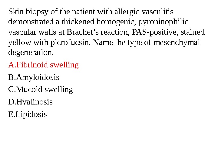
2. Fibrinoid changes • Fibrinoid swelling is deep irreversible connective tissue disorganization. • Fibrinoid is formed as a result of the main substance destruction and more increase in vascular permeability. • The appearance of the organs is changed a little. • The main signs are revealed microscopically: the bands of collagen fibers are homogenous, impregnated with plasma proteins. • Metachromasia is not marked due to GAG depolymerization of the main substance. • Fibrinoid swelling may be generalized (in systemic diseases of the connective tissue) and localized (in chronic inflammations). • The outcomes are fibrinoid necrosis, sclerosis or hyalinosis.
Skin biopsy of the patient with allergic vasculitis demonstrated a thickened homogenic, pyroninophilic vascular walls at Brachet’s reaction, PAS-positive, stained yellow with picrofucsin. Name the type of mesenchymal degeneration. A. Fibrinoid swelling B. Amyloidosis C. Mucoid swelling D. Hyalinosis E. Lipidosis
3. Hyaline changes (hyalinosis) • Hyaline changes (hyalinosis) — (greek “hyalos” — transparent, glass-like) usually refers to an alteration within cells or in the extracellular matrix, which gives a homogenous, glassy, pink appearance in routine histologic sections stained with hematoxilin and eosin. • Hyalinosis develops as a result of plasma infiltration, fibrinoid swelling, inflammation, necrosis, and sclerosis. • The outcome of hyalinosis is irreversible.
Autopsy of the man revealed the signs of rheumatic heart defect, i. e. thickened deformed cartilage-like valves with luster surface. Which process is present in the valves: A. Amyloidosis B. Fibrinoid necrosis C. Fibrinoid swelling D. Hyalinosis E. Degenerative calcification
• Amyloidosis is the term used for a group of diseases characterised by extracellular deposition of fibrillar proteinaceous substance called amyloid. “ Sago spleen” “Lardaceous spleen”
• A patient ages 42 had suffered from bronchiectasis and died of renal failure. Autopsy revealed enlarged dense pale yellow kidneys. Microscopy revealed accumulations of amorphic eosinophilic and congophilic substance in the walls of the vessels, basal membrane of the tubules, capillary loops, mesangium of the glomeruli and connective tissue of the stroma. Which of the processes is most probable? A. Secondary amyloidosis B. Idiopathic amyloidosis C. Hereditary amyloidosis D. Hyalinosis E. Senile amyloidosis
Autopsy of a man aged 56 who had suffered from fibrous-cavernous pulmonary tuberculosis revealed enlarged dense spleen. On cut section the tissue of the spleen is brown-pink, smooth with wax-like surface. Microscopy revealed diffuse congophilic staining. Which process is observed in the spleen? A. Glaze spleen B. Porphyrin spleen C. Sago spleen D. Sebaceous spleen E. Cyanotic induration of the spleen
The most frequent glycogen storage diseases or glycogenosis are • Pompe’s disease, • Mc Ardle’s disease • Gierke disease.