

2409e0383f94d387334bfec9c55dffe0.ppt
- Количество слайдов: 45
 Cancer Gene Therapy One T Cell Kills Multiple Tumor Cells David L. Liu, MD, Ph. D Instructor in Medicine, Assistant in Biology Harvard Cancer Gene Therapy Center BIDMC and Harvard Medical School
 Background Landmark of Adoptive Immunotherapy 1977, Rosenberg SA, et al. Passive Immunotherapy of cancer in animals and man 1985 Rosenberg-Lotz, LAK cells 1987 Spiess-Rosenberg, TILs 1987 Yamada, Human IL 2 gene in murine T cells 1987 Kuwana, first reported Ig. TCR chimeric receptor 1995 Treisman-Hwu, IL 2 transduced lymphocytes 2001 Maher-Sadelain, chimeric TCR/CD 28 receptor 2002 R. Junghans, Ig. CD 28 TCR (Tandem) T cells 2003 Liu, Ig. TCR-GM-CSF transduced T cells
 Classic Concept-1 Tumors are killed by LAK cells or TILs or Ig. TCR or Tandem-transduced T cells that is resulted from proliferation of injected T cells in tumor tissue. Dr. Rosenberg wrote: LAK cells or TILs traffic to and accumulate in tumor deposits. This finding led to attempts to genetically alter TILs to increase their anti-tumor activity at the tumor site.
 Classic Concept-2 In the past, Dr. Richard Junghans also wrote in application of research proposal: … He has freshed his concept because several new findings have been observed in BDL Dr. James Mier also said Proliferation of injected T cells in tumor tissue is the mechanism killing tumor, everyone says so. In 2003, an Israel research group still reported “proliferation of intratumorally injected Ig. TCR-TD T cells in tumor (Cancer Res), this is absolutely untrue.
 Classic Concept-3 In the textbook of Cellular and Molecular Immunology, Dr. Abul Abbas writes: 1. CTL killing is antigen specific. 2. CTL killing requires cell contact. 3. CTLs themselves are not injured during lysis of target cells. 4. Suppose each individual CTL is capable of sequentially killing multiple target cells, but so far no direct proof has been demonstrated.
 Classic Concept-4 v Dr. James Allison presented and demonstrated the modality how antigen presenting cells deliver a signal to T cells or to tumor cells. He was asked to answer how much long is needed for this process? v He didn’t have definite data, but we do have!
 Classic Concept-5 v Generally, it is easy to follow the hypothesis, assumption and theory that suggested by a great professor or big head. v Universally, it is hard to accept a new finding that conflicts with the traditional concept.
 Hypothesis-1 v Over the past 7 years, we examined >2000 tumor tissue slides in mice immunized with hybrid tumor vaccines, <5% of samples showed infiltration of T cells in tumor tissue. v During the past 14 months, we examined >100 tumor slides in nude mice treated with Ig. TCR or Tandem. TD T cells, no proliferation of Ig. TCR or Tandem-TD T cells was seen in tumor tissue.
 Hypothesis-2 v. A tumor at size of 6 x 6 mm in diameters (approximately 500 x 106 tumor cells) that could be cured with 50 x 106 transduced T cells in our in vivo therapy, how does one transduced T cell kill 5 to 10 tumor cells?
 Hypothesis-3 If one T cell can really kill 5 to 10 tumor cells, what modality of killing process is it? Crazed T cells? The killing process needs direct contact of TD-T cells and tumor cells, what contact modality is it? Leech Mouth-Like T Cells? What morphology changes in T cells and tumor cells will happen when kiss-death of TD-T cells and tumor cells is initiated? Both cell types?
 Materials and Methods Human PBLs from blood donor v Transduced with Ig. TCR or Ig. TCR-CD 28 viral sup. v High percentage of transduced T cells (35 -76%) v 50 x 106 CFSE labeled and WI 2 stained T cells I. v. v Ex vivo killing tests: 5, 10, 15, 30, 60, 90, 120 min. v In vivo proliferation in regressing tumor tissue v Computed digital light microscope v Invital fluoresence microscopy v Confocal microscopy v Flow cytometry v
 Stains-Double Labeling Of Cells Labeling transduced T cells: CFSE, purified WI 2 + GAM Rhodamine Labeling ECA-expressing tumor cells Purified MN 14 ab + GAM Rhodamine or PE Anti-human CD 3, CD 8 T cell Immunochemical staining
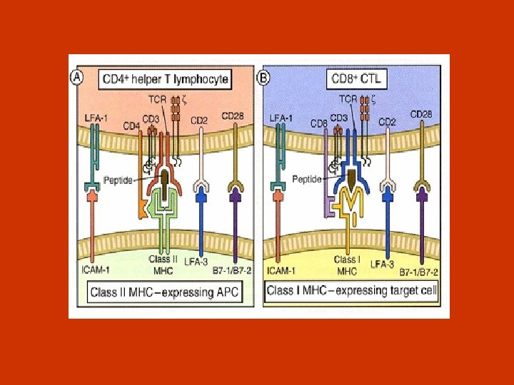
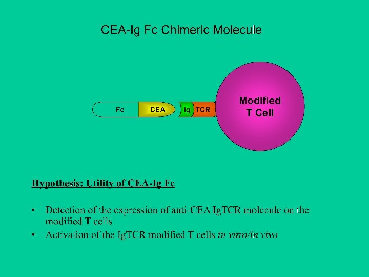
 Ig. TCR – chimeric immunoglobulin – T cell receptor Tumor Cell CEA Ig TCR Modified T Cell Advantage: Conferring the specificity of antibody to TCR and bypassing the MHC-TCR pathway for T cell cytotoxicity It is a prerequisite for a successful adoptive immunotherapy to have a large amount of T cells (1011).
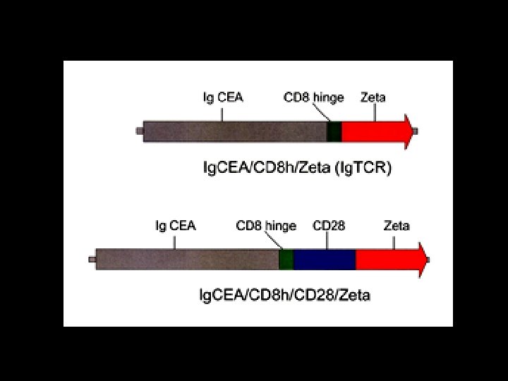
 Cloning of anti-GD 3 Ig. TCR, 2 nd generation Blp 1 Nco I LTR sc. FV (anti-GD 3) gag 1 st generation Anti-GD 3 Ig. TCR CD 8 h CD 3 z LTR Nco I gag LTR sc. FV (anti-CEA) 2 nd generation Anti-CEA Ig. CD 28 Zeta Blp 1 CD 8 h CD 28 CD 3 z LTR
 Structure of the CEA-Ig Chimera Molecule
 Constructs of Ig. TCR, Ig 28 TCR
 Production of Retrovirus Particles Transgene Transfection gag pol Genome gag pol env Packaging cell line Producer cell line Viral particle Viral RNA Target cells DNA Integration Genome Protein
 Subpopulation of T cells during transfection CD 4/CD 8 ratio: on day 7 OKT 3 2. 08 OKT 3+CD 28 2. 08 PHA 1. 51
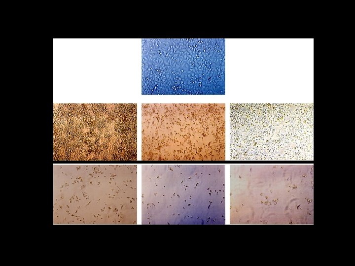
 Positively Transduced Ig. TCR or Ig. TCR-CD 28 T Cells
 76% positively transduced Ig. TCR T Cells
 EXPERIMENTAL GROUPS FIRST COURSE OF KILLING TEST T cells: Tumor cells = 3: 1 Jurkat-ATCC v Jurkat-Ig. TCR v Jurkat-Tandem v Activated T cells v Ig. TCR-TD T cells v Tandem-TD T cells v (30 x 106) + MIP-CEA (10 x 106) (30 x 106) + MIP-CEA (10 x 106)
![FIRST COURSE OF KILLING TEST [2, 3, 4 and 5 days-AICD] T Cells :](https://present5.com/presentation/2409e0383f94d387334bfec9c55dffe0/image-27.jpg "FIRST COURSE OF KILLING TEST [2, 3, 4 and 5 days-AICD] T Cells :") FIRST COURSE OF KILLING TEST [2, 3, 4 and 5 days-AICD] T Cells : Tumor Cells at 3: 1 J-ATCC J-Ig. TCR J-Tandem No killing No killing T Cells : Tumor Cells at 3: 1 Act. T Ig. TCR Tandem No Killing 40% 50% No killing No Killing 60% 75% No killing No Killing >90% 100% No killing AICD
FIRST COURSE OF KILLING TEST [2, 3, 4 and 5 days-AICD] T Cells : Tumor Cells at 3: 1 J-ATCC J-Ig. TCR J-Tandem No killing No killing T Cells : Tumor Cells at 3: 1 Act. T Ig. TCR Tandem No Killing 40% 50% No killing No Killing 60% 75% No killing No Killing >90% 100% No killing AICD
 RE-ACTIVATION OR RE-KILLING: GROUPING T cells: Tumor cells = 1: 1 Activated T cells (30 x 106) + MIP-CEA (10 x 106) Ig. TCR-TD T cells (30 x 106) + MIP-CEA (10 x 106) Tandem-TD T cells (30 x 106) + MIP-CEA (10 x 106) T cells: Tumor cells = 3: 1 Activated T cells (30 x 106) + MIP-CEA (10 x 106) Ig. TCR-TD T cells (30 x 106) + MIP-CEA (10 x 106) Tandem-TD T cells (30 x 106) + MIP-CEA (10 x 106)
, 8) T Cells : Tumor Cells at 1:") RE-KILLING TEST (Day 2, 3 (AICD), 8) T Cells : Tumor Cells at 1: 1 Act. T Ig. TCR No killing 5% No killing Tandem T Cells : Tumor Cells at 3: 1 Act. T Ig. TCR Tandem 5% No Killing 40% 50% 25% No Killing 60% 80% No killing AICD No Killing AICD No killing 40% 10% re-growing 60% 25% 10% re-growing
RE-KILLING TEST (Day 2, 3 (AICD), 8) T Cells : Tumor Cells at 1: 1 Act. T Ig. TCR No killing 5% No killing Tandem T Cells : Tumor Cells at 3: 1 Act. T Ig. TCR Tandem 5% No Killing 40% 50% 25% No Killing 60% 80% No killing AICD No Killing AICD No killing 40% 10% re-growing 60% 25% 10% re-growing
 CEA- Control CEA+ Regressed
 Human CEA+ Cancer Regressed after Gene Therapy
 Human CEA+ Cancer Regressed after Gene Therapy
 One Gene Transfected T Cell Kills Multiple Tumor Cells
 Apoptosis in Tumor Cell
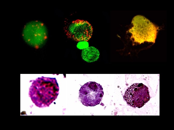
 Leech Mouth-Like T Cells
 Leech Mouth-Like T Cells
 Leech Mouth-Like T Cells
") Injected TD-T cells in Spleen (2 hs)
Injected TD-T cells in Spleen (2 hs)
") Homing or Proliferation of TD-T Cells in Spleen (Enlarged from the last slid)
Homing or Proliferation of TD-T Cells in Spleen (Enlarged from the last slid)
 Necessary Time For Change In T Cell Morphology 5 min no change in morphology 15 min no change in morphology 30 min 5 -10% T cells had change in morphology 45 min 15% T cells had change in morphology 60 min 20% T cells had change in morphology 2 hs 30% T cells had change in morphology
 Blue Color Shows Perforin Produced by Gene Transfected T Cells
 Blue Color Shows Perforins Produced by Gene Transfected T cells
 Centralized Killing Modality of T Cells
 Conclusion 1. Ig. TCR or Ig. TCR-CD 28 gene transfected T cells induced massive necrosis of CEA-expressing human colon cancer from day 1 to day 3 after treatment. 2. A new finding, leech mouth-like T cells are plunged onto surface of CEA-expressing tumor cells, is for the first time described. 3. Direct evidences of one T cell kills >5 tumor cells in vivo have been confirmed by 3 types of specific staining, HE staining, anti-h. CD 3 immunochemical staining and CFSE labeling-TD T cells. 4. The modality of centralized T cells killing tumor cells in vivo has been consistently observed. 5. Injected TD-T cells in vivo have 3 fates: reactivation and killing tumor cells inside tumor; proliferation in spleen and re-circulation into tumor; and become apoptotic or