
BYSTANDER EFFECT The bystander effect refers


- Размер: 16.1 Mегабайта
- Количество слайдов: 114
Описание презентации BYSTANDER EFFECT The bystander effect refers по слайдам
BYSTANDER EFFECT
The bystander effect refers to the induction of biological effects in cells that are not directly traversed by a charged particle. The data available concerning the bystander effect fall into two quite separate categories, and it is not certain that the two groups of experiments are addressing the same phenomenon. First, there are experiments involving the transfer of medium from irradiated cells, which results in a biological effect in nirradiated cells. Second, there is the use of sophisticated single particle microbeams, which allow specific cells to be irradiated and biological effects studied in their neighbors; in this case communication is by gap junction. Medium transfer experiments have shown a bystander effect for cell lethality, chromosomal aberrations and cell cycle delay. The type of cell, epithelial vs. fibroblast, appears to be important.
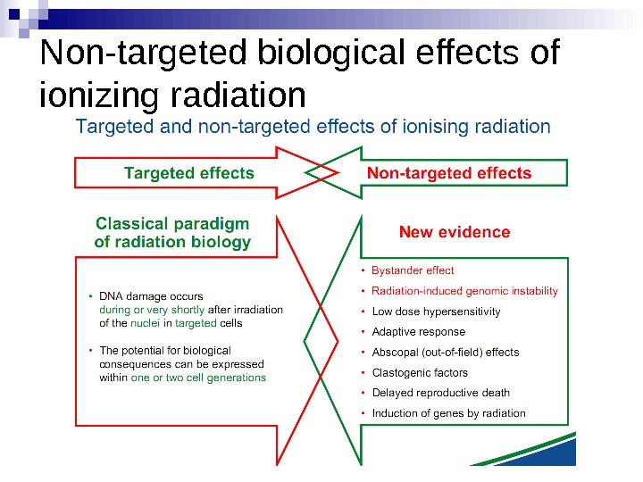
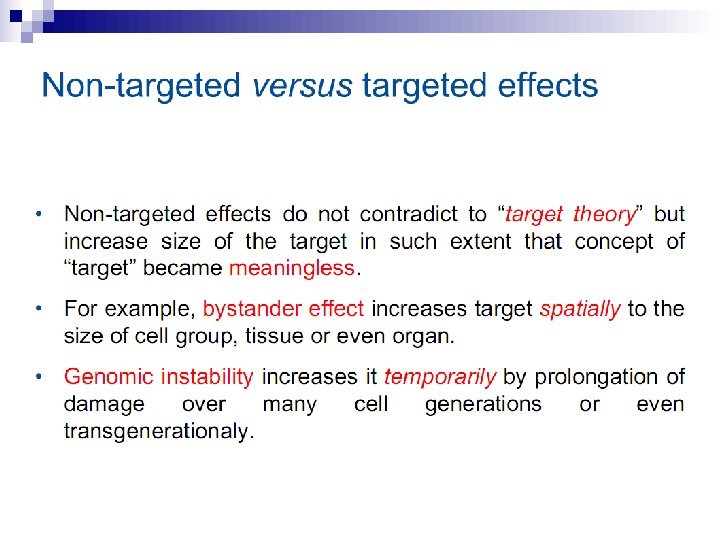
Non-targeted biological effects of ionizing radiation
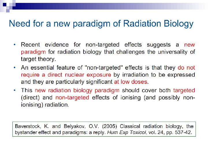
Non-targeted effects of ionizing radiation as a new paradigm of radiation biology Ward, J. (1999) New paradigms for Low-Dose Radiation Response In Proceedings of the American Statistical Association Conference on Radiation and Health. San Diego, California, USA. June 14 -17, 1998. Radiat Res, 151: 1, 92 -117.
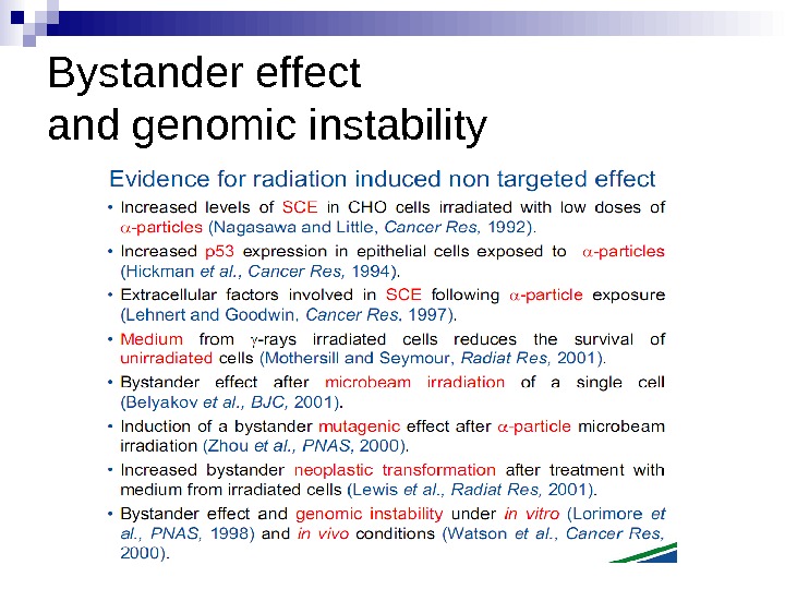
Bystander effect and genomic instability
Experiments suggest that the effect is due to a molecule secreted by irradiated cells, which is capable of transferring damage to distant cells. Use of a single microbeam has allowed the demonstration of a bystander effect for chromosomal aberrations, cell lethality, mutation, and oncogenic transformation. When cells are in close contact, allowing gap junction communication, the bystander effect is a much larger magnitude than the phenomenon demonstrated in medium transfer experiments. A bystander effect has been demonstrated for both high- and low-LET radiations but it is usually larger for densely ionizing radiation such as alpha particles. Experiments have not yet been devised to demonstrate a comparable bystander effect on a three-dimensional normal tissue. Bystander studies imply that the target for the biological effects of radiation is larger than the cell and this could make a simple linear extrapolation of radiation risks from high to low doses of questionable validity.
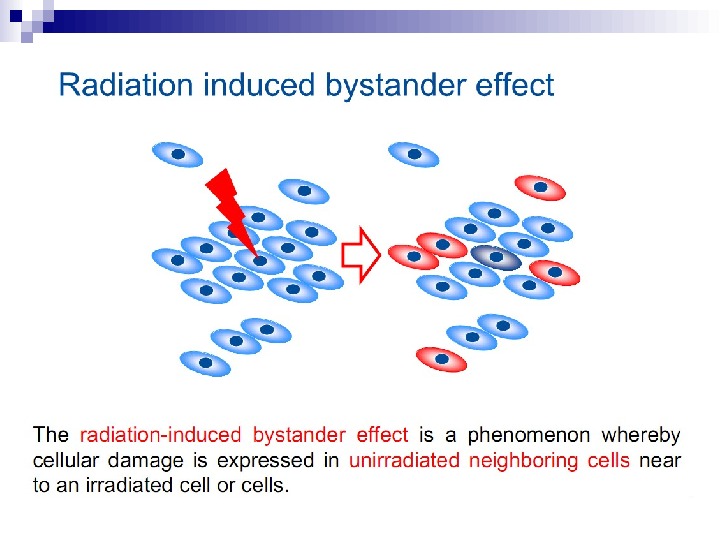
The radiation-induced bystander effect is defined as “the induction of biological effects in cells that are not directly traversed by a charged particle but are in close proximity to cells that are. ” Although these bystander effects have been demonstrated with a variety of biological endpoints in both human and rodent cell lines (as well as in 3 D tissue samples), the mechanism of the phenomenon is not known. Although gap junction communication and the presence of soluble mediator(s) are both known to play important roles in the bystander response, the precise signaling molecules have yet to be identified.
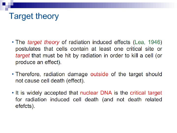
GENERATIONS OF students in radiation biology have been taught that heritable biological effects require direct damage to DNA. In fact, evidence has been available for many years that this simple statement is not strictly true. As early as the 1940’s there were reports that the inactivation of biological entities may be brought about equally by ionizations produced within the entity or by the ionization of the surrounding medium (Dale 1940, 1942, 1943; Lea et al. 1944). By 1947, Kotval and Gray had shown that alpha particles that pass close to the chromatid thread, as well as those which pass through it, have a significant probability of producing chromatid and isochromatid breaks or chromatid exchanges. The term used today to describe such phenomena is “The Bystander Effect, ” a name borrowed from the gene therapy field where it usually refers to the killing of several types of tumor cells by targeting only one type of cell within a mixed population (Cheng et al. 1999, for example).
In the radiation field, it has come to be loosely defined as the induction of biological effects in cells that are not directly traversed by a charged particle, but are in close proximity to cells that are. Interest in this effect was sparked by the report of Nagasawa and Little (1992) that, following a low dose of alpha particles, a larger proportion of cells showed biological damage than were estimated to have been hit by an alpha particle; specifically 30% of the cells showed an increase in sister chromatid exchanges even though less than 1% were calculated to have undergone a nuclear traversal. The number of cells hit was arrived at by a calculation based on the fluence of alpha particles and the cross-sectional area of the cell nucleus. The conclusion was thus of a statistical nature since it was not possible to know on an individual basis which cells were hit and which were not.
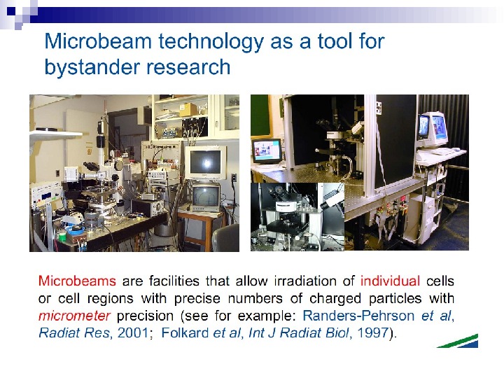
The plethora of data now available concerning the bystander effect fall into two quite separate categories, and it is not certain that the two groups of experiments are addressing the same phenomenon. First, there are experiments involving the transfer of medium from irradiated cells, which results in a biological effect in unirradiated cells. Second, there is the use of sophisticated single particle microbeams, which allow specific cells to be irradiated and biological effects studied in their neighbors (Randers-Pehrson et al. 2001).
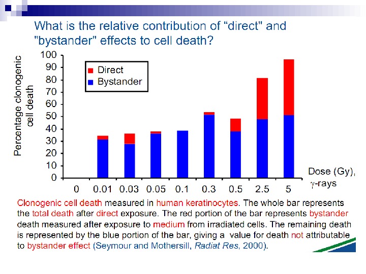
Medium transfer experiments Experiments involving the transfer of medium from irradiated to unirradiated cells have demonstrated a highly significant reduction in the plating efficiency of both normal and malignant epithelial cells— whether or not the cells were irradiated (Mothersill and Seymour 1997).
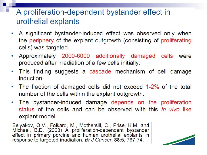
This bystander effect suggested that irradiated cells secreted a molecule into the culture medium that was capable of killing cells when that medium was transferred onto unirradiated cells. By contrast, medium irradiated in the absence of cells had no effect. Further experiments demonstrate that not all cells were capable of producing the toxic factor, nor were all cells capable of receiving the secreted signal (Mothersill and Seymour 1997, 2001). In later experiments using explants of human uroepithelium, Mothersill et al. (2001) show that there is considerable variation in the release of the bystander factor into the surrounding cell culture medium. The effect roduced by epithelial cell cultures is dependent on the cell number at the time of irradiation, can be observed as soon as 30 min post irradiation, and is still effective if taken from the irradiated cells up to 60 h after irradiation. This bystander effect can be induced by radiation doses as low as 0. 25 m. Gy and is not significantly increased up to doses of 10 Gy. Forty-eight hours after receiving irradiated medium there were many apoptotic bodies present, suggesting that apoptosis may be a prominent mechanism of cell death responsible for the reduced clonogenic survival. In addition to increased levels of cell death and reduced cloning efficiency, medium transfer experiments have shown an increase in neoplastic transformation as well as genomic instability in cells that have not themselves been irradiated.
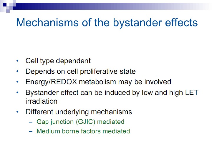
Some limited progress has been made in the search for the mechanisms involved in this bystander effect. Following exposure to radiation, the first detectable effect of transferred medium on recipient cells was a rapid calcium pulse (1– 2) followed 30– 120 min later by changes in mitochondrial membrane permeability and the induction of reactive oxygen species (Lyng et al. 2002). Gap junction communication between cells was not required to induce killing of bystander cells, but medium from cell cultures irradiated at high densities induced the greatest amount of cell death. Furthermore, the use of apoptosis inhibitors or medium from lactate dehydrogenase or glucose-6 -phosphate dehydrogenase mutant cells reduced or prevented the bystander effect. Treatment with the anti-oxidants L-lactate and l-deprenyl prevented bystander factor associated cell kill suggesting that energy/REDOX metabolism may be involved in the medium mediated bystander response.
The majority of bystander experiments involving medium transfer have utilized low-LET x or gamma rays, in contrast to microbeam experiments where alpha particles or protons have been the particles of choice. THE BYSTANDER EFFECT DEMONSTRATED BY MICROBEAM EXPERIMENTS Experiments described here involve the scoring of micronuclei, cell lethality, mutation, and oncogenic transformation.
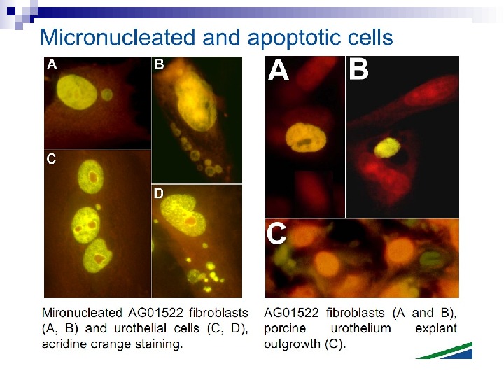
Micronuclei in normal human fibroblasts Perhaps the most direct and most dramatic demonstration of the bystander effect involves the observation of micronuclei in irradiated human fibroblasts. Cells of one population were lightly stained with cyto-orange, a cytoplasmic vital dye, while cells of another population were lightly stained blue with a nuclear vital dye. The two cell populations were mixed and allowed to attach to the culture dish, and the computer controlling the accelerator was programmed to irradiate only blue-stained cells with 10 alpha particles directed at the centroid of the nucleus. The cells were fixed and stained 48 h later, at which time micronuclei and chromosome bridges were visible in a proportion of the nonhit (i. e. , orange-stained) cells (Fig. 1). This is an astonishing emonstration of the bystander effect because the development of micronuclei implies significant chromosome damage and rearrangement, which is clearly visible in nonhit cells that have been fixed in situ.
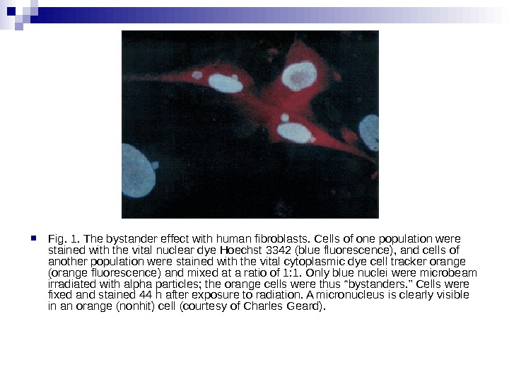
Fig. 1. The bystander effect with human fibroblasts. Cells of one population were stained with the vital nuclear dye Hoechst 3342 (blue fluorescence), and cells of another population were stained with the vital cytoplasmic dye cell tracker orange (orange fluorescence) and mixed at a ratio of 1: 1. Only blue nuclei were microbeam irradiated with alpha particles; the orange cells were thus “bystanders. ” Cells were fixed and stained 44 h after exposure to radiation. A micronucleus is clearly visible in an orange (nonhit) cell (courtesy of Charles Geard).
Cell lethality Lines of hygromycin- and neomycin-resistant V 79 cells were produced. Before exposure the hygromycinresistant cells were stained with a low concentration of a vital nuclear dye. They were then plated in micro wells in the proportion nine neomycin-resistant for every one hygromycin-resistant cell. The computer was programmed to irradiate only the 10% of cells stained with a nuclear dye with various numbers of alpha particles from 1– 16 aimed at the centroid of the nucleus. The cells were then removed and cultured for survival in the appropriate growth media, which made it possible to obtain survival curves for hit and nonhit cells.
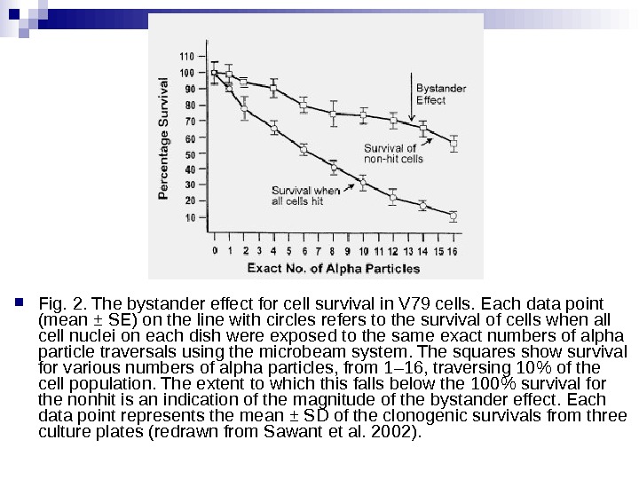
Fig. 2. The bystander effect for cell survival in V 79 cells. Each data point (mean ± SE) on the line with circles refers to the survival of cells when all cell nuclei on each dish were exposed to the same exact numbers of alpha particle traversals using the microbeam system. The squares show survival for various numbers of alpha particles, from 1– 16, traversing 10% of the cell population. The extent to which this falls below the 100% survival for the nonhit is an indication of the magnitude of the bystander effect. Each data point represents the mean ± SD of the clonogenic survivals from three culture plates (redrawn from Sawant et al. 2002).
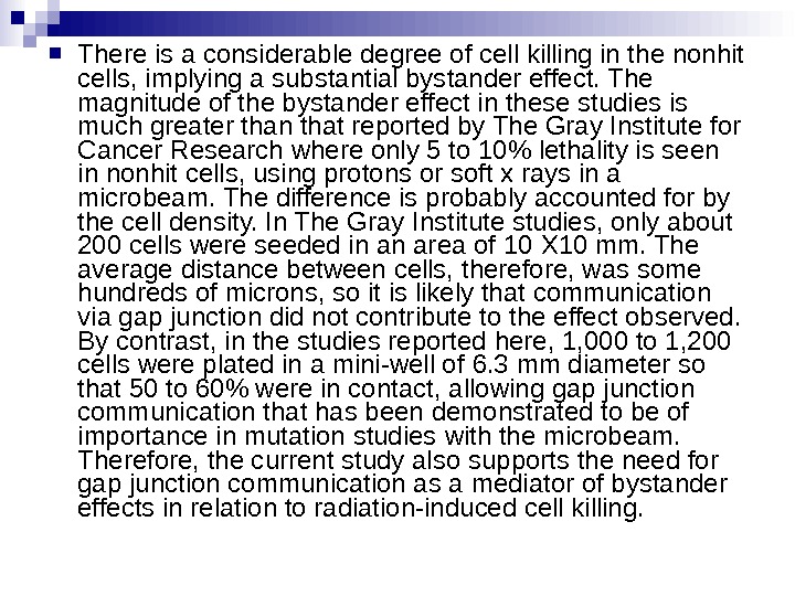
There is a considerable degree of cell killing in the nonhit cells, implying a substantial bystander effect. The magnitude of the bystander effect in these studies is much greater than that reported by The Gray Institute for Cancer Research where only 5 to 10% lethality is seen in nonhit cells, using protons or soft x rays in a microbeam. The difference is probably accounted for by the cell density. In The Gray Institute studies, only about 200 cells were seeded in an area of 10 Х 10 mm. The average distance between cells, therefore, was some hundreds of microns, so it is likely that communication via gap junction did not contribute to the effect observed. By contrast, in the studies reported here, 1, 000 to 1, 200 cells were plated in a mini-well of 6. 3 mm diameter so that 50 to 60% were in contact, allowing gap junction communication that has been demonstrated to be of importance in mutation studies with the microbeam. Therefore, the current study also supports the need for gap junction communication as a mediator of bystander effects in relation to radiation-induced cell killing.
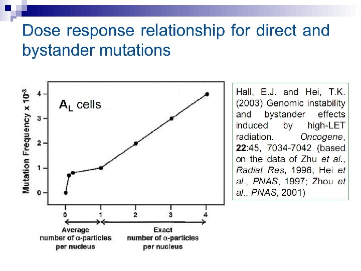
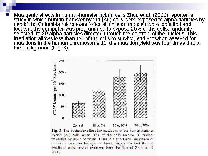
Mutagenic effects in human-hamster hybrid cells Zhou et al. (2000) reported a study in which human-hamster hybrid (AL) cells were exposed to alpha particles by use of the Columbia microbeam. After all cells on the dish were identified and located, the computer was programmed to expose 20% of the cells, randomly selected, to 20 alpha particles directed through the centroid of the nucleus. This irradiation allows less than 1% of the cells to survive, and yet when assayed for mutations in the human chromosome 11, the mutation yield was four times that of the background (Fig. 3).
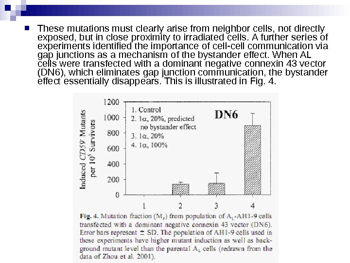
These mutations must clearly arise from neighbor cells, not directly exposed, but in close proximity to irradiated cells. A further series of experiments identified the importance of cell-cell communication via gap junctions as a mechanism of the bystander effect. When AL cells were transfected with a dominant negative connexin 43 vector (DN 6), which eliminates gap junction communication, the bystander effect essentially disappears. This is illustrated in Fig. 4.
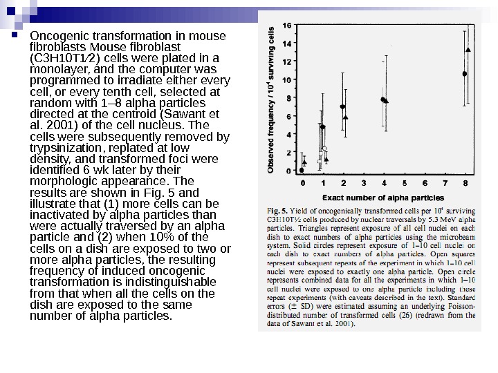
Oncogenic transformation in mouse fibroblasts Mouse fibroblast (C 3 H 10 T 1⁄2) cells were plated in a monolayer, and the computer was programmed to irradiate either every cell, or every tenth cell, selected at random with 1– 8 alpha particles directed at the centroid (Sawant et al. 2001) of the cell nucleus. The cells were subsequently removed by trypsinization, replated at low density, and transformed foci were identified 6 wk later by their morphologic appearance. The results are shown in Fig. 5 and illustrate that (1) more cells can be inactivated by alpha particles than were actually traversed by an alpha particle and (2) when 10% of the cells on a dish are exposed to two or more alpha particles, the resulting frequency of induced oncogenic transformation is indistinguishable from that when all the cells on the dish are exposed to the same number of alpha particles.
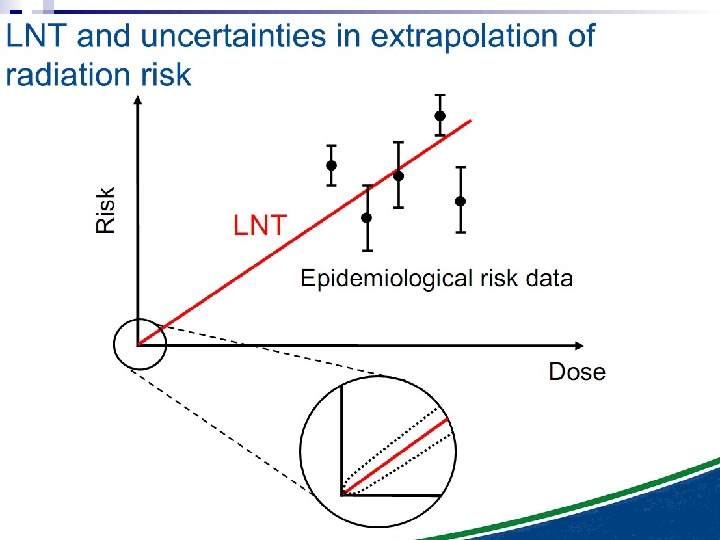
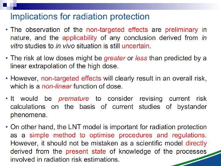
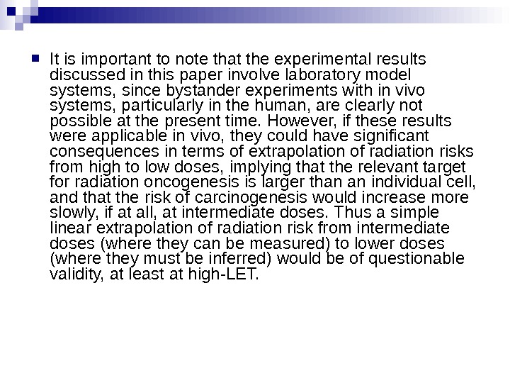
It is important to note that the experimental results discussed in this paper involve laboratory model systems, since bystander experiments with in vivo systems, particularly in the human, are clearly not possible at the present time. However, if these results were applicable in vivo, they could have significant consequences in terms of extrapolation of radiation risks from high to low doses, implying that the relevant target for radiation oncogenesis is larger than an individual cell, and that the risk of carcinogenesis would increase more slowly, if at all, at intermediate doses. Thus a simple linear extrapolation of radiation risk from intermediate doses (where they can be measured) to lower doses (where they must be inferred) would be of questionable validity, at least at high-LET.
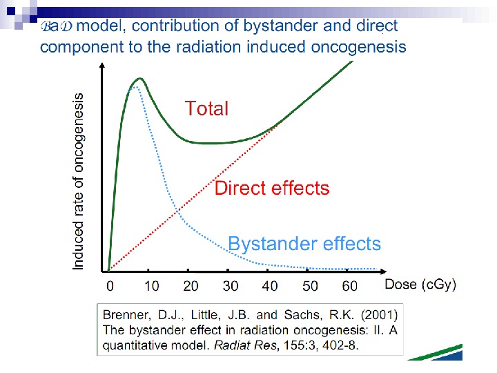
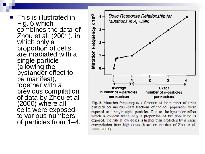
This is illustrated in Fig. 6 which combines the data of Zhou et al. (2001), in which only a proportion of cells are irradiated with a single particle (allowing the bystander effect to be manifest), together with a previous compilation of data by Zhou et al. (2000) where all cells were exposed to various numbers of particles from 1– 4.
Under these experimental conditions, it is evident that a linear extrapolation of risks from high doses to low doses (which average less than one particle per cell) would underestimate the risks at low doses. This applies, at this stage, strictly to alpha particles, and it is not known whether it would apply in an in vivo situation to, for example, radon exposure in homes and mines.
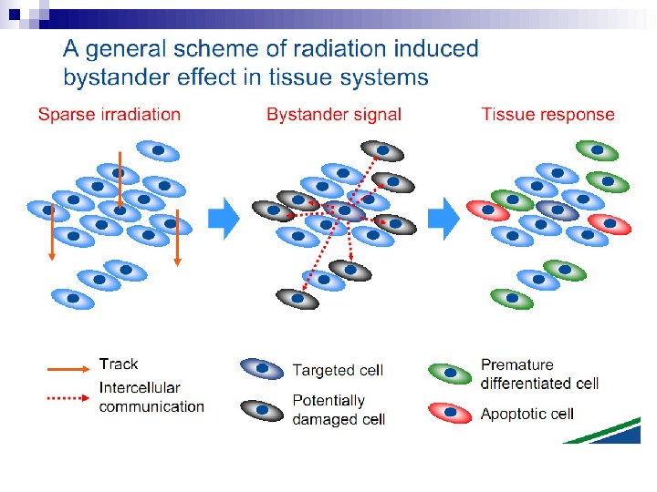
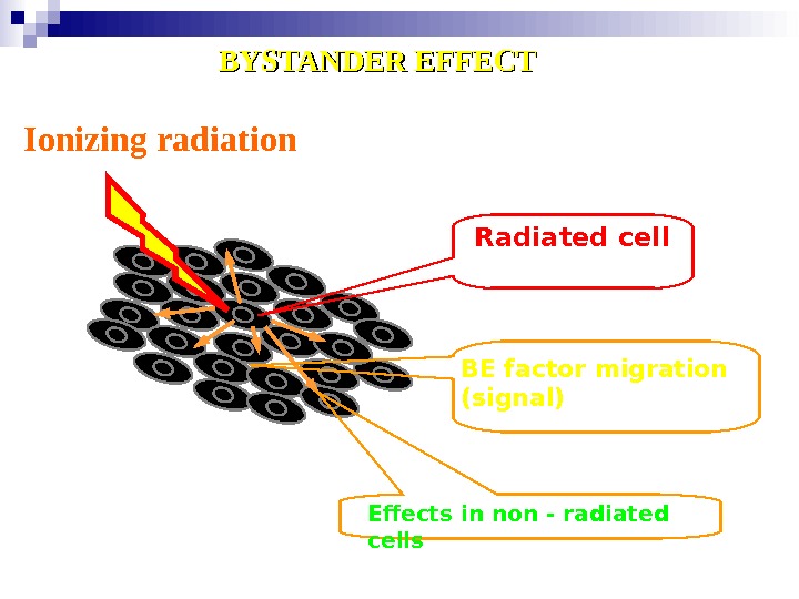
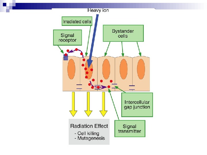
Ionizing radiation Radiated cell BE factor migration ( signal ) Effects in non — radiated cells. BYSTANDER EFFECT
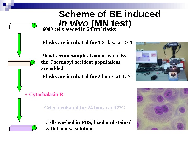
Scheme of BE induced in vivo (MN test) 6000 cells seeded in 24 cm 2 flasks Blood serum samples from affected by the Chernobyl accident populations are added Flasks are incubated for 1 -2 days at 37 ° C Flasks are incubated for 2 hours at 37 ° C + Cytochalasin B Cells incubated for 24 hours at 37 ° C Cells washed in PBS, fixed and stained with Giemsa solution
Killing Non-transduced Tumor Cells via Bystander Effect The bystander effect was first reported by Moolten (1986) showing that prodrug convertase negative cells surrounded by suicide enzyme positive cells did not survive prodrug treatment. Besides efficient killing of targeted tumor cells, neighboring, non-transduced cells are killed as well, providing an important effect in cancer treatment. Since 5 -Fluorouracil is soluble and can diffuse into adjacent cells (Huber et al. 1993) (Huber et al. 1994), the bystander effect was demonstrated using cytosine deaminase as gene of interest.
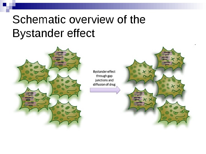
Schematic overview of the Bystander effect
Our understanding of how radiation kills normal and tumour cells has been based on an intimate knowledge of the direct induction of DNA damage and its cellular consequences. What has become clear is that, as well as responses to direct DNA damage, cell-cell signalling — known as the bystander effect — mediated through gap junctions and inflammatory responses may have an important role in the response of cells and tissues to radiation exposure and also chemotherapy agents. This Review outlines the key aspects of radiation-induced intercellular signalling and assesses its relevance for existing and future radiation-based therapies.
When ionizing radiation interacts with biological material, energy is deposited and chemical bonds are broken. In cells, the basic components of proteins, lipids and nucleic acids can all be damaged. However, a key consequence is that direct damage occurs to DNA within the nucleus, producing a range of lesions of which DNA double strand breaks (DSBs) have a pivotal role in determining whether cells survive radiation exposure. [1] If DNA damage is not correctly repaired two direct consequences can occur. Residual or unrepaired damage leads directly to chromosomal aberrations, loss of genetic material and cell death. Also, unrepaired or incorrectly repaired (misrepaired) damage can lead to mutations that might result in carcinogenesis or cell death (Figure 1).
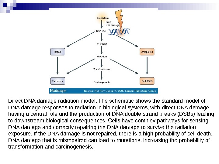
Direct DNA damage radiation model. The schematic shows the standard model of DNA damage responses to radiation in biological systems, with direct DNA damage having a central role and the production of DNA double strand breaks (DSBs) leading to downstream biological consequences. Cells have complex pathways for sensing DNA damage and correctly repairing the DNA damage to survive the radiation exposure. If the DNA damage is not repaired, there is a high probability of cell death. DNA damage that is misrepaired can lead to mutations, increasing the probability of transformation and carcinogenesis.
The mechanisms underpinning DNA damage and repair processing in irradiated cells have been extensively studied since the discovery of DNA as the genetic material by Watson and Crick over 50 years ago. This has included exhaustive study of the DNA damage sensing and signalling pathways underpinning the DNA damage response that is present in cellular systems. [2] What is clear from these efforts is that cells have multiple and complex processes for sensing and repairing changes to their genomes to enable future propagation and stability. A series of sensor, transducer and effector proteins give cells important choices in response to radiation-induced DNA damage, such as DNA repair, cell cycle delay and cell death (apoptosis). [3]
Evidence now shows that, as well as these direct DNA damage-dependent effects, irradiated cells also send signals to their neighbours. These non-irradiated cells respond to signals produced by neighbouring irradiated cells by what has been termed a bystander effect (extensively reviewed in Refs[4 -7]). The term bystander effect is not new and has been observed in response to a range of other insults including ultraviolet radiation, [8] photodynamic therapy, [9] heat[10] and chemotherapy agents. [11] Its observation in response to chemotherapy agents underpins its considerable importance in gene therapy regimens, in which not all tumour cells are targeted and indirect killing of non-targeted cells is required to ensure maximal tumour cell kill. [12]
For example, the archetypal gene therapy model is the herpes simplex virus-thymidine kinase (HSV-TK) system. In this system the HSV-TK gene is transfected into cells and these are incubated with the non-toxic agent ganciclovir, which is converted to a toxic analogue that diffuses and kills neighbouring cells. [13] This bystander effect involves direct cell-cell communication through gap junctional intercellular communication (GJIC) and requires expression and surface location of CX 43 (also known as GJC 1) gap junctions. [14] By contrast, the bystander effect mediated by the thymidine phosphorylase-5′-deoxy-5 -fluorouridine suicide gene system involves a factor released into the medium that is independent of GJIC. [15] So, the paradigm of a bystander response after radiation treatment is not new in other fields; in essence it is a manifestation of intercellular signalling that is either specific or non-specific in its mode of action.
Radiation-induced bystander responses have been observed in a range of cell types, tissue models and in vivo. Although the majority of the evidence for bystander effects has come from cellular studies, a range of other responses have been classified as bystander effects in the literature. In humans, in response to radiotherapy, longer-range effects occurring within or between tissues have also been reported and have been termed abscopal, out-of-field or distant bystander responses. [16] Radiation-induced bystander responses have been observed, not just from studies with external beam irradiation, but also from approaches using targeted radioisotopes. Several key questions emerge from these observations in terms of their relevance to cancer therapy. First, does an understanding of bystander mechanisms highlight new potential targets for cancer therapies and, if so, can this be modulated to either increase tumour cell killing or protect normal tissues? Second, do these bystander responses, especially after low-dose irradiation, contribute to increased carcinogenic risks associated with radiation exposure?
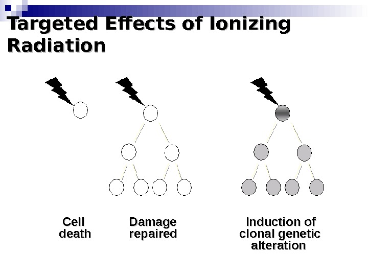
Targeted Effects of Ionizing Radiation Cell Damage Induction of death repaired clonal genetic alteration
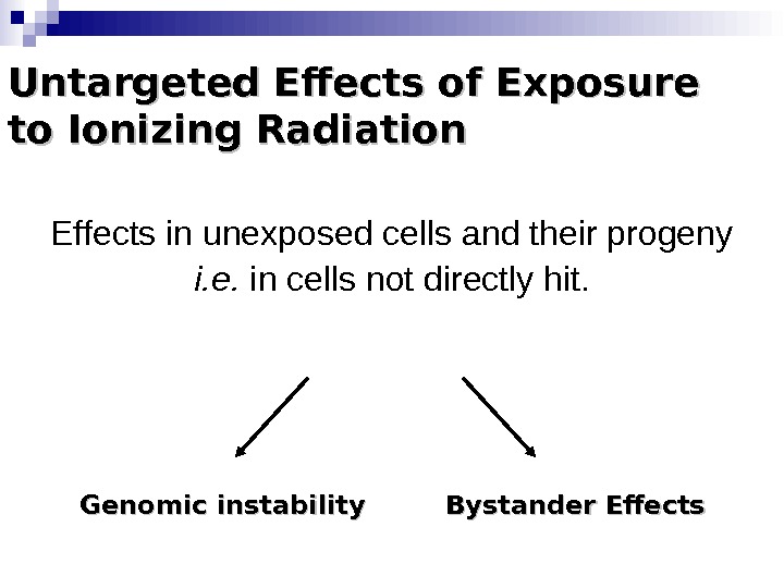
Untargeted Effects of Exposure to Ionizing Radiation Effects in unexposed cells and their progeny i. e. in cells not directly hit. Genomic instability Bystander Effects
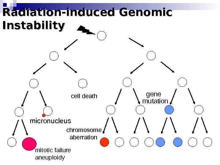
Radiation-induced Genomic Instability micronucleus chromosome aberration cell death gene mutation mitotic failure aneuploidy
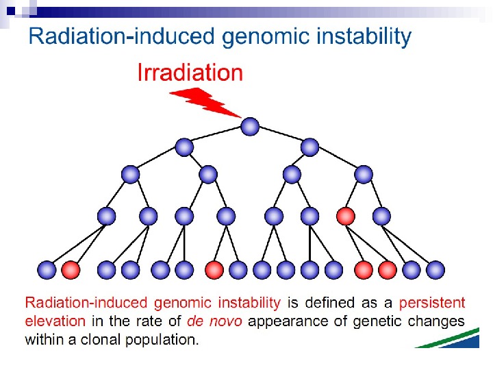
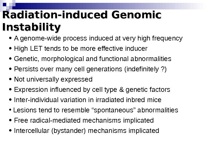
Radiation-induced Genomic Instability • A genome-wide process induced at very high frequency • High LET tends to be more effective inducer • Genetic, morphological and functional abnormalities • Persists over many cell generations (indefinitely ? ) • Not universally expressed • Expression influenced by cell type & genetic factors • Inter-individual variation in irradiated inbred mice • Lesions tend to resemble “spontaneous” abnormalities • Free radical-mediated mechanisms implicated • Intercellular (bystander) mechanisms implicated
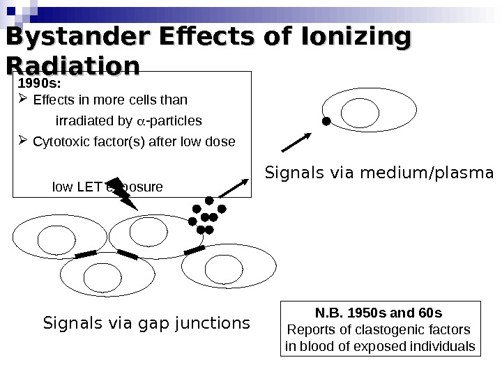
Bystander Effects of Ionizing Radiation Signals via gap junctions Signals via medium/plasma N. B. 1950 s and 60 s Reports of clastogenic factors in blood of exposed individuals 1990 s: Effects in more cells than irradiated by -particles Cytotoxic factor(s) after low dose low LET exposure
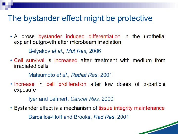
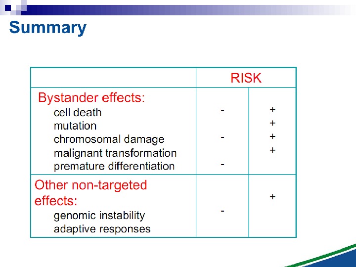
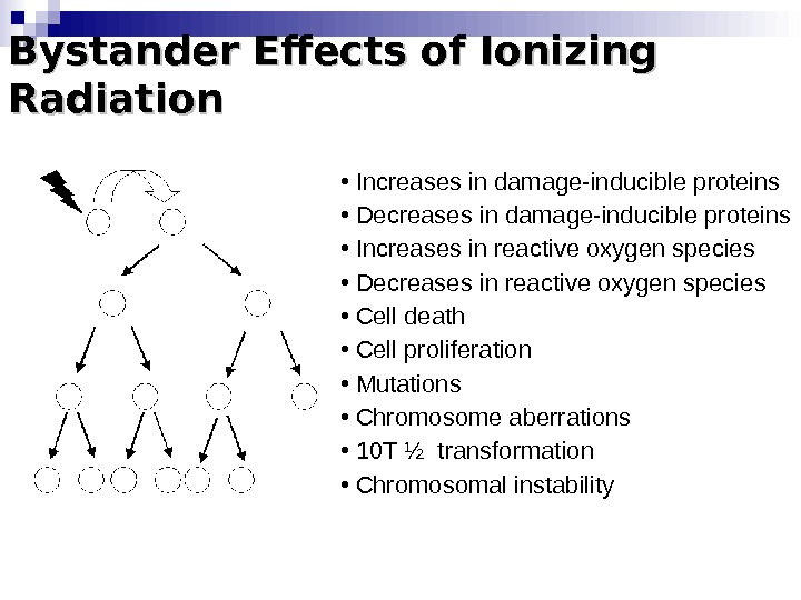
Bystander Effects of Ionizing Radiation • Increases in damage-inducible proteins • Decreases in damage-inducible proteins • Increases in reactive oxygen species • Decreases in reactive oxygen species • Cell death • Cell proliferation • Mutations • Chromosome aberrations • 10 T ½ transformation • Chromosomal instability
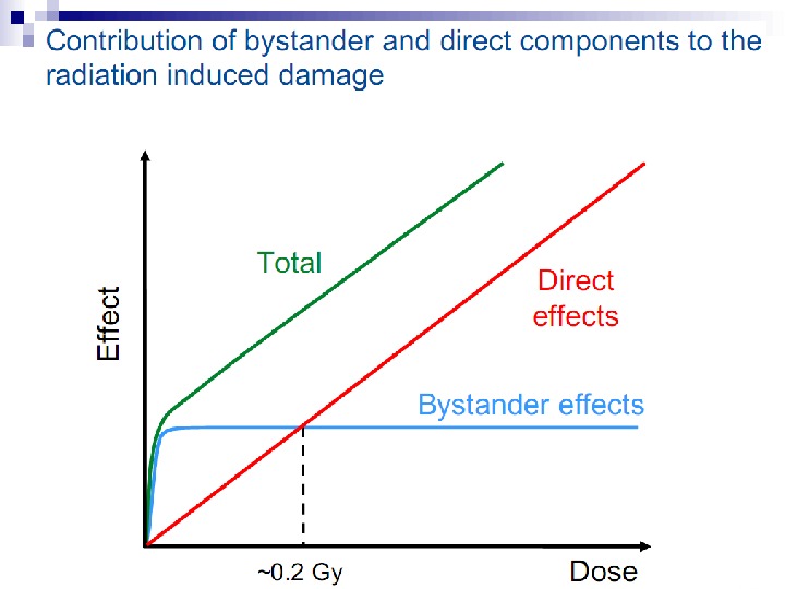
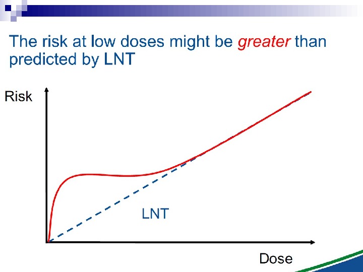
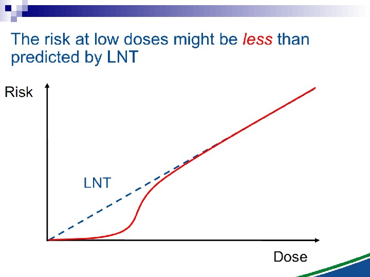
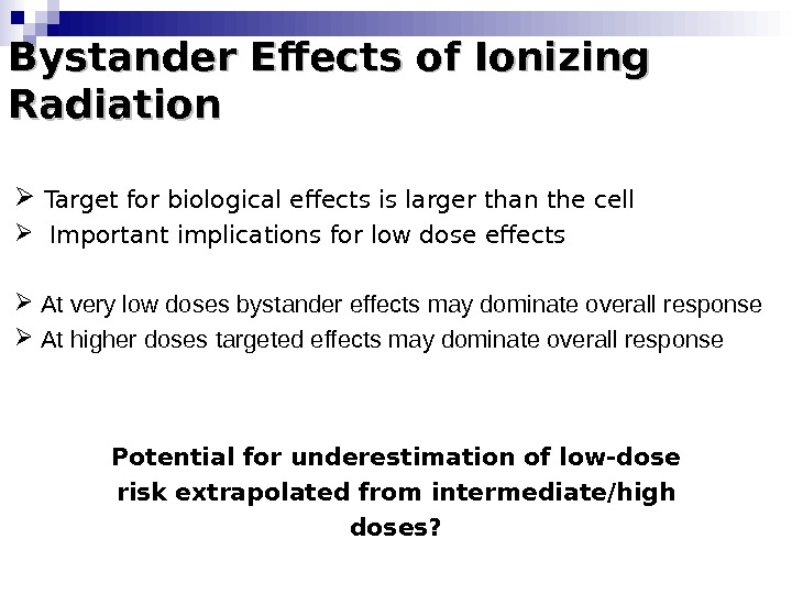
Bystander Effects of Ionizing Radiation Target for biological effects is larger than the cell Important implications for low dose effects At very low doses bystander effects may dominate overall response At higher doses targeted effects may dominate overall response Potential for underestimation of low-dose risk extrapolated from intermediate/high doses?
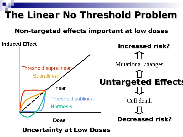
The Linear No Threshold Problem Induced Effect Dose. Threshold supralinear Supralinear Threshold sublinear Hormesis. Non-targeted effects important at low doses Untargeted Effects Mutational changes Cell death. Increased risk? Decreased risk? Uncertainty at Low Doses
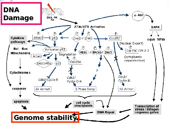
DNA Damage Genome stability
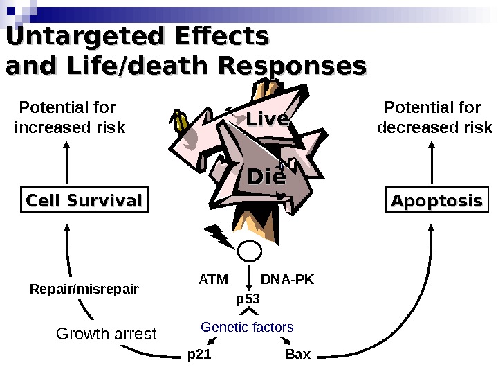
Die. Die Apoptosis Bax. DNA-PKATM p 53 p 21 Repair/misrepair. Cell Survival Growth arrest. Untargeted Effects and Life/death Responses Live. Potential for increased risk Potential for decreased risk Genetic factors