01772e51aaaf7c680e294e756a3df47b.ppt
- Количество слайдов: 59


Burn Resuscitation Bruce Potenza, MD For Trauma Conference 06/02/2009

Epidemiology of Burns 2 million people a year are burned 80% of these are less than 20% TBSA About 75, 000 patients require hospitalization Of these, 20, 000 are best cared for in a major burn center
 %TBSA 0")
LD 50 for Burns 1952 vs 1993 ________________ Age 1952 1993 (years) %TBSA 0 -14 49% 98% 15 -44 46% 72% 45 -64 27% 51% >65 10% 25%

Etiologic Types of Burns Scalds Gasoline, diesel fuel Electrical Open Flame Structural fire Car fire Smoking Natural gas explosions Chemical Lightening strike Welding Self inflicted Assault Faulty Heating equip.

Circumstances of the Burn Did the burn occur in a closed space or open space? Did the patient’s clothing catch fire? What type of flame was involved? What was the length time of the burn? What initial treatment did the patient receive?

House Fires 73% of all burn fatalities are due to house fires House fires account for only 4% of burn admissions House fires carries a 12% fatality rate of patients admitted to a burn unit 12 victims die each day due to a house fire

Automobile Fire How badly were the occupants injured? Associated blunt trauma injuries? Closed space fireinhalation injury? Combined ATLS/ABLS care

Burn Wound Depth First degree Second Degree – Superficial partial thickness – Deep partial thickness Third Degree Fourth Degree

Histology of the Skin Epidermis Dermis Appendages Subcutaneous

Depth of Burn 1 2° Wounds ° 3°

First Degree Burn Superficial burns Involve the epidermal layer of the skin Topical treatment with emollients Spontaneous resolution in days

Superficial - Partial Thickness Burns Superficial partial thickness burn The entire epidermis is involved Involves the upper portion of the dermis Spares the appendages Heals in 2 -3 weeks

Deep - Partial Thickness Burns Full epidermal involvement Deep dermal involvement Possible involvement of the dermal appendages May need grafting
 These burns include all layers")
Full Thickness Burns Full thickness burns (third degree burns) These burns include all layers of the skin and subq Excision and grafting will be needed Insensate

Burn Resuscitation

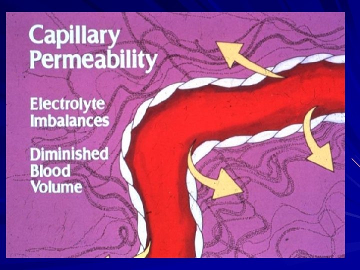
Pathophysiology of Burn Shock Hypovolemia and rapid edema formation – Microvascular permeability is increased resulting in fluid shifts – Sequestration of fluid in the burned and unburned tissue (burns >10%) Release of cellular and hormonal mediators – Examples include: histamine, bradykinin, prostaglandins, vasoactive amines and compliment

Burn Shock “Burn shock is characterized by a redistribution of blood flow from the splanchnic bed. Maintenance of cerebral and cardiac perfusion occurs at the expense of the mesenteric blood flow. ”

Burn Shock In thermal injury there is obligatory burn wound edema which is unique to burns. Burn shock is both hypovolemic shock and cellular shock. There are hemodynamic changes which include decreased vasomotor tone, tachycardia, hypotension and oliguria.

Burn Shock Maximal edema formation occurs in about 8 hours in smaller burns and in 12 -24 hours in larger burns. The rate of progression of the tissue edema is dependent upon the adequacy of resuscitation.

Burn Shock Thermal injury results in alteration of the normal capillary barrier. The change in the oncotic pressure favoring movement of solute into the interstitium combined with the increase in the hydrostatic pressure will lead to a rapid equilibration of the intravascular and interstitial compartments.

Fluid Resuscitation Goals: to maintain adequate tissue perfusion to limit the size and depth of the wound and to support the hemodynamics to the patient. Parkland formula-4 cc/kg X % TBSA burn Goal: maintain a good blood pressure, pulse and to keep urinary output 0. 5 cc/kg in and adult
 Hypertonic resus (lactated ringers plus one")
Fluid Resuscitation Crystalloid resus (lactated ringers Na= 130) Hypertonic resus (lactated ringers plus one amp of sodium bicarb or 50 m. Eq of Na. Cl results in a Na= 180) Hypertonic with 3% Na. Cl Colloid resus-should not be administered before 8 -12 hours into the burn resus due to leakage into the interstitium

Children Resuscitation Formula 3 -4 cc/kg X % TBSA burn Children have low storage of glycogen and require a constant source of glucose Continue with maintenance fluids containing dextrose Urine output 1 cc/kg child and 2 cc/kg in an infant

Fluid Resuscitation Patients with burns <15% should be given maintenance fluid requirements. Patients with burns >15% will require additional fluids to compensate for the obligatory edema and fluid loss due to the burn.

Delays in Resuscitation Delays in fully resuscitating a large burn patient can be devastating. In burns >80% a delay in only 2 hours in fluid resuscitation exponentially increase the systemic response by primed white blood cells.

Routes of Resuscitation 2 large bore I. V. lines – 8. 5 Fr peripheral introducer line Central venous catheterization – Single or triple lumen catheters – Cordis introducer Interosseous administration

Interosseous Infusion Pediatric fluid administration 180 -200 cc/hr Fluids, blood and meds can be given Watch for compartment syndrome

Actual fluid volume depends on patient response Easier to infuse more fluid than remove excess fluid Optimally, try to minimize volume & salt loading • Prevents acute renal failure • Low incidence pulmonary & cerebral edema 3/16/2018 Shock & Fluid Resuscitation 29

Cardiac output is commonly “normal” st in latter half of 1 post-burn day, IF NOT: • • Consider myocardial infarction or insufficiency Invasive monitoring may be required General patient condition reflects resuscitation adequacy Assess mental status frequently Anxiety & restlessness are early signs of hypovolemia and hypoxemia 3/16/2018 Shock & Fluid Resuscitation 30

Maintain adequate urine output Adult 0. 5 ml / kg / hr (30 – 50 cc/hr) Children weighing <30 kg 1 ml / kg / hour Indwelling urinary catheter most available & reliable resuscitation guide Incrementally or IV rate in response to UOP 3/16/2018 Shock & Fluid Resuscitation 31

Most often result of inadequate resuscitation Associated with SVR & CO Requires more rapid fluid administration Diuretics contraindicated 3/16/2018 Shock & Fluid Resuscitation 32
 High voltage electric injury or soft tissue injury")
Myoglobinuria & Hemoglobinuria (Red Pigmented Urine) High voltage electric injury or soft tissue injury due to mechanical trauma Administer fluids to maintain UOP 1. 0 -1. 5 ml / kg / hr in adults Often clears urinary heme pigments & eliminates need for diuretic 3/16/2018 Shock & Fluid Resuscitation 33
 The addition of Mannitol to IV fluid is")
Myoglobinuria & Hemoglobinuria (Red Pigmented Urine) The addition of Mannitol to IV fluid is rarely needed If used, a diuretic precludes use of UOP as guide to fluid therapy May add Na+ bicarbonate to IV fluid to maintain a slightly alkaline urine 3/16/2018 Shock & Fluid Resuscitation 34

May be misleading in burned limb Auditory signal as edema forms If used to titrate IV infusion rate, edema formation may be exaggerated 3/16/2018 Shock & Fluid Resuscitation 35

Of Limited Use Tachycardia common in adequately resuscitated adults May be related to pain as well as hypovolemia Levels of tachycardia in children dependent on normal HR 3/16/2018 Shock & Fluid Resuscitation 36

Not a reliable guide to resuscitation Whole blood or packed RBC not indicated unless • Pre-existing anemic disease • Acute blood loss from associated trauma Maintain Hct 30 – 35% Obtain baseline data for serious burns and inhalation injury Repeat based upon patient response 3/16/2018 Shock & Fluid Resuscitation 37

Inhalation Injury Inhalation injury is an acute respiratory insult caused by thermal or chemical inhalents

Thermal Injury The temperature of inhaled air is dependent upon the water content of the gas. The thermal content of steam is much higher than that of hot dry air. The supraglottic area is injured by dry heat. The infraglottic trachea is injured by steam.

Smoke Inhalation Injury

Smoke Inhalation Smoke consists of combustible products, asphyxiants and carbonaceous debris. Smoke is inhomogeneously dispersed within the airway.

Pulmonary Dysfunction with Smoke Inhalation Hypoxemia V/Q mismatch Release of pulmonary smooth muscle vasoconstrictors Increased airway resistance, pulmonary vascular resistance and decreased pulmonary compliance

Pulmonary Dysfunction With Smoke Inhalation Impaired ciliary function resulting in decreased secretory clearance. Pulmonary edema Atelectasis Sloughed epithelium and debris may result in distal airway occlusion.

Pulmonary Dysfunction With Smoke Inhalation Increased alveolar capillary permeability Generation of superoxide free radicals PMN transit is slowed through the pulmonary vascular bed Mucosal clumping of damaged cells, progressive necrosis

Clinical Signs of Inhalation Injury High index of suspicion, (ie. fire in a closed space) Concurrent oral pharyngeal burn Carbonaceous deposits in oropharynx or nares Patient with an impaired sensorium or agitation

Diagnosis of Inhalation Injury Bronchoscopy

Important Inhalation Injury Characteristics Type of material and fuel burned Type of toxic inhalant Closed space vs open space fire Duration of exposure Mental status Evidence of intoxication

Important Inhalation Injury Characteristics Size of inhaled particle determines the level of pathology. Particles >10 microns are trapped in the nasopharynx. Particles <2 microns are deposited in the airway. Particles <0. 06 microns are preferentially deposited in the alveoli.

Carbon Monoxide Poisoning Each year 10, 000 patients are injured Mortality rates as high as 31% are seen The leading cause of CO poisoning is automobile fumes Smoke inhalation is the 2 nd leading cause of CO poisoning

Pathophysiology of Carbon Monoxide Poisoning CO displaces O 2 on the hemoglobin resulting in decreased arterial O 2 content. CO binds to hemoglobin with 200 x more affinity than oxygen. CO shifts the O 2 dissociation curve to the left. CO decreases cellular respiration by binding with cytochrome a 3.

Carbon Monoxide Poisoning The mean half life of CO at room air is 320 minutes. On 100% O 2 the half life is 80 minutes. 3 ATM of hyperbaric O 2 decreases the half life to 23 minutes. The affinity of CO to myoglobin results in a prolonged clearance from cardiac and skeletal muscle. (May account for the rebound effect)

Systemic Effects of Carbon Monoxide Poisoning Systemic and cerebral hypoxemia develop. Hypotension due to decreases cardiac output as a direct result of decreased cardiac contractility. Reperfusion injury at the cellular level with the generation of free O 2 radicals. Lipid peroxidation and leukocyte sequestration occur in the brain.

Symptoms of Carbon Monoxide Poisoning CO Hb % Symptoms 10% 20% Asymptomatic, headache Dizziness, nausea, dyspnea Visual disturbances Confusion, syncope Seizures and coma Cardiopulmonary dysfunction and death 30% 40% 50% >60

Treatment of CO Poisoning Supplemental oxygen by 100% face mask or intubation Hyperbaric O 2 if available and the patient is showing physical signs or symptoms Transporting a patient to a chamber should be weighed against the patient’s condition as time is lost and transport problems may occur.

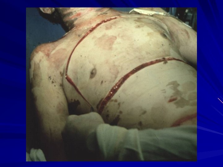
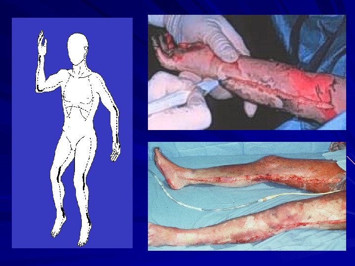
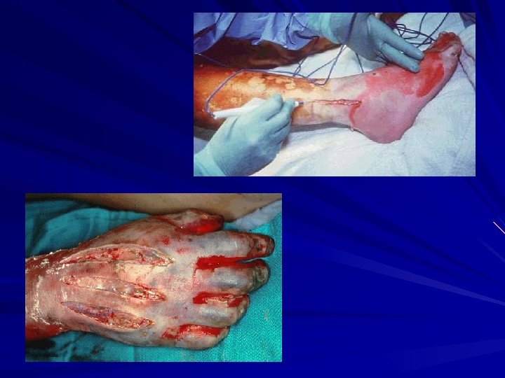
Escharotomies and Fasciotomies A circumferential partial thickness or full thickness burn may be vascular compromise This may worsen with fluid resuscitation This may occur on the extremities or on the chest and neck Early recognition of this process will prevent a compartment syndrome

Thank You
01772e51aaaf7c680e294e756a3df47b.ppt