8aae0900ad018ab2f268f5b33e70334a.ppt
- Количество слайдов: 62


Boston University School of Medicine asks all individuals involved in the development and presentation of Continuing Medical Education (CME) activities to disclose all relationships with commercial interests. This information is disclosed to CME activity participants. Boston University School of Medicine has procedures to resolve apparent conflicts of interest. In addition, presenters are asked to disclose when any discussion of unapproved use of pharmaceuticals and devices is being discussed. I, Paige Hertweck MD, have no commercial relationships to disclose. Jointly Sponsored by Boston University School of Medicine and the Society for Adolescent Health and Medicine

When the Pelvic Pain Doesn’t Go Away: A Multidisciplinary Approach to the Adolescent with Pelvic Pain Paige Hertweck, MD Chief of Gynecology, Kosair Children’s Hospital

Objectives: At the end of this session, you should be able to: • • Cite the most common causes of pelvic pain Outline the historical clues indicative of musculoskeletal pain Understand the role the musculoskeletal system may play as either a primary or secondary cause of pelvic pain Identify musculoskeletal pain on physical examination

Pelvic Pain in the Adolescent • The most common presenting complaint of adolescent women to primary care clinicians • Variety of causes: – – – – Gastrointestinal (IBDz, Celiac Dz) Genitourinary (Interstitial cystitis) Neurologic (Persistent pain after PID) Musculoskeletal Infectious (PID) Immunologic dysfunction (Endometriosis) Cognitive-psychologic (Somatization)

Acute and Sub-acute Pelvic Pain Common causes – Ovarian cysts / torsion – PID – IUD complication – Post-surgical pain – UTI or Interstitial Cystitis flare – Myofascial / musculoskeletal pain – GI pain – Many Others…

Chronic Pelvic Pain Common Causes – Endometriosis – Interstitial cystitis – Pelvic Adhesions – PID – Irritable Bowel Syndrome – Myofascial / musculoskeletal pain

Pelvic Pain Etiology: • May be single etiology – May have a single primary cause – May be exacerbation of known diagnosis • More often an overlap of more than one etiology – Example: • Endometriosis/ IBS/ Poor posture

Case: Samantha • • • 15 year old female with h/o cyclic pelvic pain Dx with endometriosis 10 weeks ago by LSC S/P excision of endo / placement Mirena IUD PMH: negative PSH: laparoscopy Social: – Sexually active one partner – Recently stopped track due to surgery and need for better grades • “After a month of crampy pain, things were fine, then I felt pretty good for 6 weeks, but the pain returned a month ago, here on my right ovary. It’s constant but flares too. ”

Case: Physical Exam/ Studies • Exam: General exam normal. • Affect: Normal, not anxious. • RLQ tenderness, no guard, no rebound • BME: AV uterus, non tender, IUS strings palpable, no abnormal discharge. Right adnexa seemed slightly tender, non enlarged. • Sono: Normal with IUS in position, Cultures GCCT and trichomonas negative.

Case: Samantha DDX: – Endometriosis flare • IF so, treatment options? ? ? – What else could it be? • • Adhesions? IC? IBS? Infection? Drug seeking? Psychiatric / Psychosomatic What else? ? ?

“When the pain doesn’t go away…” • If the diagnosis is inaccurate or incomplete, treatment will fail. • Because Musculoskeletal Dysfunction is not identified with our usual quantifiable tools … – Routine bimanual exam – Ultrasound – Cultures – Laparoscopy – CT scan . . . We may miss the true diagnosis.

Making the case for Musculoskeletal Dysfunction as a common cause of primary and secondary pelvic pain: • Prevalence of pain within the pelvis: – 3. 5%-24% of women age 15 -731 • “No gold standard diagnostic test exists for pelvic musculoskeletal problems, and the methodologic quality of available studies is low” • We must ensure we evaluate and offer treatment for these common pain syndromes. • Phys Med Rhabil Clin N Am 18 (2007) 477 -496

Abnormal musculoskeletal findings are more common in women with CPP • Asymmetric iliac crests (61% VS. 10%) • Asymmetric pubic symphysis heights (50% vs. 10%) • Positive posterior pelvic provocation testing (37% vs. 5%) • More abdominal and pelvic floor tenderness • Less able to relax pelvic floor (78% vs. 20%) AJOG, 2008; 198: 272. e 1 -272. e 7

Pelvic Pain in the Adolescent • Retrospective review 63 adolescent girls with pelvic pain • 67% had musculoskeletal pain – 11% had Endometriosis and MS pain Schroeder et al NASPAG 2000

Muscular and Myofascial Pain • Well-localized areas of pain • Trigger points: – a hyperirritable locus located in a muscle or its associated fascia, often referred from another area. – “Jump” sign when palpated. 1 • Myofascial tender point: – similar to trigger point, except the pain is not referred. 1. Reference: Source: Travell JG, Simons DG: Myofascial Pain and Dysfunction: The Trigger Point Manual, Vol. 2, Williams & Wilkins, Baltimore, 1992, p. 90.
 Adapted from: Obstet")
NEUROPATHOLOGY OF MYOFACAL PAIN ***** Trigger points Visceral dysfunction (elimination dysfunction) Adapted from: Obstet Gynecol Clin N Am 36 (2009) 699 -705 Muscle Fiber Trauma -Overt injury -Overuse / dysbehaviors (poor posture, repetitive motions, etc) Secondary pain disorder with central sensitization Sacromere shortening Endplate overactivity Neuroinflammatory mediator release Local ischemia Peripheral sensitization Afferent Overactivity Trigger Point Activation Dorsal Horn Up-regulation and Central Sensitization Hypertonic disorders / Trigger points with dysfunction and/or Pain and/or Visceral dysfunction
")
Myofascial Pain • Trigger points are found in 30 -70% of women with CPP(1) • Pelvic muscle dysfunction is seen in 5085% of patients with IC/painful bladder syndrome(2). (1) Am J Obstet Gynecol 1984; 149(5): 536 -543 J Reprod Med 1991; 36(4): 253 -259 (2)Eur J Obstet Gynecol Reprod Biol 2003; 107(2): 185 -190

Myofascial Pain • Most Common Cause: chronic repetitive stress and strain: – Faulty posture – Poor body mechanics – Poor physical conditioning
 –")
Myofascial Pain • Other causes – Direct trauma (MVC, Acute athletic injury, etc) – Can be secondary to surgical procedures (Port sites, positioning, cesarean section, urogyn procedures, etc) – Can be secondary to vaginal birth
: 773 -782")
Clinical Obstetrics and Gynecology, 2003; 46, (4): 773 -782

Diagnostic Approach • Use history / physical for accurate diagnosis – Allow patient to tell her story (therapeutic effect) – Take time with history and physical remembering other systems • Use your eyes, and hands and intuition to perform a thorough musculoskeletal exam!

CPP Evaluation: History In addition to routine history and physical, assess for the 5 major sources that might contribute to CPP – Gynecological – Gastrointestinal – Urologic – Psychological – Musculoskeletal

Pelvic Pain: History • Location: – R or LLQ vs periumbilical? • Severity: – On a 0 to 10 scale; any missed activities • Timing: – Cyclic vs noncyclic – What life events/ medical issues occurred at time of onset? – Dysmenorrhea rarely presents at menarche—instead think outflow anomaly • Modifying Factors: – What makes pain better? Worse? • Worse with movement and better with rest—think musculoskeletal pain – Same position or vary in location • Pain radiating to back is often musculoskeletal – What meds have been used to treat? Response?

Pelvic Pain History • Quality: – Cramping: more visceral – Burning: more neuropathic • Duration: – How long is hx of pain? How long each episode? • Context: – With menses? With exercise? – Recent change in habits? Heavy lifting? New sport or exercise? • Associated symptoms: – GI: Nausea/ Vomiting/ Constipation/ Better or worse with BM/ flatus – GU: Dysuria/ Frequency/ Urgency/ Nocturia

CPP History • Need complete ROS with emphasis on – – – – Bowel function Urinary tract Substance use / dependence Depression / Anxiety Sexual, physical, psychological abuse Trauma or Injury Physical activity level (or lack of activity) • Hours on computer • watching / using digital media
 overlap")
Pelvic Pain: History Difficult for patient: – Remember the CNS (T 10 -12) overlap between – Pelvic viscera (i. e. : bladder, uterus) – Somatic structures (rectus abdominus, iliopsoas) Historical Clues – Pain worse with movement/ better with rest – Pain better with curling up in fetal position – Associated low back pain

Physical Examination: Use your Eyes Observe posture – During interview – Walking to exam – Self-selected posture – Observe anterior, posterior, lateral views – Look for lateral pelvic tilt, spinal curvature

Typical Pelvic Pain Postures 1. Hyperextended knee 2. Exaggerated lumbar lordosis or Exaggerated thoracic kyphosis 3. Forward head: auditory meatus anterior to acromion Normal

Abdominal Wall: Basic Physical Exam – Assess bowel sounds – Look for abdominal distention/ ascites – Palpate for hepatosplenomegaly – Assess for abdominal masses, hernias – Check any surgical scar for hernia, tenderness

M/S Physical Examination Abdominal wall – Assess for trigger points • Areas of hyperirritability that are locally tender on compression and cause referred pain and tenderness • Common on rectus abdominus Rectus abdominus – Pain worse with head lift Pain likely in the abdominal wall

Abdominal Musculature: • Enervated by: T 7 -T 11 • Injured by: Exercise Trocars Trauma • Examination techniques: Head raise in supine position

Abdominal Wall Exam • If raising the head increases the pain, most likely abdominal wall related. • If raising the head decreases pain, most likely below the abdominal wall.
 Pain with ipsilateral")
Physical Examination Abdominal wall trigger points Psoas (lateral to the umbilicus) Pain with ipsilateral straight leg lift-pain likely psoas mm Iliacus Pain inside hollow of ASIS with straight leg lift

Hip Flexors: Psoas and Illiacus • Enervated by: L 1 -L 4 • Injured by Poor posture Exercise overuse Local inflammation • Examination techniques: Thomas Test Evaluation of trigger points

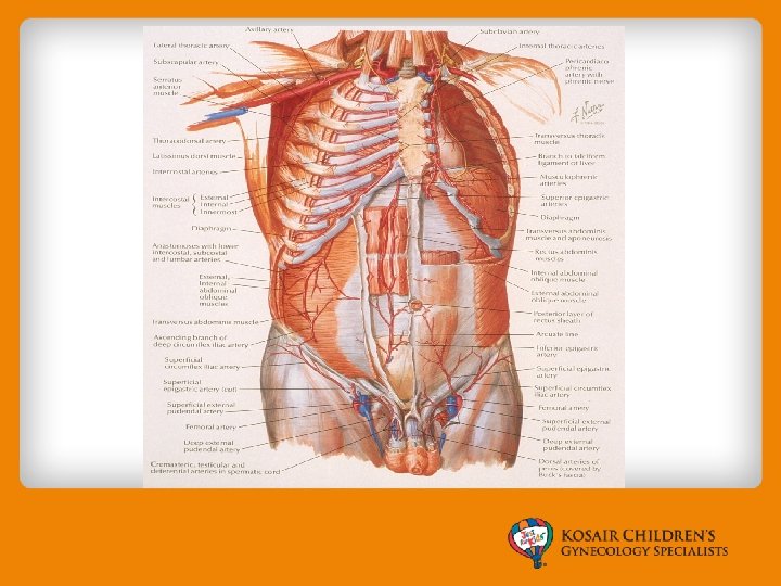
Proximity to pelvic organs

Pelvic Floor Physical Exam

Pelvic Floor • Enervated by: S 2 -S 4 • Injured with: Trauma Poor posture Exercise • Examination techniques: Palpation of tender points

Relationship of pelvic organs to pelvic musculature

Pelvic floor: view from above

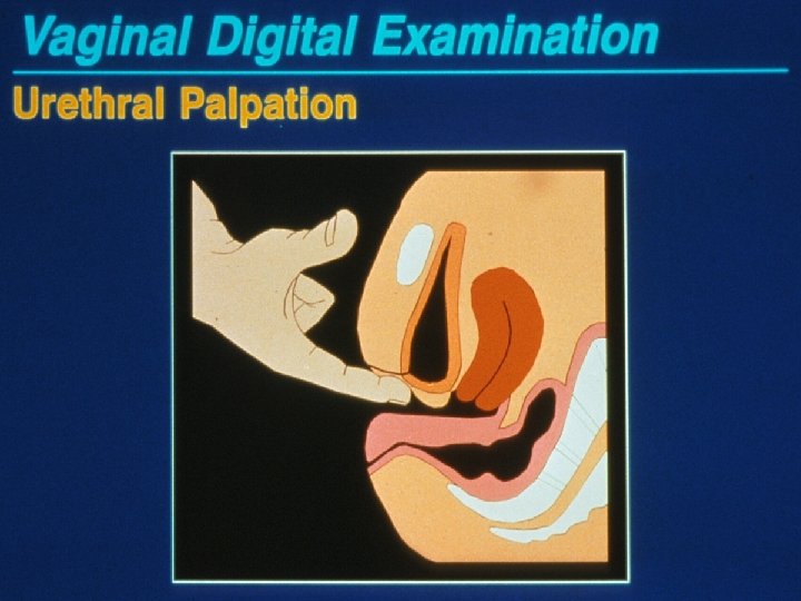
Physical Examination Visceral Exam Use single digit for exam Palpate urethra, bladder base

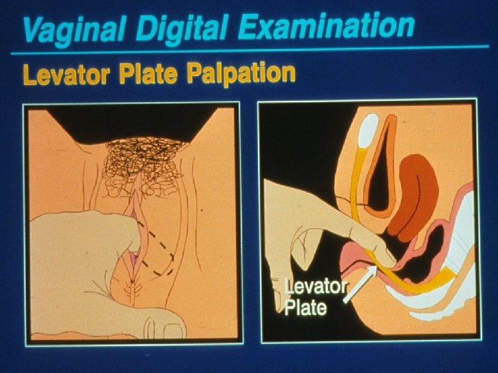
Physical Examination Vaginal, Cervix, Paracervical Tissues Single digit palpation of lateral sidewalls – Identify any trigger points – Check levator mm at 5 and 8 o’clock – Check piriformis mm at each vaginal fornix

Physical Examination Vaginal, Cervix, Paracervical Tissues Single digit palpation of lateral sidewalls Identify any trigger points Check levator mm at 5 and 8 o’clock Check piriformis mm

Piriformis muscle is found deep to the ischial spine.

Physical Examination Bimanual exam Add abdominal hand – Define uterus, adnexa – Note areas of abdominal wall vs visceral discomfort Perform rectovaginal exam
")
Examples Musculoskeletal Findings: • (Findings that can be treated with OMT/Osteopathic Manipulation or PT) Muscular: – – • Nervous System – – • Trigger points Abdominal or pelvic muscular spasm / hypertonicity Iliosoas spasm, pelvic floor spasm (vaginismus, ) piriformas syndrome etc. Others… Cutaneous nerve hypersensitivity (vulvodynia, abdominal skin paresthesia/pain) Various neuralgias / nerve impingements Visceral nerve dysfunction (voiding problems) Others… Skeletal – Leg length discrepancy (true / primary or secondary to neuro-muscular abn) – Increased lordosis, kyphosis, scoliosis – Others…

Peripheral Nerve Causes of Pain Nerve Sensory Innervation Visceral Field of Referred Pain Iliohypogastric Post. Superior gluteal Ovary and Distal Fallopian region, anterior suprapubic Tube area Ilioinguinal Medial thigh and lateral labia majora below inguinal ligament Proximal tube and uterine fundus Genitofemoral Proximal anterior thigh Proximal tube and uterine fundus Lateral Femoral Cutaneous Lateral anterior thigh Fundus and lower uterine segment Pudendal Lower uterine segment, cervix, bladder, distal ureter, upper vagina, rectum Dematomes S 2 -S 4, perineum, perianal area Data from: Steege, Metzger, Levy. Chronic Pelvic Pain, An Integrated Approach. Philadelphia: W. B. Saunders Company , 1998 Howard, Perry, Carter, et al, Pelvic Pain Diagnosis and Management. Philadelphia: Lippincott Williams and Wilkins, 2000

Accepted Physical Therapy Diagnoses • • • • 564. 6 Spasm, anus/ani sphincter 569. 7 Anal rectal pain 618. Genital prolapse 625 Pain and other symptoms associated with female genital organs - except 625. 0 Dyspareunia - not covered by ins 358. 8 Denervation 664. 4 Constipation 709. 2 Scar - painful 728 Disorders of muscle , ligaments and fascia 729 Other disorders of soft tissues 781. 3 Muscle inco-ordination. 788 Symptoms involving the urinary system 787. 6 Incontinence of feces Orthopedic codes : back pain , Si joint dysfunction , hip , sciatica

Samantha: Musculo-Skeletal Exam • Exam Findings: Right iliacus tender point • Diagnosis: Pelvic Somatic Dysfunction • Treatment Options: Physical therapy – Muscle energy – Ligamentous articular release • Lifestyle recommendations: – Stretching, exercise

So, you have made a diagnosis of musculoskeletal pain, what now? • Educate your patient on the cause(s) of pain – Validate the pain, sympathize, and empower her to take an active role Validate the pain in her treatment. (don’t say – “it’s just muscle pain. ”) – Use a pelvic model or other graphic educational material so that the patient can see the areas causing her pain. – Put a positive spin on a diagnosis of musculoskeletal pain • Treat other causes of pelvic pain, if the patient has them.

So, you have made a diagnosis of musculoskeletal pain, what now? • Refer to a Pelvic Physical therapist – Educate your patient on the expected treatment course with Physical Therapy – “Manage Up” and describe the provider as someone you trust, believe in, and respect as a partner in management. – Schedule your patient back for follow up with you, so that you can re-assess after treatment and follow up on any other aspects of her pain (Endo, IC, Anxiety, Depression, etc. )

Tips for strengthening your exam skills • Do careful muscular exams on patients without pelvic pain to identify normal. • Find an osteopathic medical student or resident to work with you in your office!

Pelvic Physical Therapy Find a physical therapist who has certification through the Herman and Wallace Institute or has Similar training on internal pelvic PT (not all physical therapist are trained and certified to do transvaginal PT) • Mission: To provide the most comprehensive, evidence-based, continuing education for the rehabilitation of the pelvic floor and pelvic girdle dysfunction, resulting in the development and certification of a skilled practitioner and the advancement of pelvic rehabilitation research. • Herman & Wallace | Pelvic Rehabilitation Institute 93 South Jackson Street #71393 Seattle, WA 98104 Phone: 646. 355. 8777 Web Address: http: //hermanwallace. com •

Physical Therapy: What to expect… • Pelvic PTs can treat: – Muscular dysfunction: levator spasm, vaginismus, dyspreunia, pelvic floor spasm, piriformas spasm syndrome, and others… – Impingement neuralgias such as pudendal nerve neuralgia, and others – Skeletal dysfunction such as: coxyx injuries, SI joint dysfunction, others – Visceral dysfunction: Bladder / Bowel dysfunction – Others: vulvodynia, IC • Initial visit is mostly assessment • Therapeutic interventions typically at follow up

Physical Therapy: Treatment Therapeutic interventions include: • Muscular facial release • Mobilization of Joints (T-spine, SI joint, Coccyx, Pubic bone) • Home vaginal stimulation • Ultrasound of the pelvic floor • Tens units for abdomen and perineum • Biofeedback Prescriptions: • Stretching exercises • Strengthening exercise • Posture recommendations • Devices to help: Therapy Balls, vaginal dilators etc.

Ashley • 21 year old college student • Stable partner for two years • 4 month history of dyspareunia, pain with penetration, unable to complete intercourse due to pain. • She describes a hypersensitivity of her vulvar skin, has had negative vaginitis assessment recently.

Ashley She has been using a crutch for 5 -6 months due to a foot injury requiring surgical intervention. • Exam: SI joint dysfunction, Levator Spasm, vulvar hypersensitivity • Assessment: Pudendal neuralgia and somatic dysfunction of pelvic floor • Plan: Pelvic Floor Rehabilitation PT

Ashley: PT evaluation EXAM FINDINGS • Abdominal trigger points along entire length or right rectus abdominis from origin to insertion. • Right Psoas trigger point. • Trigger point on central perineal body and left superficial transverse perineum • Pudendal nerve unloading in supine reveals decreased mobility with pain on the left. • Elevated sensitivity of vaginal introitus from 6: 00 -9: 00 • Pelvic floor 2/5 decreased ability to relax, quick flicks 15 in 30 seconds ASSESSMENT • Perineal muscle spasm • Pelvic floor, hip and abdominal core weakness • Abdominal wall trigger points, Right side, Right Psoas, Left perineum GOALS • Use tampons • Intercourse • Decreased pain • Increased strength PLAN: Ashley will be seen one time a week for 60 minute treatment sessions for 8 -12 visits and treatment will consist of therapeutic exercise, therapeutic activities, neuromuscular reduction and manual therapy.

Remember Muscle, Nerve, Joints • Include the Musculoskeletal System in your DDX • Make referrals often • Remember the multifactorial causes of pelvic pain will respond better to an integrative treatment plan! • Use a team approach.

Questions?
8aae0900ad018ab2f268f5b33e70334a.ppt