fcd13323c8f0a3eb71ae6337f772e9e3.ppt
- Количество слайдов: 118


, Ph. D Department of Physiology Room C 518, Block C,")
BLOOD【血液】 Qiang XIA (夏强), Ph. D Department of Physiology Room C 518, Block C, Research Building, School of Medicine Tel: 88208252 Email: xiaqiang@zju. edu. cn
 Body Fluid = 60% of Body Weight (BW) Plasma 5% of")
Internal environment (内环境) Body Fluid = 60% of Body Weight (BW) Plasma 5% of BW Extracellular Fluid 1/3, 20% of BW Interstitial Fluid 15% of BW 70 kg Male, 42 L Intracellular Fluid 2/3, 40% of BW

Plasma 5% of BW Extracellular Fluid 1/3, 20% of BW Internal Environment Interstitial Fluid 15% of BW
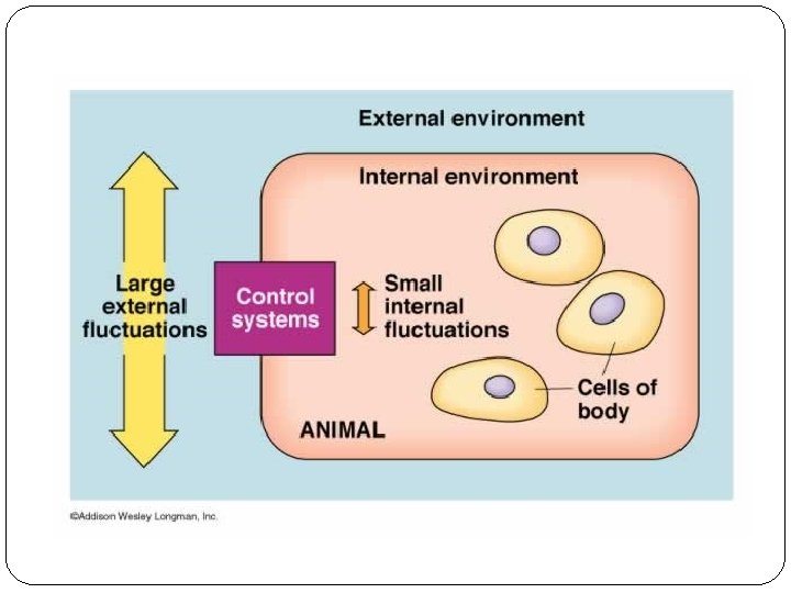
 Homeostasis (from the Greek words for “same” and “steady”): maintenance of static or")
Homeostasis(稳态) Homeostasis (from the Greek words for “same” and “steady”): maintenance of static or constant conditions in the internal environment Walter B. Cannon http: //www. harvardsquarelib rary. org/unitarians/cannon_ walter. html

Components of Homeostasis: l Concentration of O 2 and CO 2 l p. H of the internal environment l Concentration of nutrients and waste products l Concentration of salt and other electrolytes l Volume and pressure of extracellular fluid

How is homeostasis achieved? ----Regulation Body's systems operate together to maintain homeostasis: Skin system Skeletal and muscular system Circulatory system Respiratory system Digestive system Urinary system Nervous system Endocrine system Lymphatic system Reproductive system
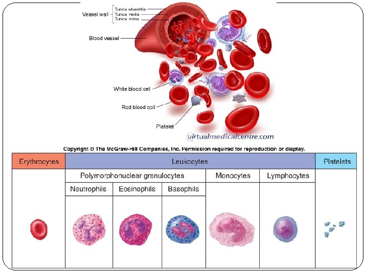
 Blood Cells Red Blood Cells (RBC) or Erythrocytes(红细胞) White Blood")
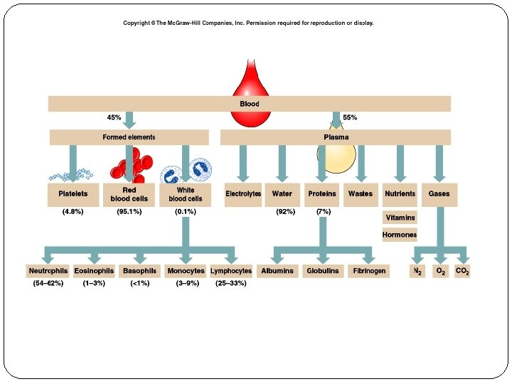
Components of blood Plasma(血浆) Blood Cells Red Blood Cells (RBC) or Erythrocytes(红细胞) White Blood Cells (WBC) or Leucocytes(白细胞) Platelets (PLT) or Thrombocytes(血小板)
 is a rapid")
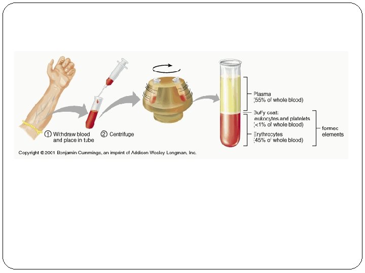
Plasma includes water, ions, proteins, nutrients, hormones, wastes, etc. The hematocrit(血细胞比容) is a rapid assessment of blood composition. It is the percent of the blood volume that is composed of RBCs (red blood cells).
 the volume of red blood cells as a percentage of")
Hematocrit(packed cell volume, 血细胞比容) the volume of red blood cells as a percentage of centrifuged whole blood M: 40~50% F: 37~48% International Council for Standardization in Haematology (ICSH) Recommendations for "Surrogate Reference" Method for the Packed Cell Volume
 Depending on hematocrit & protein")
Physical & chemical properties of blood 1. Specific Gravity(比重) Depending on hematocrit & protein composition Whole blood: 1. 050~1. 060 Plasma: 1. 025~1. 035 Red blood cells: 1. 090
 relative viscosity of whole blood 4~5 depending on hematocrit relative viscosity of")
2. Viscosity(粘度) relative viscosity of whole blood 4~5 depending on hematocrit relative viscosity of plasma 1. 6~2. 4 related to the protein composition of the plasma
 The osmotic pressure of a solution depends on the number of")
3. Osmotic Pressure(渗透压) The osmotic pressure of a solution depends on the number of solute particles in the solution, NOT on their chemical composition and size
 Crystalloid Osmotic Pressure(晶体渗透压) Pressure generated by all")
Plasma osmotic pressure (~300 m. Osm/L) Crystalloid Osmotic Pressure(晶体渗透压) Pressure generated by all crystal substances, particularly electrolytes Important in maintaining fluid balance across cell membranes Colloid Osmotic Pressure(胶体渗透压) Osmotic pressure generated by plasma proteins, particularly albumin. Approximately 25 mm. Hg, but important in fluid transfer across capillaries
: Na. HCO 3/H")
4. Plasma p. H Normal range: 7. 35~7. 45 Buffer systems(缓冲系统): Na. HCO 3/H 2 CO 3, Pro-Na/Pro, Na 2 HPO 4/Na. H 2 PO 4 Hb-K/Hb, Hb. O 2 -K/Hb. O 2, K 2 HPO 4/KH 2 PO 4, KHCO 3/H 2 CO 3
")
Functions of blood Transportation O 2 and CO 2 Nutrients (glucose, lipids, amino acids) Waste products (e. g. , metabolites) Hormones Regulation p. H Body temperature Protection Blood coagulation Immunity
 Plasma 5% of BW Extracellular")
Plasma Body Fluid = 60% of Body Weight (BW) Plasma 5% of BW Extracellular Fluid 1/3, 20% of BW Interstitial Fluid 15% of BW 70 kg Male, 42 L Intracellular Fluid 2/3, 40% of BW
 serves as transport medium; carries heat Proteins (6~8%")
u Composition Water (92% of plasma) serves as transport medium; carries heat Proteins (6~8% of plasma) Inorganic constituents (1% of plasma) e. g. , Na+, Cl-, K+, Ca 2+… Nutrients glucose, amino acids, lipids & vitamins Waste products e. g. , nitrogenous wastes like urea Dissolved gases O 2 & CO 2 Hormones

Plasma proteins
(60 -80% of plasma proteins) • most important in maintenance of")
• Albumins (白蛋白)(60 -80% of plasma proteins) • most important in maintenance of osmotic balance • produced by liver • Globulins (球蛋白)( 1 -, 2 -, -) • important for transport of materials through the blood (e. g. , thyroid hormone & iron) • clotting factors • produced by liver except -globulins which are immunoglobulins (antibodies) produced by lymphocytes • Fibrinogen(纤维蛋白原) • important in clotting • produced by liver
 (红细胞)")
Red blood cells (Erythrocytes) (红细胞)

Structure Biconcave No nucleus Few organelles Small Hemoglobin molecules

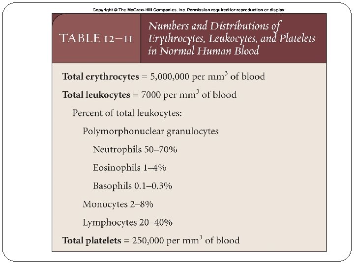
u Count RBC count M: 4. 0~5. 5× 10¹²/L F: 3. 5~5. 0× 1012/L Hemoglobin(血红蛋白) M: 120~160 g/L F: 110~150 g/L
")
u Physiological properties Plastic deformability (可塑变形性)
 Erythrocyte Sedimentation Rate (ESR)(红细胞沉降率) The distance that red blood cells settle")
d Suspension stability(悬浮稳定性) Erythrocyte Sedimentation Rate (ESR)(红细胞沉降率) The distance that red blood cells settle in a tube of blood in one hour Normal value [Westergren method(魏氏法,国际血液学标准化委员 会推荐魏氏法为标准法)]: M: 0~15 mm/h,F: 0~20 mm/h An indication of inflammation which increases in many diseases, such as tuberculosis & rheumatoid arthritis… International Council for Standardization in Haematology (ICSH)
")
红细胞叠连(rouleaux formation)
 the susceptibility of a red blood cell to break apart when")
Osmotic fragility (渗透脆性) the susceptibility of a red blood cell to break apart when exposed to saline solutions of a lower osmotic pressure than that of the human cellular fluid

Notice that hemolysis begins in the 0. 45% tube and is complete in the 0. 35% tube.

Only substances which act as impermeant molecules can be used to make isotonic solutions (等张溶液). E. g. cells placed in an isosmotic solution (等渗溶液) of urea (1. 9%), a permeant molecule, will swell and bust. Solutions which have the same calculated osmotic pressure are said to be ISOSMOTIC but are not necessarily ISOTONIC

u Function of RBCs 1. Transport of O 2 and CO 2 2. Buffering
")
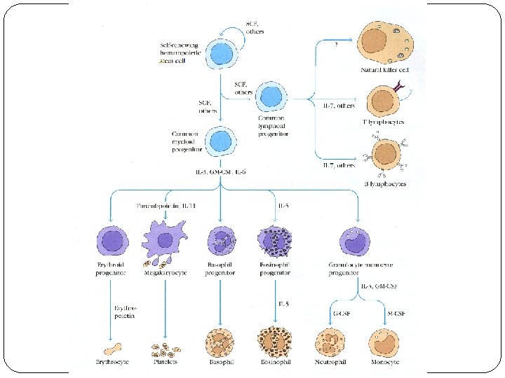
u Production of RBC (Erythropoiesis)

Hemocytoblast stem cell Stem cell becomes committed Early erythroblasts have ribosomes Erythroblasts accumulate iron and hemoglobin Normoblasts eject organelles Released as erythrocyte

Nutritional Requirements for Erythropoiesis 1. Many vitamins, minerals, and proteins are necessary for normal RBC production 2. Clinically, folic acid(叶酸), Vit. B 12, and iron (铁) are the most important. Deficiencies of these factors lead to characteristic anemias(贫血)
. Iron")
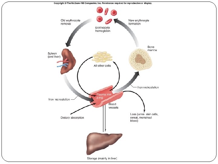
Diagram of iron kinetics from iron stores to developing red blood cell (RBC). Iron stores include the bone marrow, reticuloendothelial system (liver and spleen) and RBCs. Transferrin (total iron-binding capacity [TIBC]) transports iron (Fe) to developing erythrocytes. Iron is deposited in the RBC, and transferrin returns to storage sites to bind more Fe for transport. Lactoferrin is a competitor of transferrin; it takes Fe that is free and returns it to storage sites. Lactoferrin levels are elevated in anemia of chronic disease. Increases in interleukin-1 increase the sequestration of Fe in storage sites. (Hb=hemoglobin)
 2. Hormones: Androgen(雄激素) Others Hypoxia-inducible factor 1, HIF-1")
u Regulation of Erythropoiesis 1. Erythropoietin(促红细胞生成素) 2. Hormones: Androgen(雄激素) Others Hypoxia-inducible factor 1, HIF-1

Erythropoiesis is hormonally regulated: decreased oxygen delivery to the kidney causes the secretion of erythropoietin, which activates receptors in bone marrow, leading to an increase in the rate of erythropoiesis.

u Destruction of RBC Macrophages engulf old RBCs Iron is salvaged Heme degrades into bilirubin average lifespan = about 120 days
 Anemia is defined as a qualitative or quantitative deficiency of hemoglobin, a protein")
Anemia(贫血) Anemia is defined as a qualitative or quantitative deficiency of hemoglobin, a protein found inside red blood cells (RBCs) The three main classes of anemia: excessive blood loss (acutely such as a hemorrhage or chronically through low-volume loss) excessive blood cell destruction (hemolysis) deficient red blood cell production (ineffective hematopoiesis)
")
Iron deficiency anemia (缺铁性贫血)
")
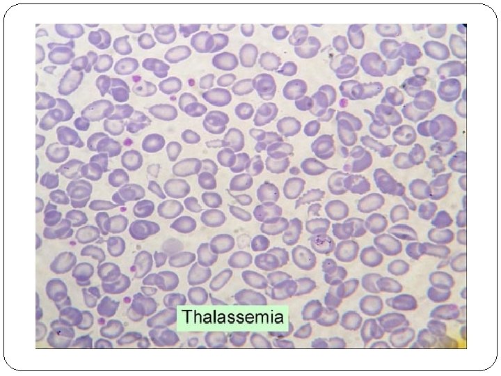
巨幼红细胞性贫血(megaloblastic anemia)
 Red blood cells without (left and middle) and with (right) hemolysis. Note that")
Hemolysis(溶血) Red blood cells without (left and middle) and with (right) hemolysis. Note that the hemolyzed sample is transparent, because there are no cells to scatter light.
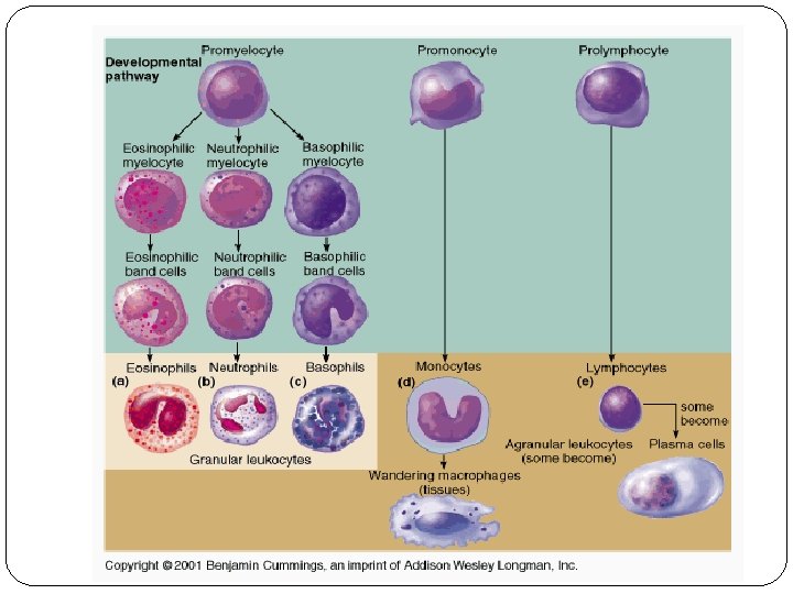
 (白细胞) u Types of WBC")
White blood cells (Leucocytes) (白细胞) u Types of WBC
")
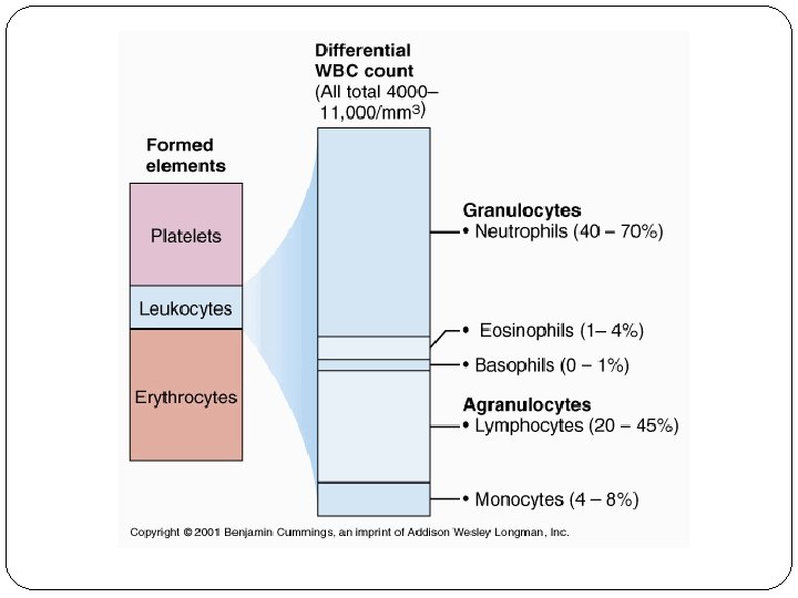
u WBC count WBC Granulocytes Neutrophils Eosinophils Basophils Monocytes Lymphocytes Total Count ( 109/L) % 2. 0~7. 0 50~70 0. 02~0. 5~5 0~0. 1 0~1 0. 12~0. 8 3~8 0. 8~4. 0 20~40 4~10

Leukopoiesis Myeloblasts become all of the granular leukocytes Monoblasts become monocytes Lymphoblasts become lymphocytes
 u u u Formed in the bone marrow from cells called megakaryocytes")
Platelets (Thrombocytes) u u u Formed in the bone marrow from cells called megakaryocytes Without nucleus, but can secrete a variety of substances normal value: (100~300) x 109/L Average lifespan=7~14 days Play an important role in hemostasis

u Physiological properties of platelets 1. Adhesion Platelets adhere to the vessel wall at the site of injury von Willebrand factor, v. WF
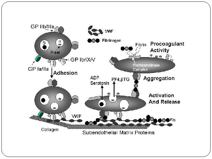

Unifying model of platelet adhesion to collagen at arterial shear. Two different pathways by which human and mouse platelets firmly adhere to collagen at arterial shear are illustrated. In both, the majority of platelets are initially tethered to collagen via GP Ib/IX/V interacting with collagen-bound VWF (left), although a minority of platelets interact directly with collagen independently of VWF/GP Ib/IX/V. In the first pathway (upper), signaling from GP VI first leads to activation of integrins α 2β 1 (GP Ia/IIa) and αIIbβ 3 (GP IIb/IIIa). Activated integrins then firmly attach the platelet to collagen, either directly (α 2β 1) or via collagen-bound VWF (αIIbβ 3) (right). In the second pathway (lower), platelets first adhere to collagen via integrin α 2β 1, before GP VI engages collagen and induces activation. These two pathways are likely to reinforce each other and the events of thrombus formation. Release of secondary mediators (ADP and Tx. A 2) would further potentiate these events (right). (Redrawn from Auger JM, Kuijpers MJ, Senis YA: Adhesion of human and mouse platelets to collagen under shear: a unifying model. FASEB J 2005; 19: 825 -827. )

2. Aggregation Platelets adhere to one another
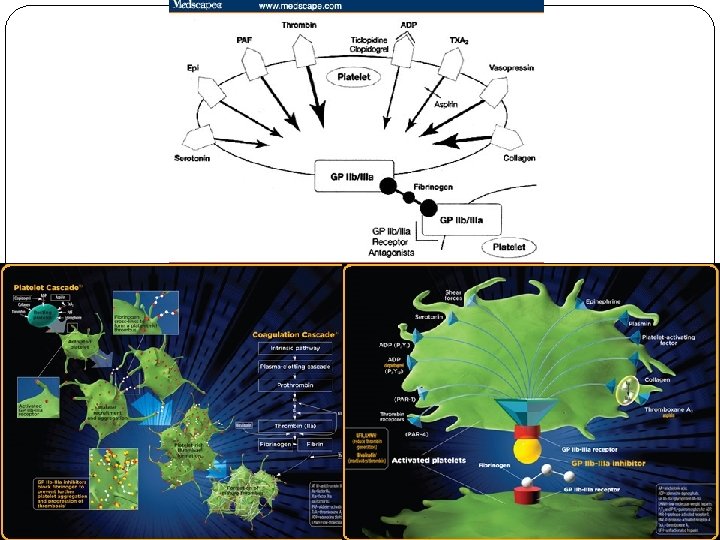

Platelet Aggregation Pathway Platelet activation and coagulation normally do not occur within an intact blood vessel. After vessel wall injury, platelet-plug formation is initiated by the adherence of platelets to subendothelial collagen. In high shear arterial blood, platelets are first slowed down from their blood flow velocity by interacting with the collagen-bound von Willebrand factor (VWF) and subsequently stopped by binding directly to collagen via their glycoprotein receptor complex. The activation of these collagen receptors on platelets following their binding to collagen activates phospholipase C (PLC)-mediated cascades. This results in a mobilization of calcium from the dense tubula system. An increase in intracellular calcium is associated with activation of several kinases necessary for morphological change, the presentation of the procoagulant surface, the secretion of platelet granular content, the activation of glycoproteins, and the activation of Phospholipase A 2 (PLA 2). Activation of PLA 2 releases arachidonic acid (AA), which is a precursor for TBXA 2 synthesis. PTGS 1 catalyzes the first step in the formation of TBXA 2 from AA. This reaction is irreversibly blocked by aspirin, which also leads to the blockage of platelet aggregation These processes result in the local accumulation of molecules like thrombin, TBXA 2, and ADP, which are important for the further recruitment of platelets as well as the amplification of activation signals as described above. The secreted agonists activate their respective G protein coupled receptors: thrombin receptor (F 2 R), thomboxane A 2 receptor (TBXA 2 R), and ADP receptors (P 2 RY 1 and P 2 RY 12). The P 2 RY 12 receptor couples to Gi, and when activated by ADP, inhibits adenylate cyclase. This interaction counteracts the stimulation of c. AMP formation by endothelial-derived prostaglandins, which alleviates the inhibitory effect of c. AMP on IP 3 -mediated calcium release. Thienopyridines, a class of oral antiplatelet agents, permanently inhibit P 2 RY 12 signaling, which is sufficient to block platelet activation. F 2 R, TBXA 2 R and P 2 RY 1 couple to the Gq-PLC-IP 3 -Ca 2+ pathway, inducing shape change and platelet aggregation. In addition, receptor signaling through G 12/13 (F 2 R; TBXA 2 R) contributes to morphological changes through activation of kinases. Platelet adhesion, cyotoskeletal reorganization, secretion, and amplification loops are all different steps towards the formation of a platelet-plug. These cascades result in the activation of the Fibrinogen Receptor expressed on platelet cells. This activation develops binding sites for fibrinogen, which are not available in inactive platelets. The binding of fibrinogen results in the linkage of activated platelets through fibrinogen bridges, thereby mediating aggregation. Inhibition of this receptor through Glycoprotein IIb/IIIa inhibitors blocks platelet aggregation induced by any agonist.

Inducers of platelet aggregation ADP Low dose 1 st reversible phase High dose 2 nd irreversible phase Thromboxane A 2 (TXA 2) Collagen Thrombin

Phospholipid Phospholipase A 2 Arachidonic Acid Cyclo-oxygenase PGG 2 & PGH 2 Thromboxane synthase Prostacyclin synthase (Platelets) (Vascular endothelium) TXA 2 Aggregation Contraction PGI 2 Anti-aggregation Relaxation

Platelet interactions with agonists and antagonists of platelet aggregation, the vessel wall, other platelets, and adhesive macromolecules. Agents in parentheses prevent the formation or inhibit the function of the adjacent agonists of platelet aggregation. ADP = adenosine diphosphate, VWF = von Willebrand factor, c. AMP = cyclic adenosine monophosphate, GP = glycoprotein.

3. Release or secretion: Platelets contain alpha and dense granules Dense granules: containing ADP or ATP, calcium, and serotonin α-granules: containing platelet factor 4, PDGF, fibronectin, Bthromboglobulin, v. WF, fibrinogen, and coagulation factors V and XIII
, showing its alpha and dense granules and")
Schematic drawing of the platelet (top figure), showing its alpha and dense granules and canalicular system. The bottom figure illustrates the platelet's major functions, including secretion of stored products, as well as its attachment, via specific surface glycoproteins (GP), to denuded epithelium (bottom) and other platelets (left). VWF: von Willebrand factor; TSP: thrombospondin; PF 4: platelet factor 4; PDGF: platelet derived growth factor; -TG: beta thromboglobulin; ADP: adenosine diphosphate; ATP: adenosine triphosphate.

A schematic representation of selected platelet responses to activation and the congenital disorders of platelet function. AC = adenylyl cyclase; BSS = Bernard–Soulier syndrome; CO = cyclooxygenase; DG = diacylglycerol; G = GTP-binding protein; IP 3 = inositol trisphosphate; MLC = myosin light chain; MLCK = myosin light chain kinase; P 2 Y 1, P 2 Y 12 = G-protein-coupled ADP receptors; PAF = platelet activating factor; PGG 2/PGH 2 = prostaglandin arachidonic pathway intermediates; PIP 2 = phosphatidylinositol bisphosphate; PKC = protein kinase C; PLA 2 = phospholipase A 2; TK = tyrosine kinase; PLC = phospholipase C; TS = thromboxane synthase; Tx. A 2 = thromboxane A 2; v. WD = von Willebrand disease; v. WF = von Willebrand factor. The Roman numerals in the circles represent coagulation factors and yellow Ps indicate phosphorylation. (Modified with permission from Rao AK: Congenital disorders of platelet function: disorders of signal transduction and secretion. Am J Med Sci 1998; 316: 69 -76. )
 血块回缩试验: 30~ 60分钟开始回缩,24小时完全回缩")
4. Contraction Clot retraction (血块回缩) 血块回缩试验: 30~ 60分钟开始回缩,24小时完全回缩

5. Adsorption Clotting factors: I, V, XIII
 Formation Large multinucleated cells that pushes against the wall of")
Production of Platelets (Thrombocytes) Formation Large multinucleated cells that pushes against the wall of the capillary Cytoplasmic extensions stick through and separate
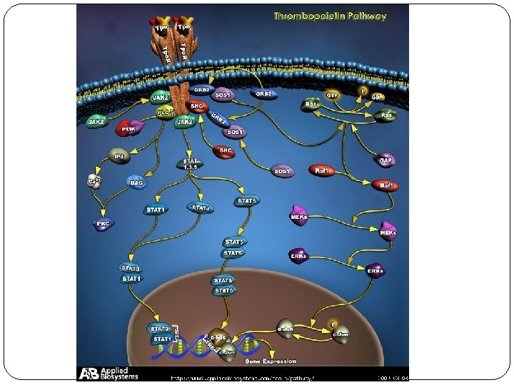
, is a glycoprotein hormone")
Thrombopoietin (leukemia virus oncogene ligand, megakaryocyte growth and development factor), is a glycoprotein hormone produced mainly by the liver and the kidney that regulates the production of platelets by the bone marrow It stimulates the production and differentiation of megakaryocytes, the bone marrow cells that fragment into large numbers of platelets
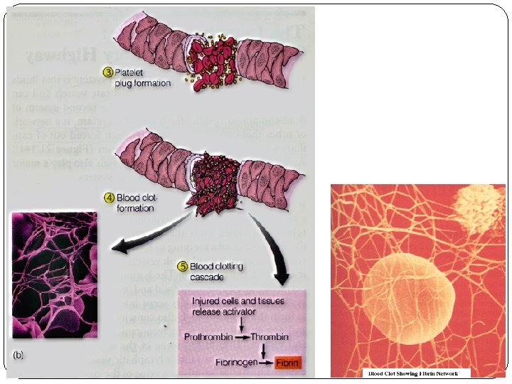
 The arrest of bleeding following injury and the result of 3 interacting, overlapping")
Hemostasis(止血) The arrest of bleeding following injury and the result of 3 interacting, overlapping mechanisms: Vascular spasm(血管收缩) Formation of a platelet plug(血小板血栓形成) Blood coagulation (clotting)(血液凝固) Bleeding time (出血时间):<9 min
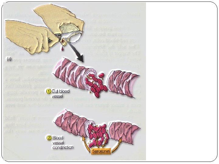

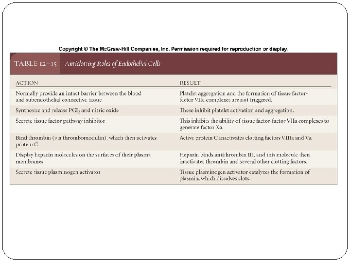
Role of vascular endothelium in hemostasis o Vasoconstriction: reduced blood flow facilitates contact activation of platelets and coagulation factors o Exposure of sub-endothelial basement membrane and collagen o Release of tissue thromboplastins (组织因子) o Synthesis of basement membrane components, tissue factor (组织因子), v. WF, plasminogen activator (纤溶酶原激活物), antithrombin III (抗凝血 酶III), thrombomodulin (血栓调节蛋白)

Signaling mediates responses to damage in a blood vessel: adjacent endothelial cells are a source of signals that influence platelet aggregation and alter blood flow and clot formation at the affected site.

Role of platelets in hemostasis Release of vasoconstricting substances Formation of the "platelet plug" Promotion of blood clotting Clot retraction

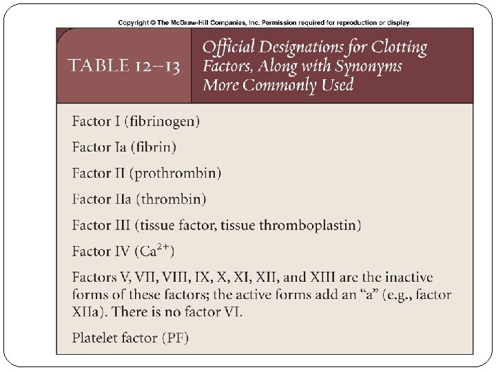
u Blood coagulation Clotting factors Clotting factor Synonyms I II IV V VIII IX X XI XIII fibrinogen纤维蛋白原 prothrombin凝血酶原 tissue thromboplastin组织因子 Ca 2+ proaccelerin前加速素易变因子 proconvertin前转变素稳定因子 antihemophilic factor抗血友病因子 plasma thromboplastin component血浆凝血活酶 Stuart-Prower factor plasma thromboplastin antecedent血浆凝血活酶前质 contact factor接触因子 fibrin-stabilizing factor纤维蛋白稳定因子

The liver plays a critical role in producing and modifying blood-borne proteins, including those used in the clotting pathway. Moreover, bile salts from the liver facilitate the absorption of lipids in the diet, including vitamin K, which is required for the synthesis of prothrombin.
 II (prothrombin) Tissue factor")
Coagulation factors and related substances Number and/or name I (fibrinogen) II (prothrombin) Tissue factor Calcium Function Forms clot (fibrin) Its active form (IIa) activates I, V, VIII, XIII, protein C, platelets V (proaccelerin, labile factor) Co-factor of VIIa (formerly known as factor III) Required for coagulation factors to bind to phospholipid (formerly known as factor IV) Co-factor of X with which it forms the prothrombinase complex VI VII (stable factor) VIII (antihemophilic factor) IX (Christmas factor) X (Stuart-Prower factor) XI (plasma thromboplastin antecedent) XII (Hageman factor) XIII (fibrin-stabilizing factor) von Willebrand factor prekallikrein high-molecular-weight kininogen (HMWK) fibronectin antithrombin III heparin cofactor II Unassigned – old name of Factor Va Activates IX, X Co-factor of IX with which it forms the tenase complex Activates X: forms tenase complex with factor VIII Activates II: forms prothrombinase complex with factor V Activates IX Activates factor XI and prekallikrein Crosslinks fibrin Binds to VIII, mediates platelet adhesion Activates XII and prekallikrein; cleaves HMWK Supports reciprocal activation of XII, XI, and prekallikrein Mediates cell adhesion Inhibits IIa, Xa, and other proteases; Inhibits IIa, cofactor for heparin and dermatan sulfate ("minor antithrombin") protein C protein S Protein Z-related protease inhibitor (ZPI) Inactivates Va and VIIIa Cofactor for activated protein C (APC, inactive when bound to C 4 b-binding protein) Mediates thrombin adhesion to phospholipids and stimulates degradation of factor X by ZPI Degrades factors X (in presence of protein Z) and XI (independently) plasminogen alpha 2 -antiplasmin tissue plasminogen activator (t. PA) urokinase plasminogen activator inhibitor-1 (PAI 1) plasminogen activator inhibitor-2 (PAI 2) cancer procoagulant Converts to plasmin, lyses fibrin and other proteins Inhibits plasmin Activates plasminogen Inactivates t. PA & urokinase (endothelial PAI) Inactivates t. PA & urokinase (placental PAI) Pathological factor X activator linked to thrombosis in cancer protein Z

Exploration of the details of the clotting pathway has yielded detailed information about the sequence, only a portion of which is represented here. Note thrombin’s influence in three different directions.

Knowledge that thrombin plays a central role in clotting has generated detailed studies of the possible pathways resulting in its formation: the extrinsic pathway is the more important of the two under most circumstances.

Coagulation cascade 3 processes 2 pathways
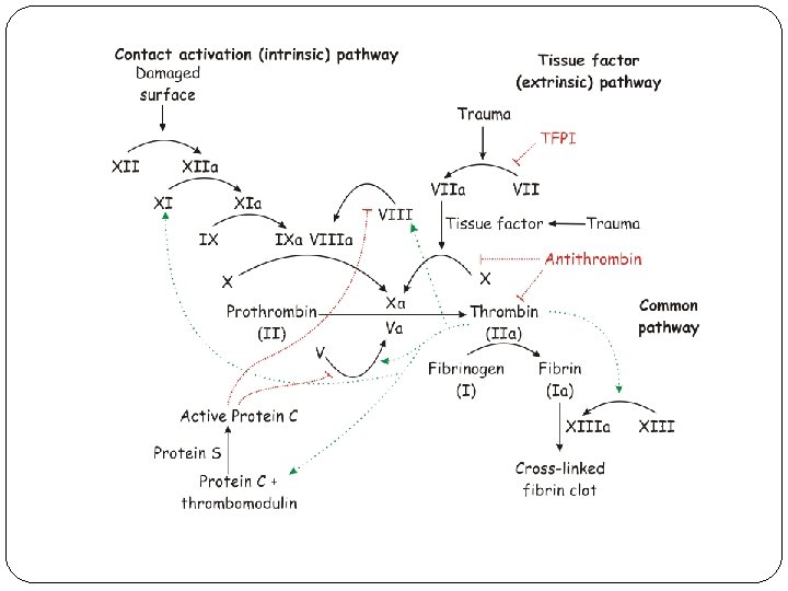
 Following damage to the blood vessel, endothelium Tissue Factor (TF)")
Tissue factor pathway (extrinsic) Following damage to the blood vessel, endothelium Tissue Factor (TF) is released, forming a complex with FVII and in so doing, activating it (TF-FVIIa). TF-FVIIa activates FIX and FX. FVII is itself activated by thrombin, FXIa, plasmin, FXII and FXa. The activation of FXa by TF-FVIIa is almost immediately inhibited by tissue factor pathway inhibitor (TFPI). FXa and its co-factor FVa form the prothrombinase complex, which activates prothrombin to thrombin. Thrombin then activates other components of the coagulation cascade, including FV and FVIII (which activates FXI, which, in turn, activates FIX), and activates and releases FVIII from being bound to v. WF. FVIIIa is the co-factor of FIXa, and together they form the "tenase" complex, which activates FX; and so the cycle continues. ("Tenase" is a contraction of "ten" and the suffix "-ase" used for enzymes. )
 The contact activation pathway begins with formation of the primary")
Contact activation pathway (intrinsic) The contact activation pathway begins with formation of the primary complex on collagen by high-molecular-weight kininogen (HMWK), prekallikrein, and FXII (Hageman factor). Prekallikrein is converted to kallikrein and FXII becomes FXIIa converts FXI into FXIa. Factor XIa activates FIX, which with its co-factor FVIIIa form the tenase complex, which activates FX to FXa. The minor role that the contact activation pathway has in initiating clot formation can be illustrated by the fact that patients with severe deficiencies of FXII, HMWK, and prekallikrein do not have a bleeding disorder.

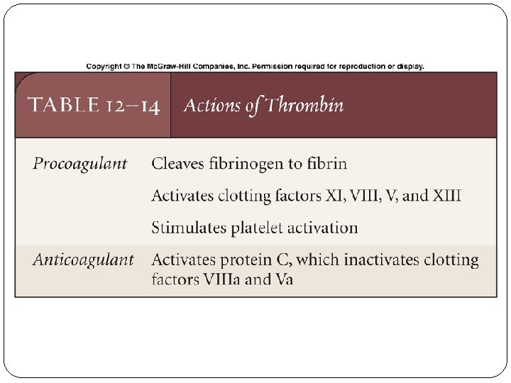
Final common pathway Thrombin has a large array of functions Its primary role is the conversion of fibrinogen to fibrin, the building block of a hemostatic plug. In addition, it activates Factors VIII and V and their inhibitor protein C (in the presence of thrombomodulin), and it activates Factor XIII, which forms covalent bonds that crosslink the fibrin polymers that form from activated monomers. Following activation by the contact factor or tissue factor pathways, the coagulation cascade is maintained in a prothrombotic state by the continued activation of FVIII and FIX to form the tenase complex, until it is downregulated by the anticoagulant pathways.

Structure of Fibrinogen Fibrin Polymerization

A deficiency of a clotting factor can lead to uncontrolled bleeding. Vitamin K is a cofactor needed for the synthesis of factors II, VII, IX, & X in the liver. So a deficiency of Vitamin K predisposes to bleeding.

Hemophilia & Bolshevik Revolution Rasputin http: //en. wikipedia. org/wiki/Grigori_Ras putin
 serum = plasma – fibrinogen and some of the other clotting factors")
Serum (血清) serum = plasma – fibrinogen and some of the other clotting factors + substances released by vascular endothelial cells and platelets Clotting time (凝血时间): 4 -12 min
 o Serine Protease Inhibitor Antithrombin III(抗凝血酶III) inhibiting all serine proteases of")
u Anticoagulants(抗凝物质 ) o Serine Protease Inhibitor Antithrombin III(抗凝血酶III) inhibiting all serine proteases of the blood coagulation system, including: o thrombin o factor IXa, XIa, XIIa
 Protein C, thrombomodulin, Protein S… o Tissue factor pathway inhibitor")
o Protein C system(蛋白C系统) Protein C, thrombomodulin, Protein S… o Tissue factor pathway inhibitor (TFPI)(组织因子途径抑制物)

In an uninjured vessel, thrombin bound to thrombomodulin activates protein C, which blocks the clotting response.
 Ø A polysaccharide produced by the tissue mast cells and the basophils")
o Heparin(肝素) Ø A polysaccharide produced by the tissue mast cells and the basophils of circulating blood Ø Interfering primarily with the action of thrombin after combining with antithrombin III
 o 2 processes o Activation of plasminogen o Degradation of fibrin o 4")
Fibrinolysis(纤维蛋白溶解) o 2 processes o Activation of plasminogen o Degradation of fibrin o 4 components of plasma fibrinolysis system o Plasminogen(纤维蛋白溶解酶原) o Plasmin(纤维蛋白溶解酶) o Plasminogen activator o Plasminogen inhibitor

Following tissue repair, fibrin clots are dissolved in a process mediated by plasmin; synthetic plasminogen activators can be used immediately after a stroke or heart attack to help dissolve clots and restore blood flow.
")
o 2 pathways of plasminogen activation Fibrin Degradation Products (FDP)
 Urokinase o Plasminogen inhibitor Plasminogen")
o Extrinsic Plasminogen activator Tissue-type plasminogen activator (t. PA) Urokinase o Plasminogen inhibitor Plasminogen activator inhibitor type-1 (PAI-1) 2 -antiplasmin Antithrombin III

Blood group o Erythrocytes carry on their surfaces many antigens, but the most important and commonly recognized are the A and B substances and the Rhesus (Rh) factors

ABO group
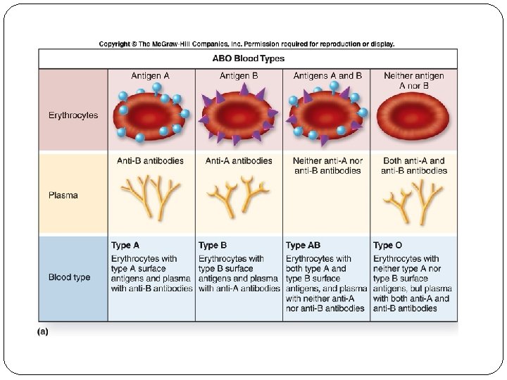

Blood type Antigen Antibody A A anti-B B B anti-A AB A & B neither O neither anti-A & anti-B

O AB A B

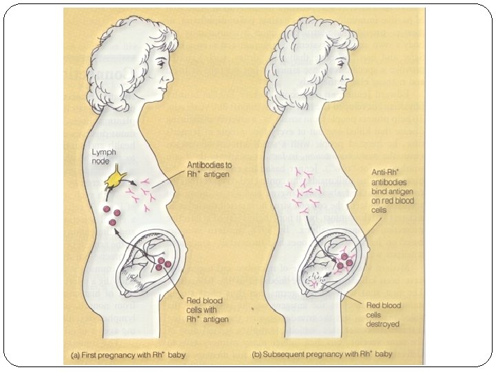
Rh group inherited independent of ABO system Rh positive = antigen present (mainly D antigen) & no antibodies Rh negative = no antigen & antibodies will be produced if exposure occurs

Blood volume & blood transfusion Blood volume The total blood volume is 7 ~ 8% of body weight. For a 70 Kg male, it is 5. 0 ~ 5. 5 L.

u Blood transfusion Transfusion is the process of replacing blood or blood component which a body has lost in surgery, through an accident or as a result of medical treatment such as chemotherapy. Sterility, Viability, Quantity, Safety & Quality

Risk from Transfusion 1. Allergic reactions to the blood or one of its components 2. Hemolytic reaction 3. Diseases transmission, such as HIV, Hepatitis B, C virus

Basic principles 1. Unexpected, emergency blood transfusion is rarely required. It is needed only in situations of massive hemorrhage like severe trauma, gynecologic and obstetric emergency, or gastrointestinal bleeding. 2. In many cases, resuscitation can be achieved by use of colloid or crystalloid plasma expanders instead of blood. 3. Blood transfusion is not free of risk, even in the best of conditions.

Guideline 1. Ensuring that transfusion recipients and donors have compatible blood group 2. Cross-match test 3. Tests screening for Hepatitis virus, HIV… in blood donated

Donor Cross-match Test Recipient RBC Plasma

Blood transfusion options Option Definition Advantage Disadvantage Preoperative Autologous Donation A patient's blood is collected and stored until needed Disease transmission and allergic reactions are eliminated Must be planned in advance May delay surgery Certain medical conditions disqualify Perioperative Autologous Transfusion Blood lost during or after surgery is collected, processed and returned Disease transmission and allergic reactions are eliminated Must be planned in advance Certain medical conditions disqualify Volunteer Blood Donation Blood voluntarily donated to a community blood center Availability in emergencies Risk of disease transmission and allergic reaction Directed Donor Blood Donation Patient selects blood Patient feels safe donor with donors selected May be higher risk of disease transmission and allergic reaction Blood type must be compatible or identical Must be planned in advance Some hospitals will not accept

v Transfusion of whole blood v Transfusion of blood components Ø Your blood is sent to the lab to determine blood type and to check for viral diseases. Ø It is sent to the blood component lab to be divided up into plasma, platelets and red blood cells. Ø It goes to hospital services to be distributed to the Hospital blood bank. Ø Finally it goes to a thankful recipient.

Thank you for your attention!
fcd13323c8f0a3eb71ae6337f772e9e3.ppt