f430e91def64b13ac0640ec6034a3a31.ppt
- Количество слайдов: 20


Blinded Light by the The irradiation of lenses during routine CT head examinations across a NHS Trust; a quality improvement project Dr Jack Roberts, Dr Hebah Taufik Supervisors: S Perrio, A Alam

Background § The lens of the eye is sensitive to ionising radiation. § Radiation induced cataractogenesis is recognised at § repeated exposure. CT head examinations are increasing in frequency with many patients experiencing repeated examination. [1] § 3. 57% of all radiological exposures in 2008 vs. 2. 23% in 1997 § The International Commission on Radiological Protection § (ICRP) has recently reduced 8 fold the threshold absorbed dose limit for the lens to 0. 5 Gy. (ICRP, 2011) The typical dose to the lens during a CT head (excluding the lens) is as much as 0. 05 Gy. [2, 3]

The Problem § Exposure of the lens is of no diagnostic benefit. § The standard is that all CT head scans should aim to exclude the lenses. (Ionising Radiation (Medical Exposure) Regulations 2000). § Radiologists had subjectively noted that the rate of lens § inclusion at the trust was higher than the acceptable standard. Our aim was to measure compliance with a preliminary audit and look to improve practice if required.

Lens

Initial Audit Method § A retrospective sample of 50 consecutive CT head scans were chosen at both trust sites. § The patient demographics, examination indication and the exposure of both, one or neither lens were collected from review of the PACS and CRIS systems. § Those patients scanned whilst in a neck collar were excluded from the audit.

Results Site 1 Site 2 Number Av Age % Both 33 69. 6 70% 15 70. 8 30% Single 6 58. 3 13% 11 74. 5 22% Neither 8 42. 4 17% 24 65. 9 48% Total 47 62. 7 100 50 69. 2 100% • At Site 1, at least one lens was exposed in 83% of scans (39/47) • At Site 2, at least one lens was exposed in 52% of scans (26/50) • Across the entire trust, at least one lens was exposed in 67% (65/97)

Results § As a trust, we were failing to meet the irradiation § § standard that patient lenses should be avoided during CT head examinations. Scanning at Site 2 was closer to the standard than at Site 1. This may have been related to the types of cases scanned. At both sites, the lenses of younger patients were scanned less often than those of older patients.

Problem Analysis § The most significant reason for the inclusion of lenses on § CT head examinations is incorrect patient positioning. The patient should be positioned with the neck flexed and CT gantry tilted.

Gantry Scout film https: //www. med-ed. virginia. edu/courses/rad/headct/technique. html

Problem Analysis § The most significant reason for the inclusion of lenses on § CT head examinations is incorrect patient positioning. The patient should be positioned with the neck flexed and CT gantry tilted. § Responsible factors: § Patient immobilisation (these patients were excluded) § Patient inability (with increasing age some patients are unable to comply, this may explain the age differential) § Inadequate equipment § Inadequate planning

Delivery Plan 1. A replacement head sponge to be ordered as a matter of urgency. a) In the interim, the use of a rolled up towel/gown was suggested. 2. The importance of correct positioning in view of the recent ICRP press release and IRMER legislation was reinforced with an email to the CT radiographers and reminder posters within the CT control rooms. 3. Re-audit in 3 months.

Delivery Plan § Team Members on board: § CT Superintendent § CT Radiographers § Radiologists – CT and Audit Lead § Costs: § Minimal! § Replacement head sponge approx. £ 50

Recommendations in use

Measuring Quality Improvement Success: RE-AUDIT

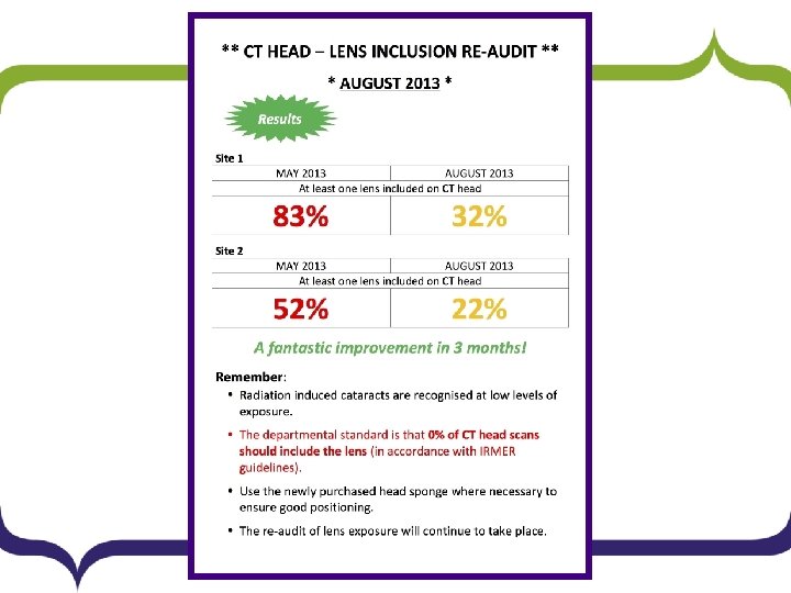
Results Site 1 Site 2 Number Av Age % Both 11 79. 1 22% 1 68 2% Single 5 61 10% 10 76. 7 20% Neither 33 54. 9 67% 39 70. 7 78% Total 49 60. 5 100% 50 71. 9 100% • At Site 1, at least one lens was exposed in 32% of patients (16/49) vs. 83% previously. • At Site 2, at least one lens was exposed in 22% (11/50) vs. 52% previously. • Across the entire trust, at least one lens was exposed in 27% (27/99) vs. 67% previously.

Conclusions § Compliance has improved. § Positive steps have been made – there remains § scope for improvement. Scanning at the Site 2 remains closer to the standard than at Site 1.

Further Recommendations § The positive results of the audit have been emailed to the radiographers and continued compliance is to be encouraged in departmental newsletters. § A further poster displaying the results of the audit has been displayed in the CT control room and staff room. § Re-audit will be performed in 9 months to monitor longterm compliance and the project has been added to the annual audit list.

Acknowledgements and References § With thanks to Dr Stephen Perrio and Dr Adeeb Alam who supervised this project. § References: 1. Frequency and Collective Dose for Medical and Dental X-ray Examinations in the 2. 3. UK, 2008 D Hart, B F Wall, M C Hillier and P C Shrimpton, HPA-CRCE-012 A Comparison of reduction in CT dose through the use of gantry angulations or bismuth shields, 2006, D. E. Heaney, C. A. J. Norvill, Australasian Physics & Engineering Sciences in Medicine Volume 29, Issue 2 , pp 172 -178 https: //rpop. iaea. org/RPOP/RPo. P/Content/Information. For/Health. Professionals/6_Ot her. Clinical. Specialities/radiation-cataract-patient-protection. htm

Thank you Any comments or questions?
f430e91def64b13ac0640ec6034a3a31.ppt