эндоскопическая хирургия (ин).ppt
- Количество слайдов: 152

Bases of endoscopic surgery

“Grosse Chirurge machen grosse Schnitte”

Endoscopic surgery it is area of the surgery, allowing to execute radical operations or diagnostic procedures without a wide dissection of integument or through dot punctures of tissues (laparoscopic, thoracoscopic, rhinoscopic, arthroscopic operations), or through natural physiological apertures (FGDS, colonoscopy, bronchoscopy, cystoscopy, etc. )

Development of endoscopic surgery u u u Hippocrat (460 -375 up to AD) - has described carrying out of the proctoscopy; Abdul Quasim (936 -1013) - investigated neck of uterus using a glass mirror reflector; R. P. Arnaud (1651 -1723) - has created the first extracorporal source of light for the medical purposes; Phillip Bozini (1773 -1809) - has created endoscope which design has been named "LICHTLEITER"; John Fisher, 1827 – has created one of the first endoscops; Gustave Trouve in 1873 in has designed "polyscope", intended for gastroscopy and cystoscopy, brightness of a luminescence of a platinum wire in which was adjusted with a help of a rheostat.

u u u u u George Kelling (1901) – for the first time has made a laparoscopy in experiment on a dog; D. O. Ott (1901) – has informed about "ventroscopy" inspection of a abdominal cavity by means of a candle, a frontal mirror and a tube; Heinz Kalk (1928 г) - has developed a technique laparoscopic puncture biopsy of a liver, and in 1939 has published the work based on research of 200 patients; Janos Veress (1938) - has invented a needle with spring mandrin. For today is most widely used tool for imposing of pneumoperitoneum; Raul Palmer (1947) - has offered ways of definition of position of a needle widely used now for insufflation (Palmer-test); Kurt Semm - with the colleagues and pupils have developed methodics of the majority laparoscopic interventions on organs of a small pelvis, have created enormous amount laparoscopic tools and devices De Kok in 1977 for the first time has executed laparoscopic appendectomy; E. Muhe (1985 г) - has executed the first laparoscopic cholecystectomy; U. I. Gallinger (1991 г) for the first time in Russia has executed laparoscopic cholecystectomy in the Science Centre of surgery of Russian Academy of Medical Science.

Light source of Arno

Philipp Bozzini (1773 -1809)

Эндоскоп Джона Фишера (1827)

Trouve’s “polyscope”

Эндоскоп Антонина Жана Дезормо (Antonin Jean Desormeaux) 1853

Гастроскоп Иохана Микулича (Johann Mikulicz) 1881

Гастроскоп Иохана Микулича (Johann Mikulicz) 1881

Георг Келлинг (George Kelling) "Осмотр пищевода и желудка с помощью гибких инструментов" 23 сентября 1901

Эндоскопический инструментарий Максимилиана Нитце (Max Nitze) и Жозефом Ляйтером (Josef Leiter) 1883

Цистоскоп Максимилиана Нитце с инструментальным каналом 1897

Ханс Кристиан Джакобеус – профессор медицины Стокгольмского университета

Хайнц Кальк (Heinz Kalk) -основатель немецкой лапароскопической школы

Raul Palmer

Карл Шторц (Karl Storz)

Первый экстракорпоральный источник холодного света фирмы Карл Шторц (Karl Storz) 1964

Курт Земм

Экспериментальная лапароскопическая холецистэктомия на животном, 1985

Филипп Мурэ (Phillipe Mouret) 1987

Advantages of endosurgery in comparison with traditional operations u u u Slight trauma of tissues Short hospital period Decrease of disability terms Cosmetic effect Decrease of frequency and weight of complications Economic efficiency

Complications General lethality come to 0, 5 %, and frequency of complications – 10 %; u Wound infection – meets in 1 -2 % of cases; u Damage of internal organs; u Pneumomediastinum, subcutaneous emphysema; u Pneumothorax; u Development of a gas embolism u Electrosurgical damages; u Cardiovascular collapse; u Postoperative pain in a right shoulder; u Damage of vessels and nerves of a forward belly wall; u

Relative contraindications u u u Heavy accompanying pathology of cardiovascular and respiratory systems - Obstructive diseases of lungs - Cardiovascular insufficiency of 2 -3 degrees - Old myocardial infarction - The transferred operations on heart and large vessels - The congenital and acquired heart diseases Diffuse peritonitis Heavy coagulopathy Adiposity of 3 -4 degrees Late terms of pregnancy Portal hypertensia

The minimal set for carrying out endoscopic operations a) Needles for imposing pneumoperitoneum; b) trocars with clamps and adapters; c) Tools for suture of trocar apertures; d) Manipulators: dissectors, cissors, clips, retractors; e) The equipment for irrigation and aspiration; f) Tools for coagulation; i) Suture materials and tools for endoscopic suture; j) Devices for ligation vessels and ducts.

The general requirements to endoscopic tools а) Handiness: the handle of the tool should not complicate manipulations, at long operation there should not be a weariness of a wirst; u б) Sensitivity: the tool should provide the maximal sensitivity as the surgeon is deprived at endoscopic manipulations of tactile sensitivity; u в) Electroisolation: isolating layer should reach up to branches of the tool and to be strong enough; u г) Presence of the rotary mechanism providing rotation of a working part of the tool on 360 degrees around of a longitudinal axis. u

Essentially the complex will consist of the following blocks: a) A videocamera; b) A video monitor; c) The illuminator - the electronic device having a powerful lamp (xenon or halogen); d) Laparoscope with an optical path; e) Insufflator - it is intended for submission of carbonic gas in a abdominal cavity at imposing and maintenance of pneumoperitoneum; f) Aquapurator - it is intended for washing and evacuation of liquid contents of a abdominal cavity; i) Electrocoagulator; j) The rack - handcart.


Харольд Хопкинс (Harold H. Hopkins)


Введение первого троакара

Ревизия брюшной полости

Введение первого троакара

Срабатывания защиты троакара

Положение Тренделенбурга

Положение Фаулера

Положение на боку


Традиционная холецистэктомия


Лапароскопическая холецистэктомия

ОСНОВНЫЕ ЭТАПЫ ЛАПАРОСКОПИЧЕСКОЙ ОПЕРАЦИИ u I. Оперативный доступ (1. Наложение пневмоперитонеума (карбоксиперитонеума), введение первого троакара и ревизия брюшной полости; 2. Экспозиция). u II. Оперативный прием (3. Операция). u III. Выход из операции (4. Окончание операции).

Положение пациента и хирургической бригады при лапароскопической холецистэктомии: А –французский вариант; Б – американский вариант


Грыжесечение

Грыжа - выход внутренностей, покрытых париетальной брюшиной, из полости живота через слабые места в мышечно-апоневротическом слое брюшной стенки под кожу. Эвентрация - выход внутренностей из брюшной полости через разрыв париетальной брюшины. Элементы грыжи: 1. Грыжевые ворота - щель или отверстие в брюшной стенке, через которое выходят органы брюшной полости. 2. Грыжевой мешок - париетальный листок брюшины, выталкиваемый выходящими из брюшной полости внутренностями. Состоит из шейки, тела и дна. - Шейка - участок брюшины на уровне грыжевых ворот, являющийся анатомической границей между полостью брюшины и полостью грыжевого мешка. 3. Содержимое грыжевого мешка (чаще большой сальник и петли тонкой кишки).


Пластика передней стенки пахового канала по Мартынову

Пластика передней стенки пахового канала по Girard

Способ Marcy

Способ Bassini

Способ Bassini

Способ Shouldice

Способ Shouldice

Способ Postempski

Способ Postempski

Безнатяжная пластика

Способ Lichtenstein

Способ Lichtenstein

Способ Lichtenstein

Способ Gilbert

Способ Rutcov - Robbins

Способ Trabucco

Способ Trabucco


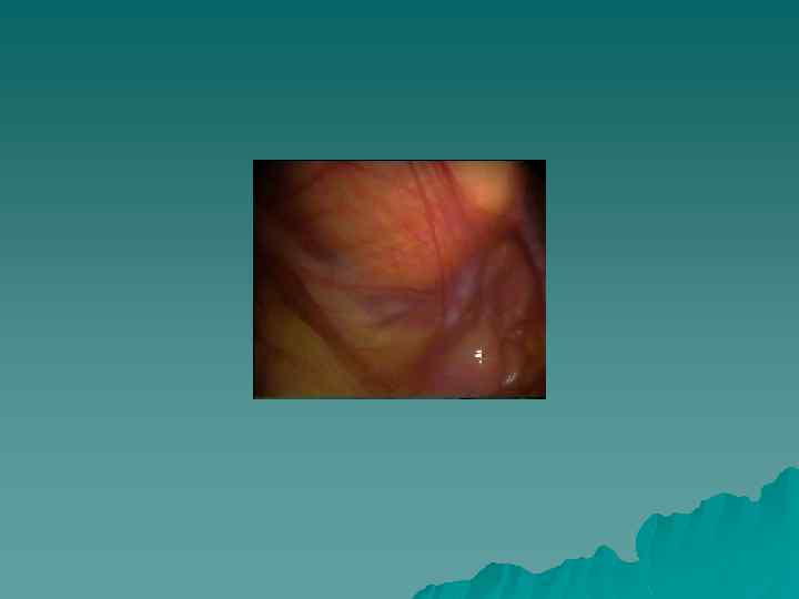
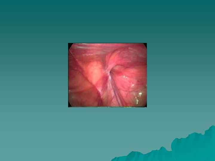



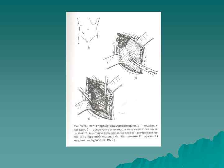
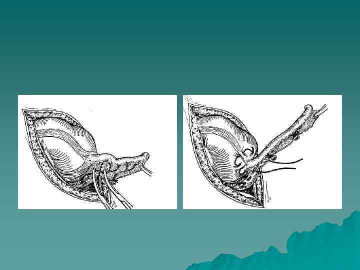
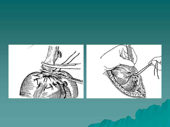
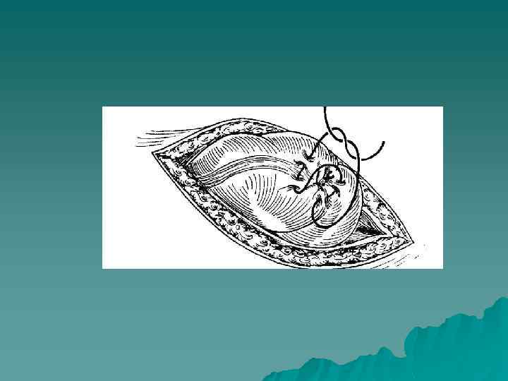
Аппендэктомия
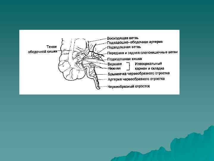
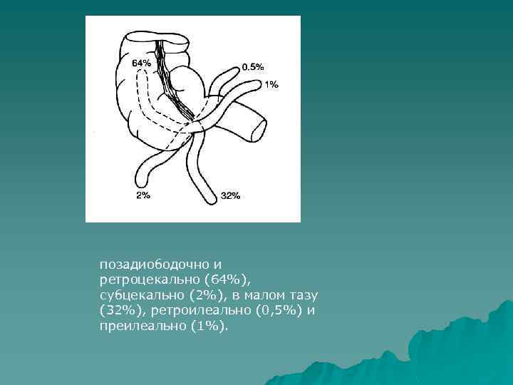
позадиободочно и ретроцекально (64%), субцекально (2%), в малом тазу (32%), ретроилеально (0, 5%) и преилеально (1%).


эндоскопическая хирургия (ин).ppt