Ankylosing Spondylitis RUS 2015.pptx
- Количество слайдов: 77


Ankylosing Spondylitis Gleb Slobodin MD 2017
. \"Steifigkeit der Wirbelsaule und ihre Verkrummung als besondere")
Discovery 1890 s Bechterew W. (1893). "Steifigkeit der Wirbelsaule und ihre Verkrummung als besondere Erkrankungsform". Neurol Centralbl 12: 426– 434 Strumpell A. (1897). "Bemerkung Marie P. (1898). "Sur la spondylose uber die chronische ankylosirende rhizomelique". Rev Med 18: 285– 315. Entzundung der Wirbelsaule und der Huftgelenke". Dtsch Z Nervenheilkd 11 (3– 4): 338– 342

Description 20 th century

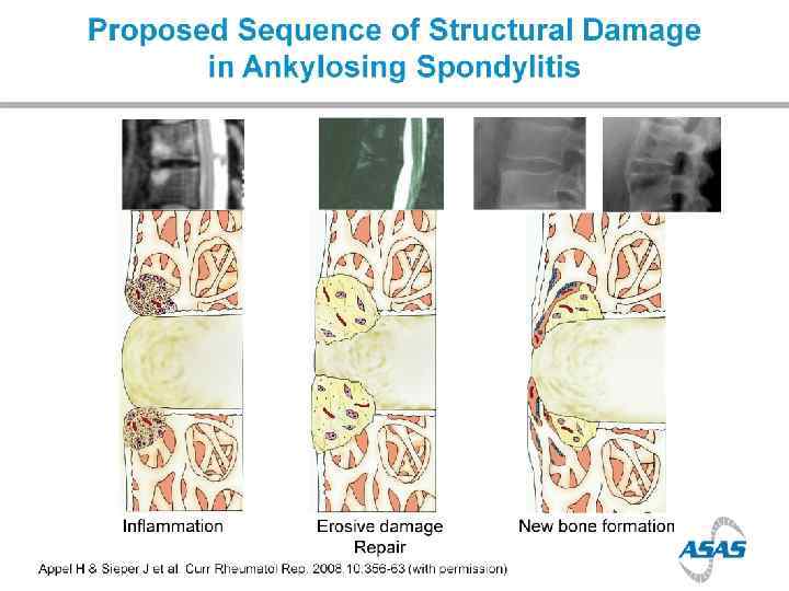
Bone formation in AS


Spine

BASMI

Sacroiliac joints

Spine

Patient’s symptoms One half comes from inflammation, the second half comes from structural changes

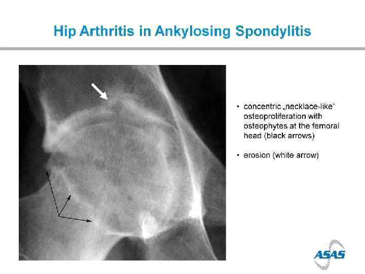
Arthritis

Enthesopathy
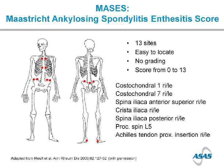
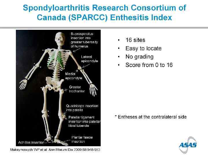

Enthesopathy
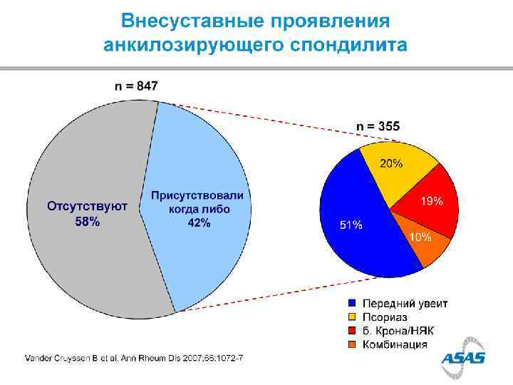

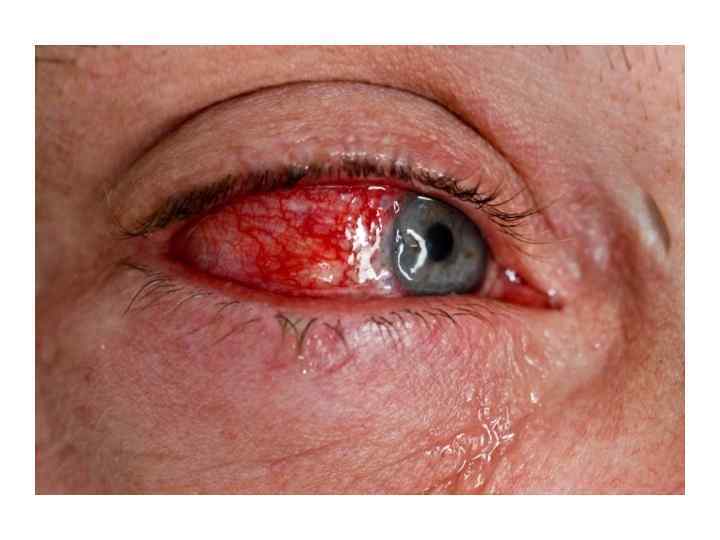
Uveitis

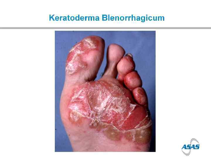
Diarrhea

Aortitis

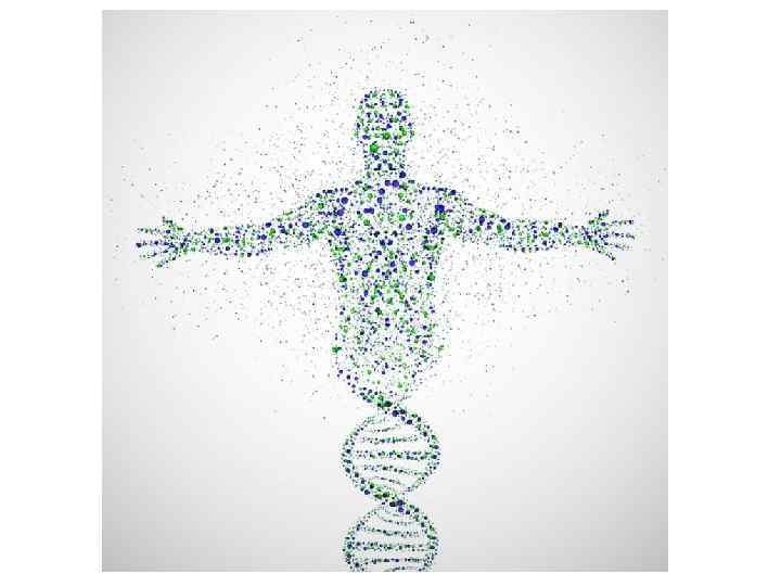
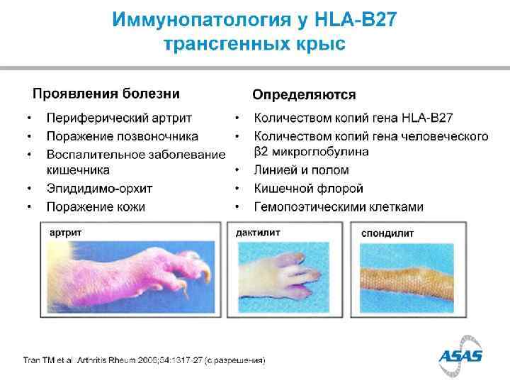
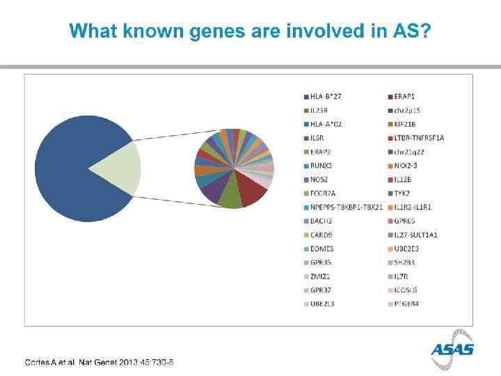
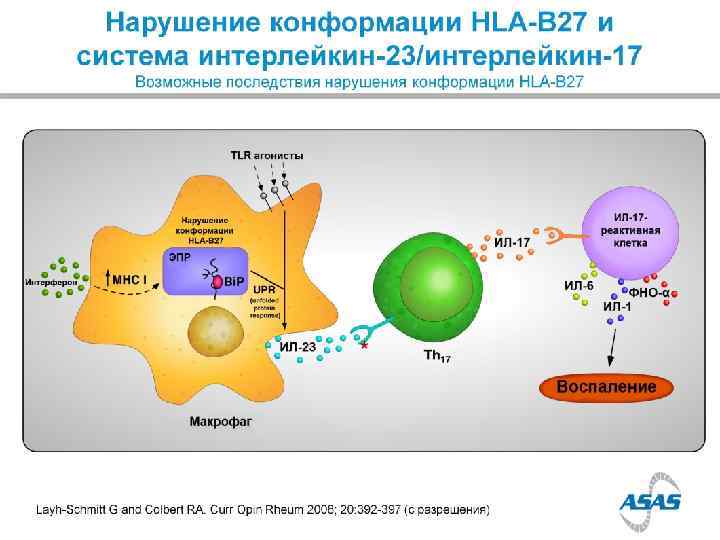
HLA B 27 1970 s HLA B 27 + in 90% of Caucasian AS pt Only 5% of HLA B 27+ persons will have AS HLA B 27 represents only about 25% of ‘genetic risk’ for AS

Non X-ray imaging Computed tomography Better comparing to X-rays But not validated and radiation-related

Non X-ray imaging of SIJ Bone scan • Sensitivity is about 50% Specificity is about 50%

Non X-ray imaging of SIJ MRI • 22 yo F with right LBP

Non X-ray imaging of SIJ MRI • 16 yo M with 6 months LBP

Non X-ray imaging of SIJ MRI • 33 yo M with Psoriasis & LBP

Spine - MRI
 chronic back pain in 20%")
Prevalence of Axial Sp. A in US (NHANES study) chronic back pain in 20% of population IBP in 7% of population; > in younger adults Ax. Sp. A (including AS) in 1. 4% AS in 0. 5% AS nr. Ax. Sp. A MALES=FEMALES (Ax. Sp. A); M: F=2: 1 (AS) 12% in 2 years

Disease progression

Disease progression > in males > in smokers > in those with high CRP >>> in those with syndesmophytes at baseline

Disease progression

TNF – i. TNF 1990 s-2000 s

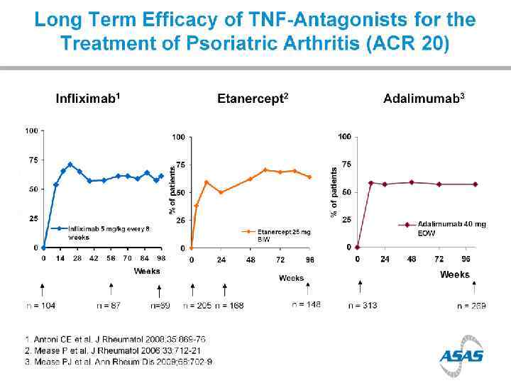
Efficacy of TNFi in AS

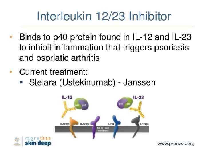
Other biologics: anti IL-17

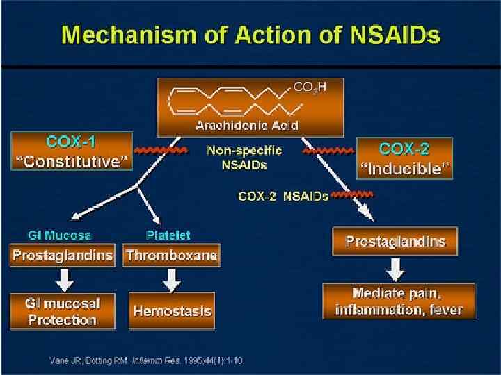
Anti-TNF & NSAIDS

A ROYAL FAMILY of rheumatic diseases

Q 1 A female with daily inflammatory low back pain during the last two years presents for evaluation. You do not find anything wrong on her examination. Your next step will be to order everything but HLA B 27. 1 X-ray of her pelvis. 2 MRI of her pelvis. 3 C-reactive protein. 4

Q 2 HLA B 27 is negative, and CRP is normal. Pelvis Xrays are normal as well. Your next step will be MRI of sacroiliac joints. 1 Technetium bone scan. 2 Computed tomography of the whole spine. 3 Follow up after 6 months. 4

Q 3 The MRI shows Enthesitis. 1 Bone marrow edema. 2 Capsulitis. 3 Erosion. 4

Q 4 MRI of sacroiliac joints is reported as abnormal, with bone marrow edema around SIJ on both sides. The diagnosis is Non specific low back pain. 1 Ankylosing spondylitis. 2 Undifferentiated spondyloarthritis. 3 Non-radiographic axial spondyloarthritis. 4

Q 5 Non-radiographic axial spondyloarthritis is An early phase of ankylosing spondylitis. 1 A variant of osteitis condensanse ilii. 2 A new name for DISH. 3 A variant of reactive arthritis. 4

Q 6 Recommended treatment will be NSAIDs. 1 Physical therapy. 2 TNF-alpha blockade. 3 Surgery. 4

The great A case from 1200 s BC

The great A case from 1200 s BC

DISH vs AS

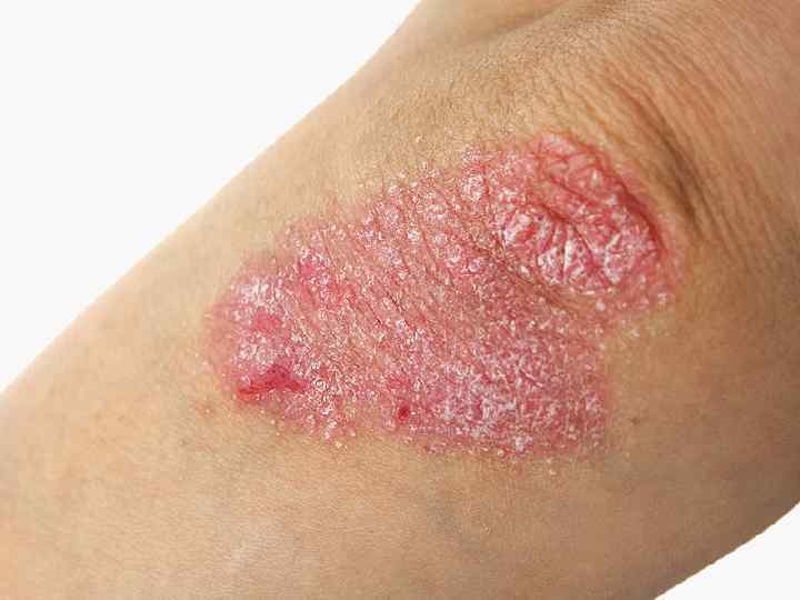
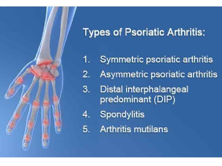
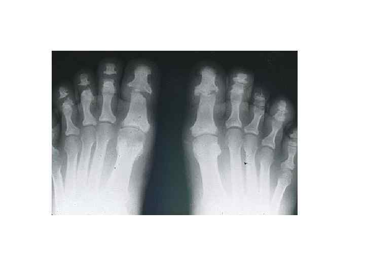
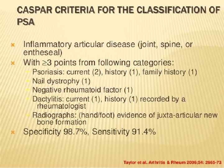
Psoriatic arthropathy

Psoriatic arthropathy

Psoriasis

Enthesopathy

Uveitis

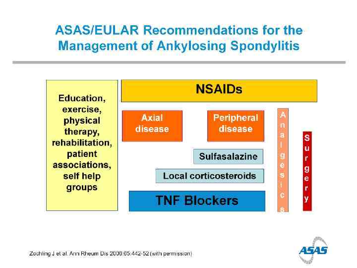
Treatment

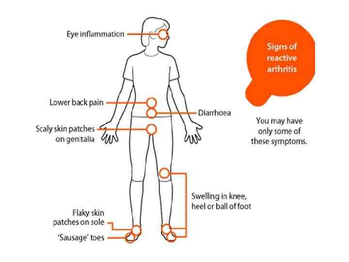
Reactive arthritis
Ankylosing Spondylitis RUS 2015.pptx