Anatomy of the Head & Neck The Facial

5194-the_skull-_maxilla_and_mandible_l4.ppt
- Количество слайдов: 47
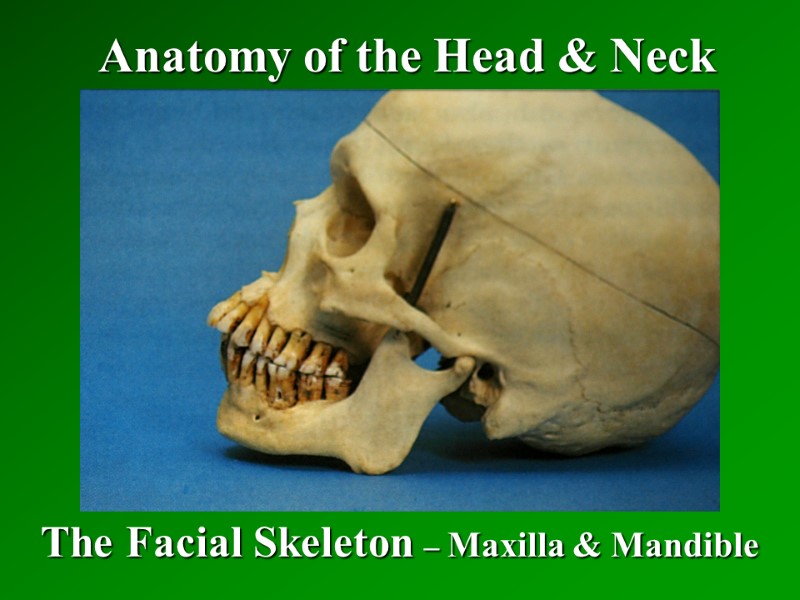
 Anatomy of the Head & Neck The Facial Skeleton – Maxilla & Mandible
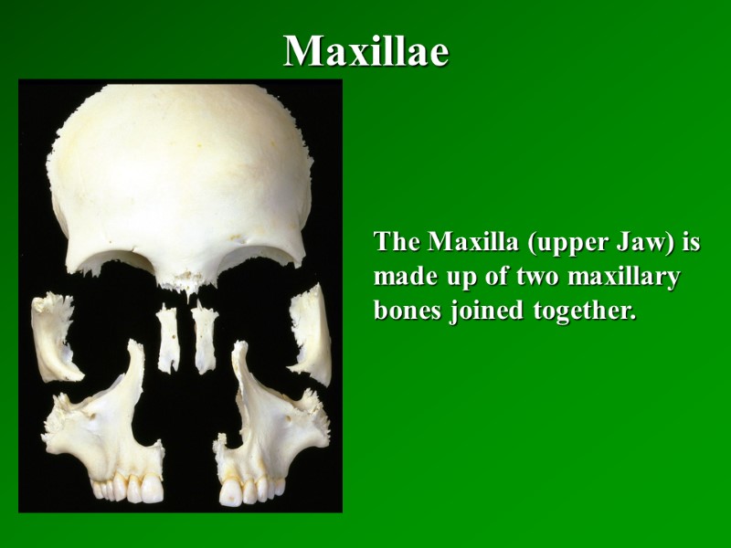
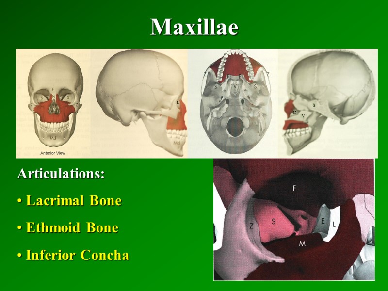
 Maxillae The Maxilla (upper Jaw) is made up of two maxillary bones joined together.
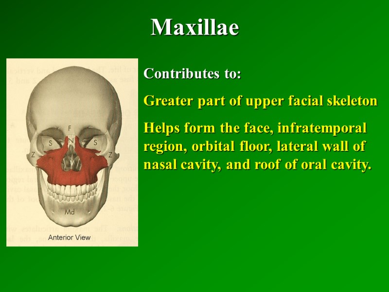
 Maxillae Contributes to: Greater part of upper facial skeleton Helps form the face, infratemporal region, orbital floor, lateral wall of nasal cavity, and roof of oral cavity.
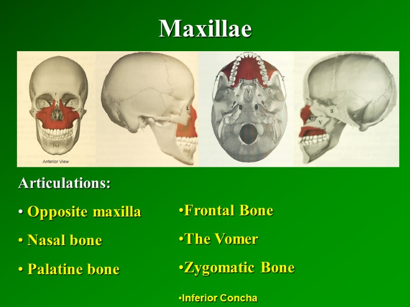
 Maxillae Articulations: Opposite maxilla Nasal bone Palatine bone Frontal Bone The Vomer Zygomatic Bone Inferior Concha
 Maxillae Articulations: Lacrimal Bone Ethmoid Bone Inferior Concha
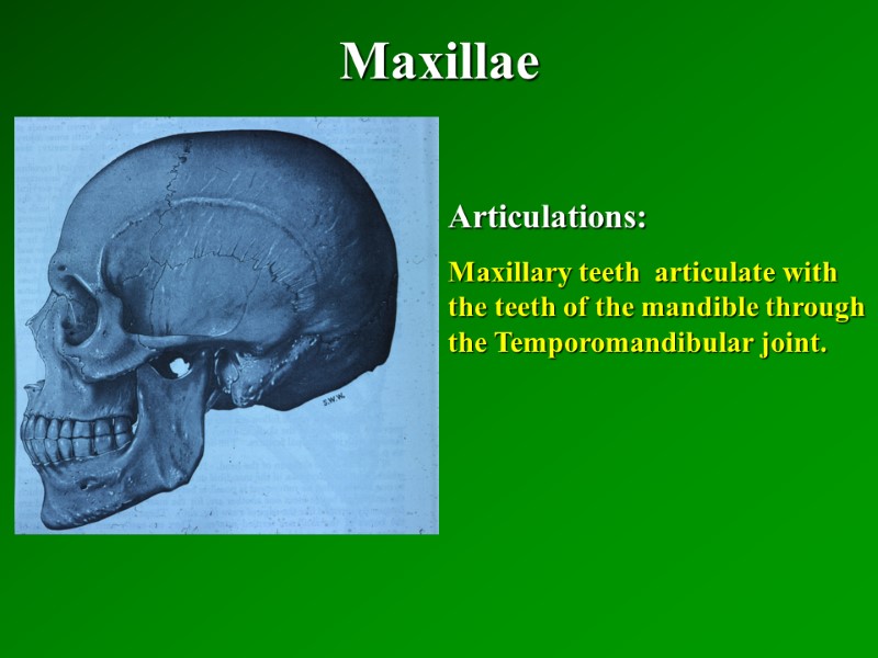
 Maxillae Articulations: Maxillary teeth articulate with the teeth of the mandible through the Temporomandibular joint.
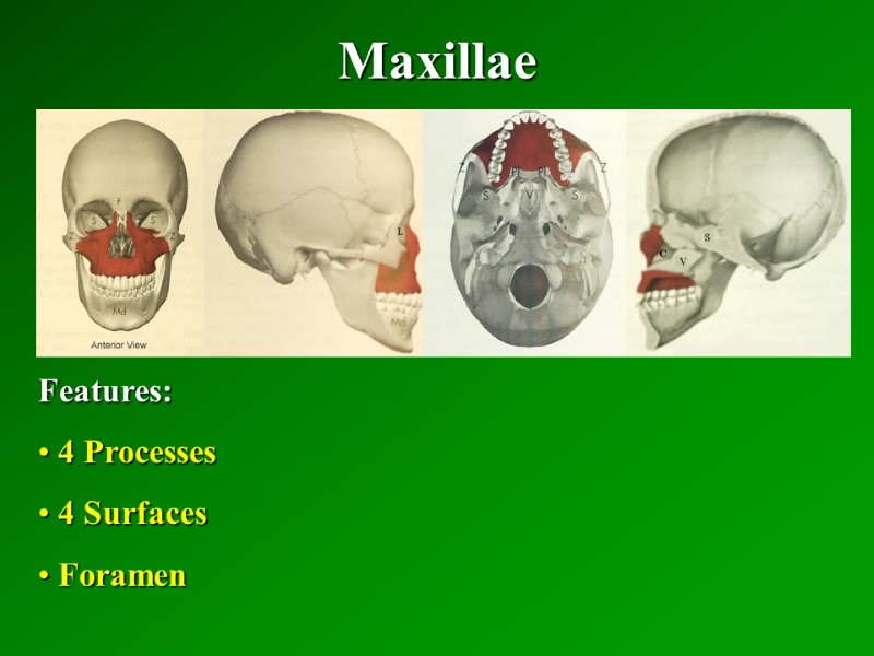
 Maxillae Features: 4 Processes 4 Surfaces Foramen
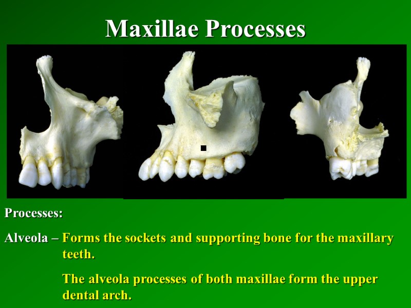
 Maxillae Processes Processes: Alveola – Forms the sockets and supporting bone for the maxillary teeth. The alveola processes of both maxillae form the upper dental arch. .
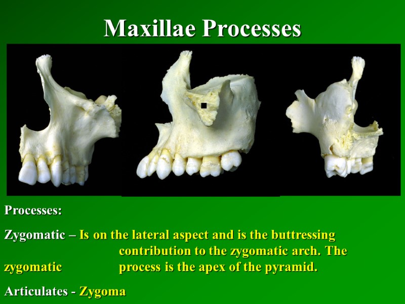
 Maxillae Processes Processes: Zygomatic – Is on the lateral aspect and is the buttressing contribution to the zygomatic arch. The zygomatic process is the apex of the pyramid. Articulates - Zygoma .
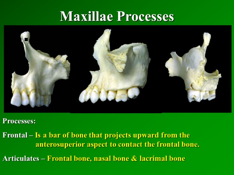
 Maxillae Processes Processes: Frontal – Is a bar of bone that projects upward from the anterosuperior aspect to contact the frontal bone. Articulates – Frontal bone, nasal bone & lacrimal bone .
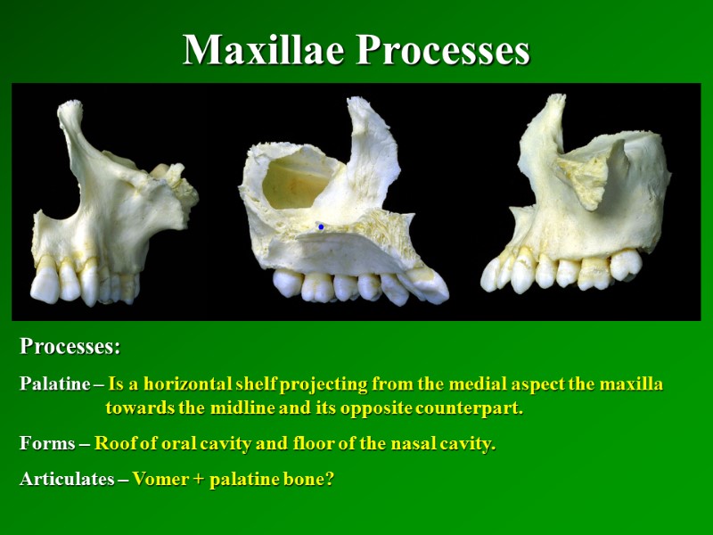
 Maxillae Processes Processes: Palatine – Is a horizontal shelf projecting from the medial aspect the maxilla towards the midline and its opposite counterpart. Forms – Roof of oral cavity and floor of the nasal cavity. Articulates – Vomer + palatine bone?
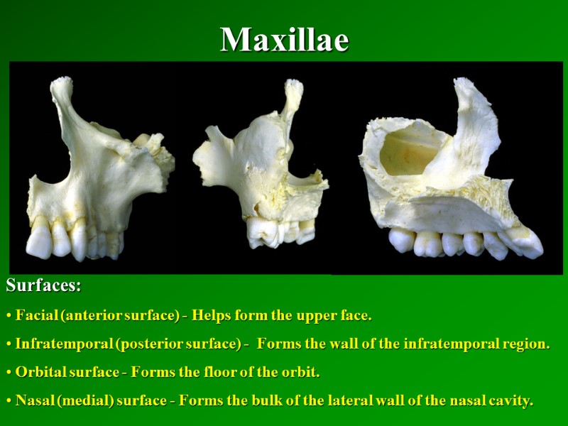
 Maxillae Surfaces: Facial (anterior surface) - Helps form the upper face. Infratemporal (posterior surface) - Forms the wall of the infratemporal region. Orbital surface - Forms the floor of the orbit. Nasal (medial) surface - Forms the bulk of the lateral wall of the nasal cavity.
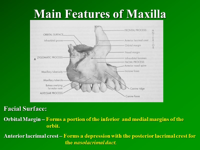
 Main Features of Maxilla Facial Surface: Orbital Margin – Forms a portion of the inferior and medial margins of the orbit. Anterior lacrimal crest – Forms a depression with the posterior lacrimal crest for the nasolacrimal duct.
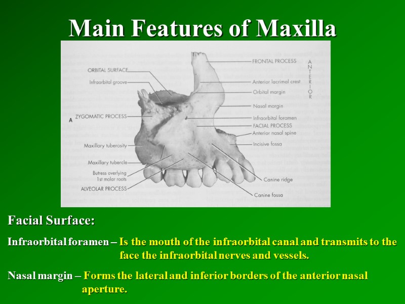
 Main Features of Maxilla Facial Surface: Infraorbital foramen – Is the mouth of the infraorbital canal and transmits to the face the infraorbital nerves and vessels. Nasal margin – Forms the lateral and inferior borders of the anterior nasal aperture.
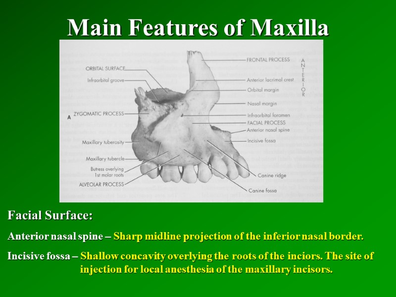
 Main Features of Maxilla Facial Surface: Anterior nasal spine – Sharp midline projection of the inferior nasal border. Incisive fossa – Shallow concavity overlying the roots of the inciors. The site of injection for local anesthesia of the maxillary incisors.
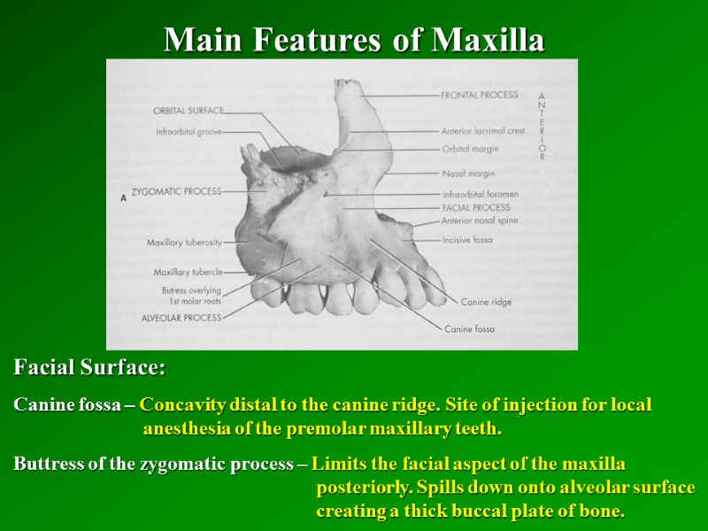
 Main Features of Maxilla Facial Surface: Canine fossa – Concavity distal to the canine ridge. Site of injection for local anesthesia of the premolar maxillary teeth. Buttress of the zygomatic process – Limits the facial aspect of the maxilla posteriorly. Spills down onto alveolar surface creating a thick buccal plate of bone.
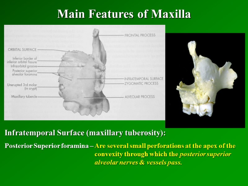
 Main Features of Maxilla Infratemporal Surface (maxillary tuberosity): Posterior Superior foramina – Are several small perforations at the apex of the convexity through which the posterior superior alveolar nerves & vessels pass.
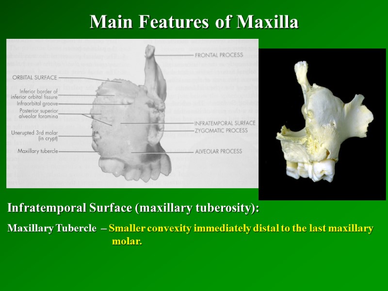
 Main Features of Maxilla Infratemporal Surface (maxillary tuberosity): Maxillary Tubercle – Smaller convexity immediately distal to the last maxillary molar.
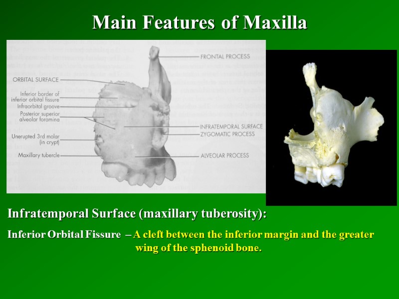
 Main Features of Maxilla Infratemporal Surface (maxillary tuberosity): Inferior Orbital Fissure – A cleft between the inferior margin and the greater wing of the sphenoid bone.
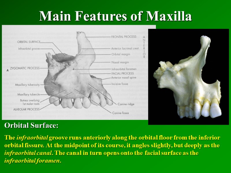
 Main Features of Maxilla Orbital Surface: The infraorbital groove runs anteriorly along the orbital floor from the inferior orbital fissure. At the midpoint of its course, it angles slightly, but deeply as the infraorbital canal. The canal in turn opens onto the facial surface as the infraorbital foramen.
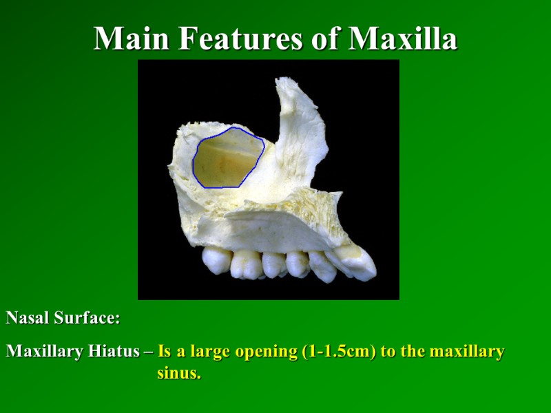
 Main Features of Maxilla Nasal Surface: Maxillary Hiatus – Is a large opening (1-1.5cm) to the maxillary sinus.
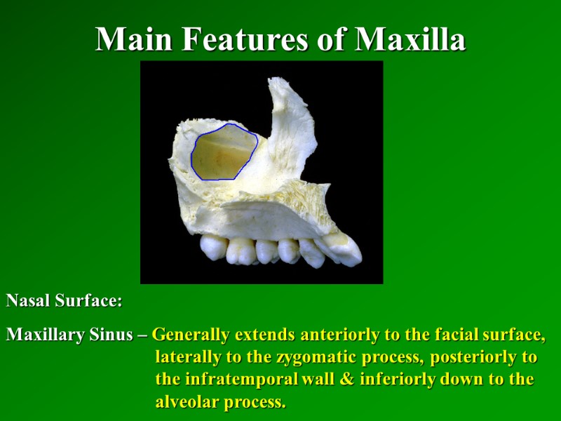
 Main Features of Maxilla Nasal Surface: Maxillary Sinus – Generally extends anteriorly to the facial surface, laterally to the zygomatic process, posteriorly to the infratemporal wall & inferiorly down to the alveolar process.
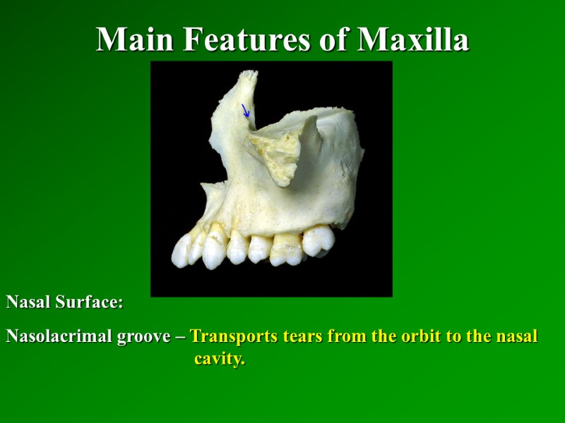
 Main Features of Maxilla Nasal Surface: Nasolacrimal groove – Transports tears from the orbit to the nasal cavity.
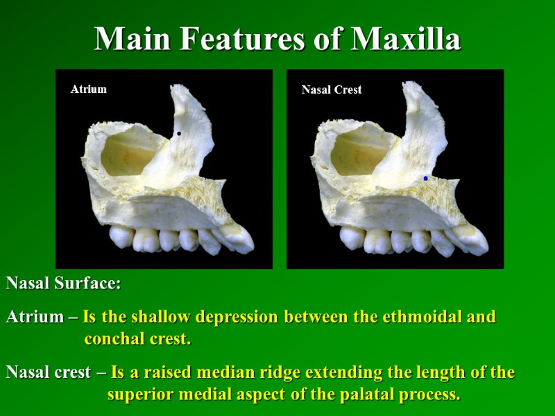
 Main Features of Maxilla Nasal Surface: Atrium – Is the shallow depression between the ethmoidal and conchal crest. Nasal crest – Is a raised median ridge extending the length of the superior medial aspect of the palatal process. Atrium Nasal Crest
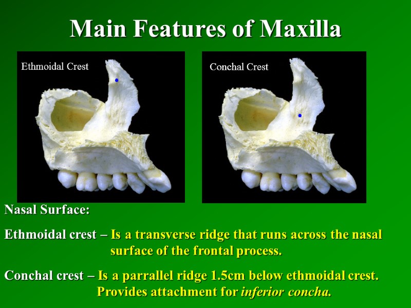
 Main Features of Maxilla Nasal Surface: Ethmoidal crest – Is a transverse ridge that runs across the nasal surface of the frontal process. Conchal crest – Is a parrallel ridge 1.5cm below ethmoidal crest. Provides attachment for inferior concha. Ethmoidal Crest Conchal Crest
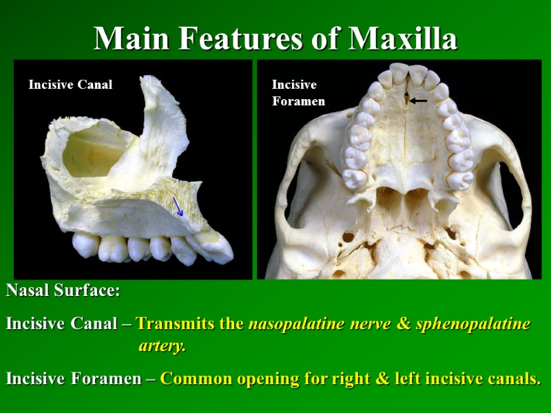
 Main Features of Maxilla Nasal Surface: Incisive Canal – Transmits the nasopalatine nerve & sphenopalatine artery. Incisive Foramen – Common opening for right & left incisive canals. Incisive Canal Incisive Foramen
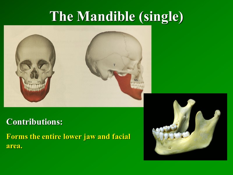
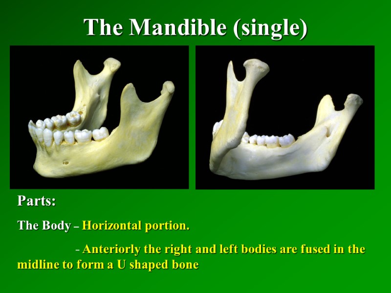
 The Mandible (single) Contributions: Forms the entire lower jaw and facial area.
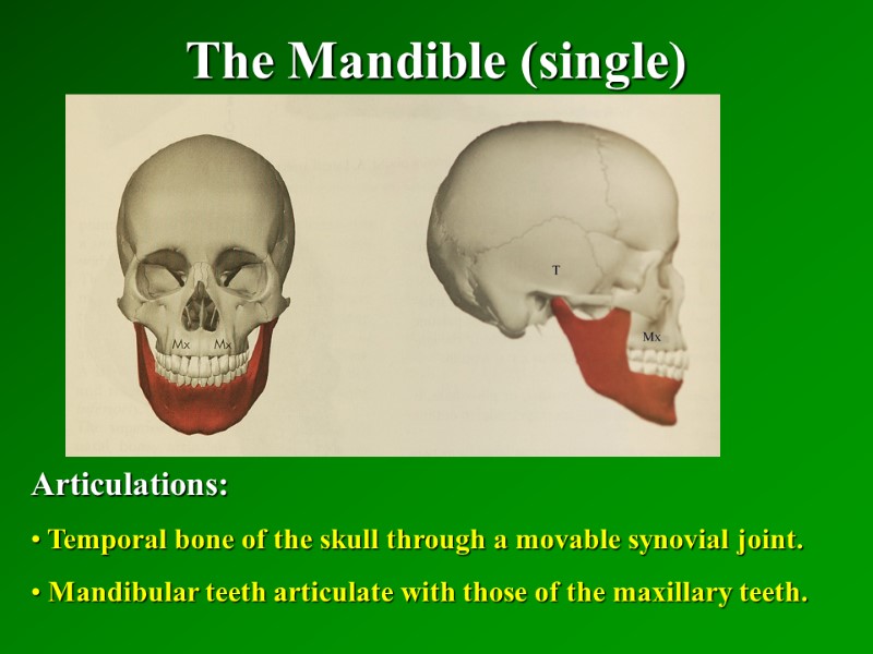
 The Mandible (single) Articulations: Temporal bone of the skull through a movable synovial joint. Mandibular teeth articulate with those of the maxillary teeth.
 The Mandible (single) Parts: The Body – Horizontal portion. - Anteriorly the right and left bodies are fused in the midline to form a U shaped bone
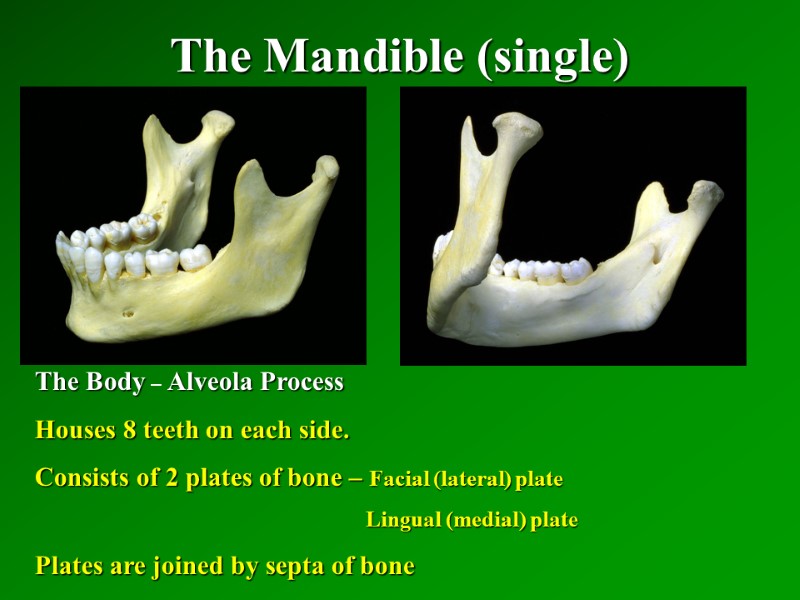
 The Mandible (single) The Body – Alveola Process Houses 8 teeth on each side. Consists of 2 plates of bone – Facial (lateral) plate Lingual (medial) plate Plates are joined by septa of bone
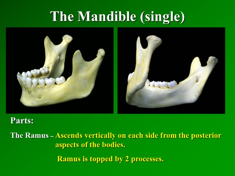
 The Mandible (single) Parts: The Ramus – Ascends vertically on each side from the posterior aspects of the bodies. Ramus is topped by 2 processes.
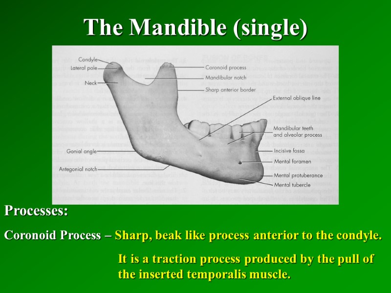
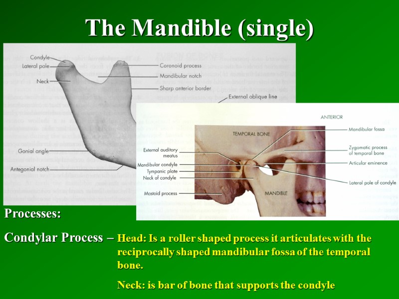
 The Mandible (single) Processes: Coronoid Process – Sharp, beak like process anterior to the condyle. It is a traction process produced by the pull of the inserted temporalis muscle.
 The Mandible (single) Processes: Condylar Process – Head: Is a roller shaped process it articulates with the reciprocally shaped mandibular fossa of the temporal bone. Neck: is bar of bone that supports the condyle
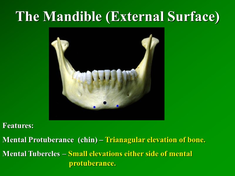
 The Mandible (External Surface) Features: Mental Protuberance (chin) – Trianagular elevation of bone. Mental Tubercles – Small elevations either side of mental protuberance.
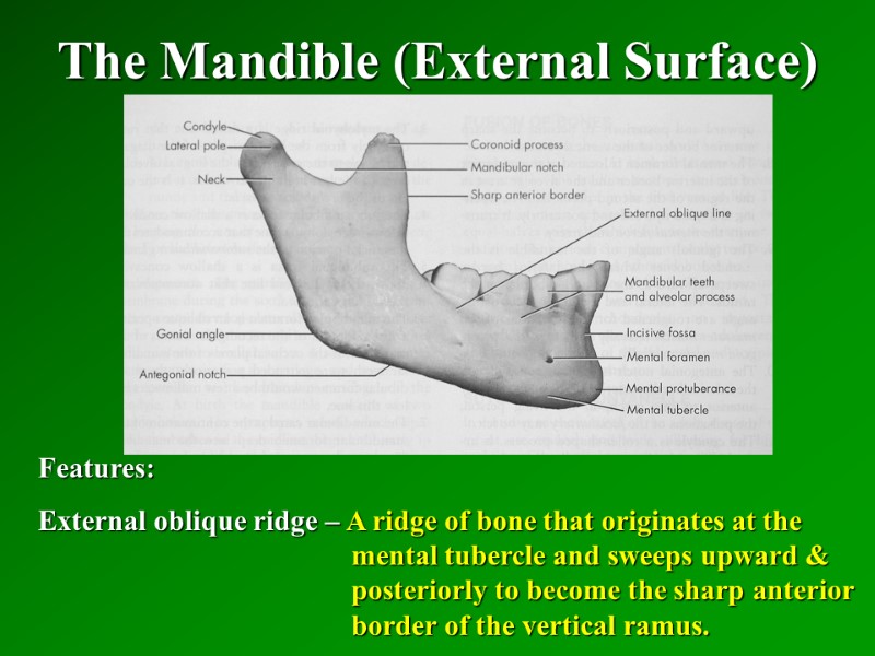
 The Mandible (External Surface) Features: External oblique ridge – A ridge of bone that originates at the mental tubercle and sweeps upward & posteriorly to become the sharp anterior border of the vertical ramus.
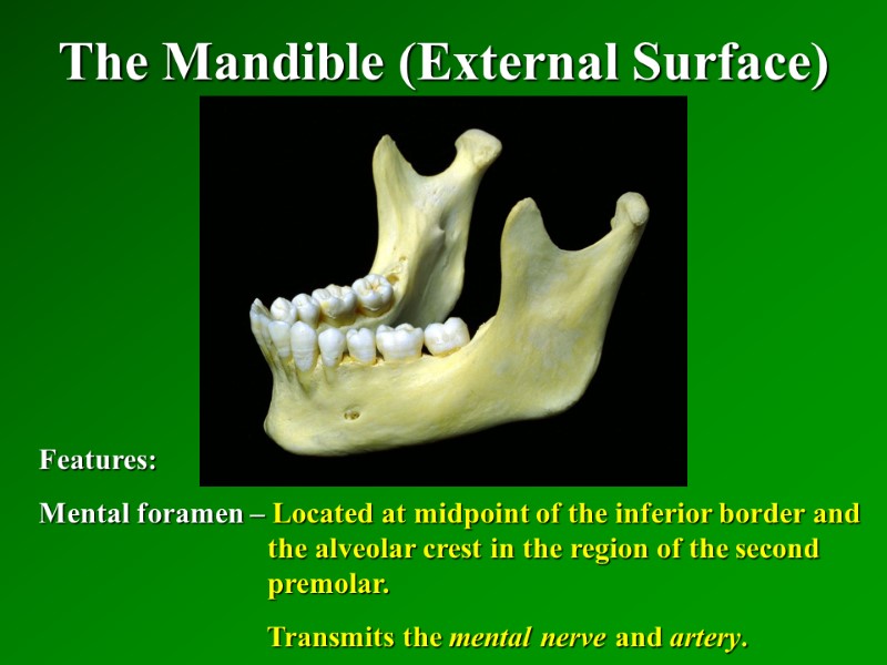
 The Mandible (External Surface) Features: Mental foramen – Located at midpoint of the inferior border and the alveolar crest in the region of the second premolar. Transmits the mental nerve and artery.
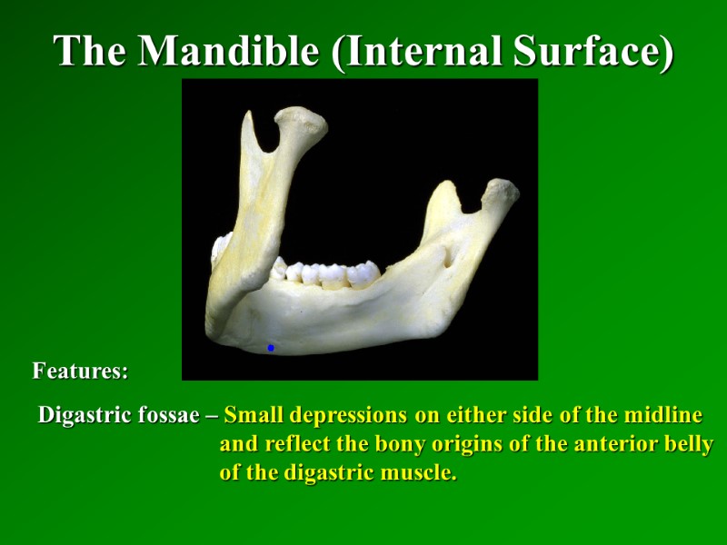
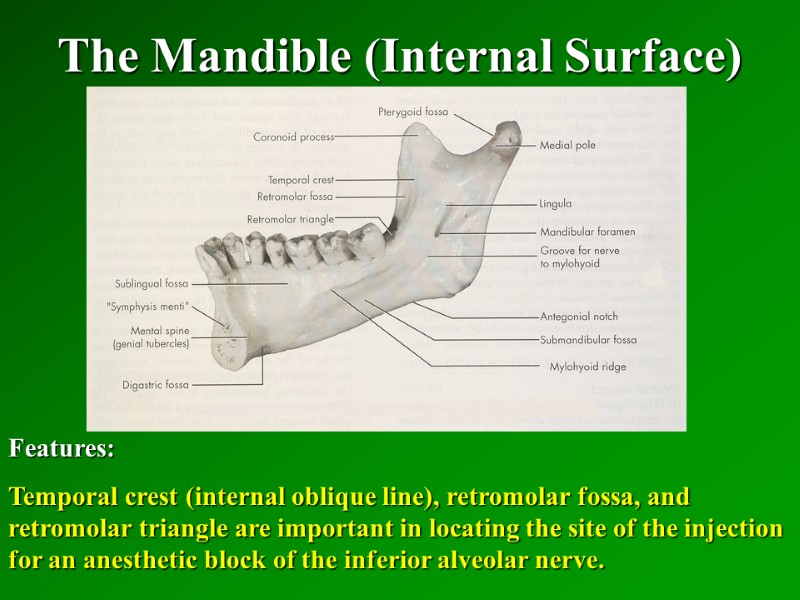
 The Mandible (Internal Surface) Features: Digastric fossae – Small depressions on either side of the midline and reflect the bony origins of the anterior belly of the digastric muscle.
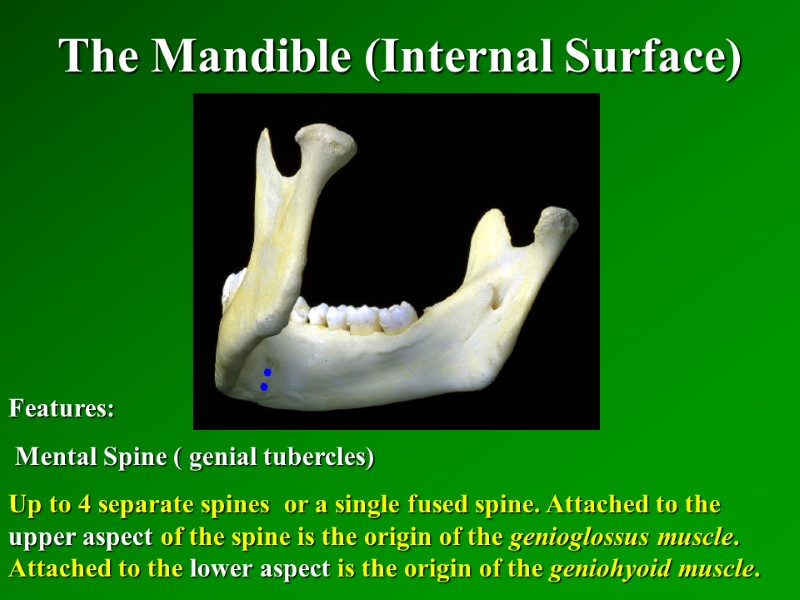
 The Mandible (Internal Surface) Features: Mental Spine ( genial tubercles) Up to 4 separate spines or a single fused spine. Attached to the upper aspect of the spine is the origin of the genioglossus muscle. Attached to the lower aspect is the origin of the geniohyoid muscle.
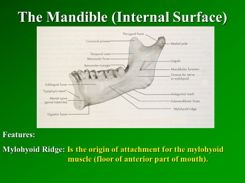
 The Mandible (Internal Surface) Features: Mylohyoid Ridge: Is the origin of attachment for the mylohyoid muscle (floor of anterior part of mouth).
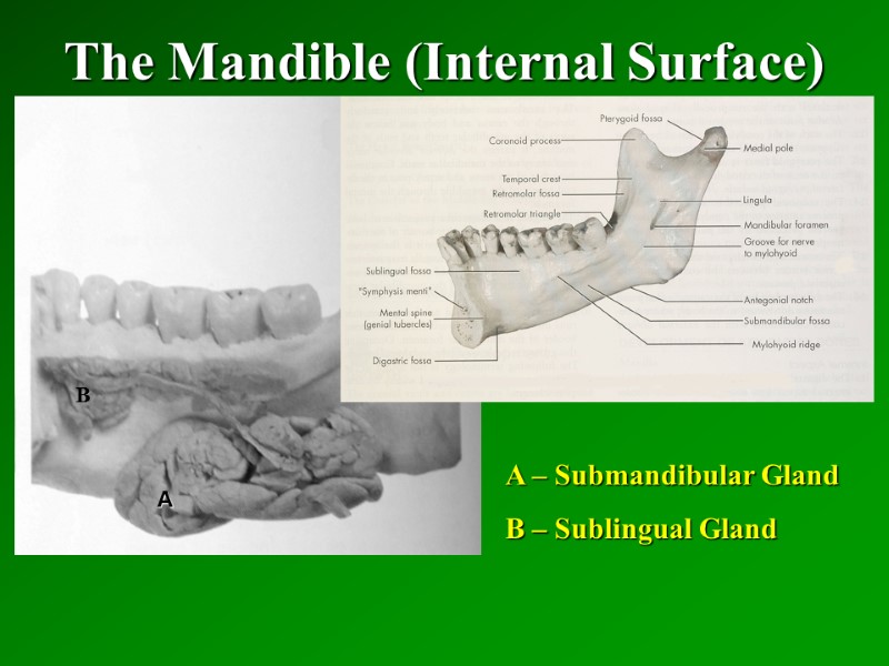
 The Mandible (Internal Surface) A B A – Submandibular Gland B – Sublingual Gland
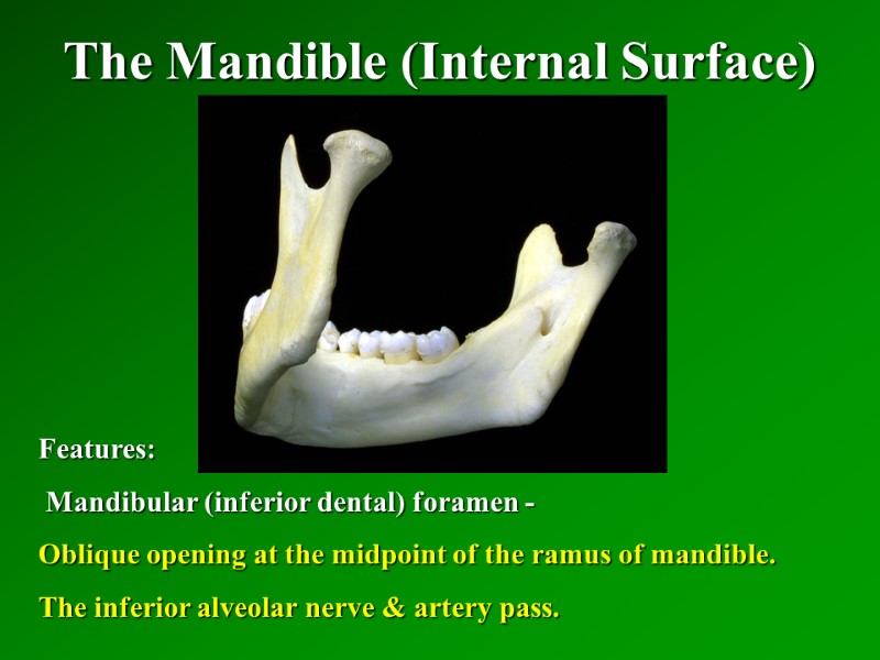
 The Mandible (Internal Surface) Features: Mandibular (inferior dental) foramen - Oblique opening at the midpoint of the ramus of mandible. The inferior alveolar nerve & artery pass.
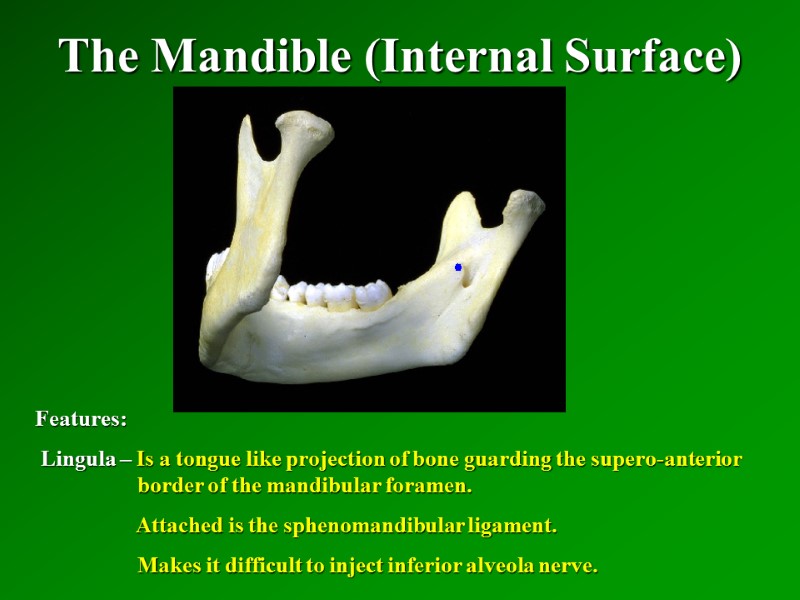
 The Mandible (Internal Surface) Features: Lingula – Is a tongue like projection of bone guarding the supero-anterior border of the mandibular foramen. Attached is the sphenomandibular ligament. Makes it difficult to inject inferior alveola nerve.
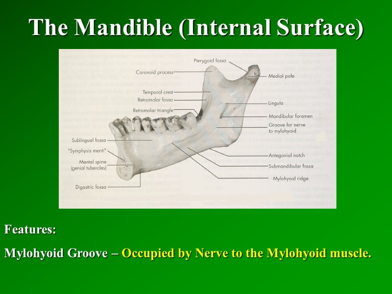
 The Mandible (Internal Surface) Features: Mylohyoid Groove – Occupied by Nerve to the Mylohyoid muscle.
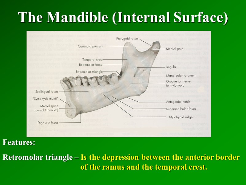
 The Mandible (Internal Surface) Features: Retromolar triangle – Is the depression between the anterior border of the ramus and the temporal crest.
 The Mandible (Internal Surface) Features: Temporal crest (internal oblique line), retromolar fossa, and retromolar triangle are important in locating the site of the injection for an anesthetic block of the inferior alveolar nerve.
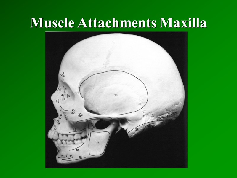
 Muscle Attachments Maxilla
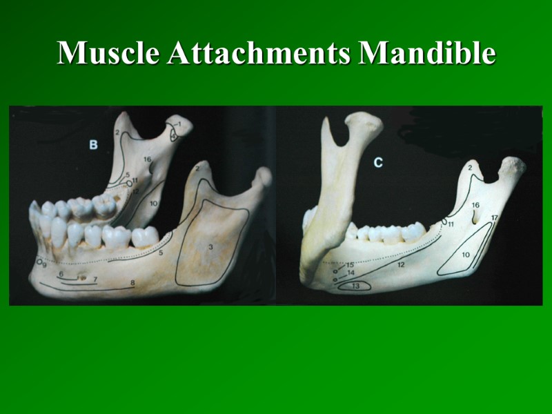
 Muscle Attachments Mandible

