Anatomy of lungs.pptx
- Количество слайдов: 37
 Anatomy of lungs
 Anatomy of lungs Lungs are a pair of respiratory organs situated in thoracic cavity. Right and left lungare separated by mediastinum. Texture-Spongy Color-brown(young) mottled black(adults) Weight-R. L. -600 gr L. L. -550 gr
 of airways and") Together, the lungs contain approximately 2, 400 kilometres (1, 500 mi) of airways and 300 to 500 million alveoli. The total surface area of lungs vary from 3050 square metres up to 70 -100 square metres in adults
Together, the lungs contain approximately 2, 400 kilometres (1, 500 mi) of airways and 300 to 500 million alveoli. The total surface area of lungs vary from 3050 square metres up to 70 -100 square metres in adults
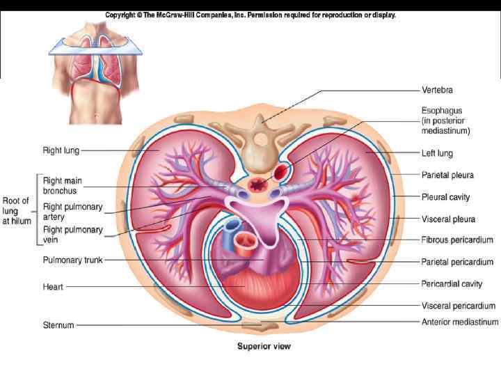
 Structure of Lungs Each lung is conical in shape and is contained in its own pleural sac. The lungs are separated from each other by the heart and great vessels in the middle mediastinum. The lungs are attached to the heart and trachea by the structures in the root of the lungs and to the pericardium by the pulmonary ligaments. Each lung has an apex, base, and hilum(hilus)
 There are three surfaces to the lungs: the costal surface is the outer, thoracic surface which is smooth and convex. This surface area is large and corresponds to the form of the thoracic cavity, being deeper at the back than at the front. The costal surface is in contact with the costal pleura and in specimens that have been hardened in situ, slight grooves are visible which correspond to the overlying ribs. The mediastinal surface of the lung is in contact with the mediastinal pleura and presents the cardiac impression. The diaphragmatic surface of lung is the portion of the lung which borders on the thoracic diaphragm.
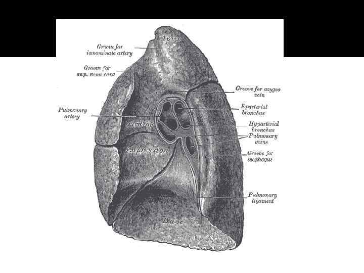
 The apex of the Lung The rounded, tapered superior end or apex of the lung extends through the superior thoracic aperture into the root of the neck. Here, it lies in close contact to the dome or the cupula of the pleura. The apex of the lung is crossed by the subclavian artery, which produces a groove in the mediastinal surface. The artery, however, is separated from the cupula by the suprapleural membrane.
 The Base of the lung This is the concave diaphragmatic surface of the lung, which is related to the dome of the diaphragm. The base of the right lung is deeper because the right dome rises to a more superior level. Its inferior border is thin and sharp where it enters the costodiaphragmatic recess.
 The Root of the Lung The root serves as the attachment of the lung and is the "highway" for the transmission of the structures entering and leaving the lung at the hilum. It connects the medial surface of the lung to the heart and trachea and is surrounded by the reflection of parietal to visceral pleura.
 The Hilum of the Lung This is the where the root is attached to the lung. It contains the main bronchus, pulmonary vessels (one artery and two veins), bronchial vessels, lymph vessels, and nerves entering and leaving the lung
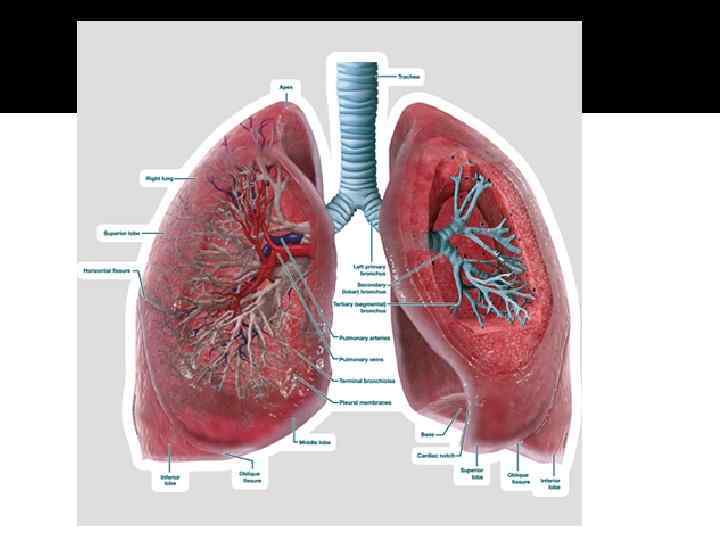
 Right lung The right lung is divided into three lobes (as opposed to two lobes on the left), superior, middle, and inferior, by two interlobular fissures: The right lung has a higher volume, total capacity and weight, than that of the left lung. Although it is 5 cm shorter due to the diaphragm rising higher on the right side to accommodate the liver, it is broader than the left lung due to the cardiac notch of the left lung.
 Left lung The left lung is divided into two lobes, an upper and a lower, by the oblique fissure, which extends from the costal to the mediastinal surface of the lung both above and below the hilum. The left lung, unlike the right does not have a middle lobe. However the term lingula is used to denote a projection of the upper lobe of the left lung that serves as the homologue. This area of the left lobe - the lingula, means little tongue(in Latin) and is often referred to as the tongue in the lung. There are two bronchopulmonary segments of the lingula: superior and inferior. It is thought that the lingula of the left lung is the remnant of the middle lobe, which has been lost through evolution
 Difference between R. L. and L. L. Right lung has three lobes, whereas left lung has only two. Right lung is shorter and wider, whereas the left lung is narrower and more oblong. Anterior border of the left lung is marked by a deep cardiac notch, while that of the right lung is straight. Therefore, left lung is much smaller than right lung (heart occupies space on the left side). Due to the compression of the liver, the volume of the right lung is higher than that of the left lung. Hence, the right lung is heavier than left lung. The base of the right lung is more concave, while the base of left lung is less concave. At the hilums of both lungs, the right lung possesses two bronchi and the left lung possesses only one bronchus. Left lung has only oblique fissure, while right lung has horizontal and oblique fissure.
 Lobes and Fissures of the Lungs
 and inferior (lower) lobes by") The Left Lung This is divided into superior (upper) and inferior (lower) lobes by a long deep oblique fissure. This extends from its costal to its medial surface. The superior lobe has a large cardiac notch on its anterior border, where the lung is deficient owing to the bulge of the heart. The anteroinferior part of the superior lobe has a small tongue-like projection called the lingula. The inferior lobe of the left lung is larger than the superior lobe and lies inferoposterior to the oblique fissure.
The Left Lung This is divided into superior (upper) and inferior (lower) lobes by a long deep oblique fissure. This extends from its costal to its medial surface. The superior lobe has a large cardiac notch on its anterior border, where the lung is deficient owing to the bulge of the heart. The anteroinferior part of the superior lobe has a small tongue-like projection called the lingula. The inferior lobe of the left lung is larger than the superior lobe and lies inferoposterior to the oblique fissure.
, middle, and inferior (lower) lobes") The Right Lung This is divided into superior (upper), middle, and inferior (lower) lobes by horizontal and oblique fissures. The horizontal fissure separates the superior and middle lobes. The oblique fissure separates the inferior lobe from the superior and middle lobes. The superior lobe is smaller than in the left lung, and the middle lobe is wedge-shaped
The Right Lung This is divided into superior (upper), middle, and inferior (lower) lobes by horizontal and oblique fissures. The horizontal fissure separates the superior and middle lobes. The oblique fissure separates the inferior lobe from the superior and middle lobes. The superior lobe is smaller than in the left lung, and the middle lobe is wedge-shaped
 Surfaces of the lung
 The Costal Surface of the Lung This surface is large, smooth, and convex. It is related to the costal pleura, which separates it from the ribs, their costal cartilages, and the innermost intercostal muscles. The posterior part of this surface is related to the thoracic vertebrae.
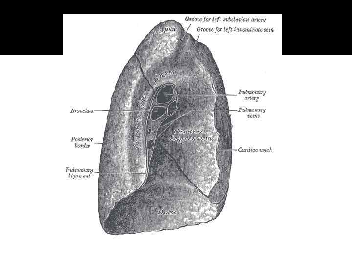
 The Mediastinal Surface This medial surface is concave because it is related to the middle mediastinum. Because 2/3 of the heart is to the left, the pericardial concavity is deeper in the left lung. The mediastinal surface of the embalmed lung shows a cardiac impression produced by the heart and the great vessels. This surface also contains the root of the lung, around which the pleura forms a "sleeve" or covering. The pulmonary ligament hangs inferiorly from the pleural sleeve around the root of the lung.
 The Diaphragmatic Surface This is a deeply concave surface, often referred to as the base of the lung. It rests on the convex dome of the diaphragm. The concavity is deeper in the right lung because of the higher position of the dome. Laterally and posteriorly, the diaphragmatic surface is bound by a thin sharp margin that projects into the costodiaphragmatic recess.
 Topography
 Apex of the Lung This is represented by a line drawn superolaterally from the sternoclavicular joint to a point 2. 5 cm superior to the medial 1/3 of the clavicle and then inferolaterally to thejunction of the middle and lateral thirds of the clavicle.
 Anterior Border of the Right Lung This corresponds to the anterior border of the right pleura. Between the level of the 2 nd and 4 th cartilages, its anterior border is near the median plane. Inferior to the 4 th costal cartilage, the surface of the right lung gradually diverges from this plane and leaves the sternum posterior to the 6 th costal cartilage.
 The Anterior Border of the Left Lung This corresponds to the anterior border of the left pleura as far as the level of the 4 th costal cartilage. Here, the anterior border deviates laterally to a point about 2. 5 cm lateral to the left edge of the sternum to form the cardiac notch. It then turns inferiorly and slightly medially to the 6 th costal cartilage.
 The Inferior Borders of the Lungs These are indicated by a line drawn from the inferior end of the line representing the anterior border that crosses the 6 th rib at the midclavicular line, the 8 th rib in the midaxillary line and the 10 th rib in the midscapular line. These borders end about 2. 5 cm lateral to the spinous process of T 10 vertebra. They lie two ribs superior to the pleura on each of three vertical lines just mentioned.
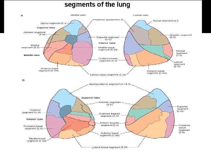
 The Bronchi, Roots, and Bronchopulmonary Segments of the Lungs
 The principal or main bronchus, one for each lung, passes inferolaterally from the bifurcation of the trachea. The bronchus accompanies the pulmonary artery into the wedge-shaped hilum of the lung, where it subdivides. The right main bronchus is wider, shorter and more vertical (significant in inhaled obstructions) than the left one. It is about 2. 5 cm long and passes directly into the root of the lung. The left main bronchus is about 5 cm long and passes inferolaterally, inferior to the arch of aorta, and anterior to the oesophagus and the descending thoracic aorta. Within each lung, the bronchi divide in a constant fashion and in constant directions so that each branch supplies a clearly defined sector of the lung. Each main bronchus divides into secondary or lobar bronchi (two for left, three for right), each of which supplies a lobe of the lung. Each lobar bronchus divides into tertiary or segmental bronchi, which supply specific segments of the lung, called bronchopulmonary segments.
 Innervation of Lungs The parasympathetic and sympathetic innervations of the lungs originate in a nerve plexus in the thoracic area. In stage 13 (32 days) 13 immigrated neural crest cells form an extrapulmonary nerve plexus originating at the vagus nerve (parasympathetic) and the cervical and upper thoracic ganglia of the symphathetic trunk. With the growth of the lung anlagen the trachea and the whole bronchial tree become increasingly supplied from proliferating and fully-grown neural crest cells. They form the intramural plexus of the lungs. At the end of the embryonic period anouter nerve plexus has formed at the tracheal and bronchial periphery, around the cartilage anlage. An inner peribronchial nerve plexus develops somewhat later in the lamina propria under the epithelium. It accompanies the formation of thebronchial smooth musculature and the glands of the bronchial mucosa. Further distal in the bronchioli, the inner and outer nerve plexus merge to form aperibronchial plexus.
 1 Right jugular vein 2 Right jugular and auxiliary lymph 3 plexus 4 Subclavian vein 5 Superior caval vein 6 Right thoracic duct 7 Left jugular vein Left jugular and auxiliary lymph 8 plexus 9 Left subclavian vein 10 Left thoracic duct (atrophied) 11 Cisterna chyli 12 Inguinal lymph nodes Thoracic duct
 Broncho pulmonary segments After the trachea divides and the left and right main stem bronchi are formed, they enter the lung substance and divide. This initial division is into secondary bronchi, but subsequent divisions give rise to smaller and smaller bronchi and bronchioles until the smallest bronchioles connect to the innumerable alveoli. Each segment has its own pulmonary arterial branch and thus, the bronchopulmonary segment is a portion of lung supplied by its own bronchus and artery. Each segment is functionally and anatomically discrete meaning that a single segment can be surgically removed without affecting its neighbours.
 Left Lung left upper lobe apicoposterior segment anterior segment superior lingular inferior lingular superior segment anteromedial segment lateral segment posterior segment left lower lobe
 Right Lung right upper lobe apical segment posterior segment anterior segment right middle lobe lateral segment medial segment right lower lobe superior segment anterior segment medial segment lateal segment posterior segment