
Agglutination and Precipitation Reactions Antigen An


practice_agglutination_amp_precipitation.ppt
- Размер: 6 Mегабайта
- Количество слайдов: 40
Описание презентации Agglutination and Precipitation Reactions Antigen An по слайдам
Agglutination and Precipitation Reactions
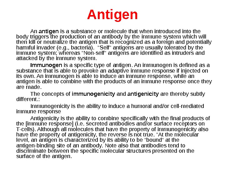
Antigen An antigen is a substance or molecule that when introduced into the body triggers the production of an antibody by the immune system which will then kill or neutralize the antigen that is recognized as a foreign and potentially harmful invader (e. g. , bacteria). «Self» antigens are usually tolerated by the immune system; whereas «Non-self» antigens are identified as intruders and attacked by the immune system. Immunogen is a specific type of antigen. An immunogen is defined as a substance that is able to provoke an adaptive immune response if injected on its own. An immunogen is able to induce an immune response, while an antigen is able to combine with the products of an immune response once they are made. The concepts of immunogenicity and antigenicity are thereby subtly different. : Immunogenicity is the ability to induce a humoral and/or cell-mediated immune response Antigenicity is the ability to combine specifically with the final products of the [immune response] (i. e. secreted antibodies and/or surface receptors on T-cells). Although all molecules that have the property of immunogenicity also have the property of antigenicity, the reverse is not true. «At the molecular level, an antigen is characterized by its ability to be «bound» at the antigen-binding site of an antibody. Note also that antibodies tend to discriminate between the specific molecular structures presented on the surface of the antigen.
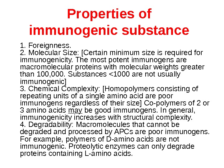
Properties of immunogenic substance 1. Foreignness. 2. Molecular Size: [Certain minimum size is required for immunogenicity. The most potent immunogens are macromolecular proteins with molecular weights greater than 100, 000. Substances <1000 are not usually immunogenic] 3. Chemical Complexity: [Homopolymers consisting of repeating units of a single amino acid are poor immunogens regardless of their size] Co-polymers of 2 or 3 amino acids may be good immunogens. In general, immunogenicity increases with structural complexity. 4. Degradability: Macromolecules that cannot be degraded and processed by APCs are poor immunogens. For example, polymers of D-amino acids are not immunogenic. Proteolytic enzymes can only degrade proteins containing L-amino acids.
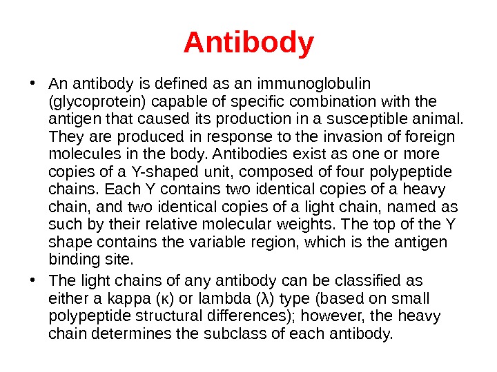
A ntibody • An antibody is defined as an immunoglobulin (glycoprotein) capable of specific combination with the antigen that caused its production in a susceptible animal. They are produced in response to the invasion of foreign molecules in the body. Antibodies exist as one or more copies of a Y-shaped unit, composed of four polypeptide chains. Each Y contains two identical copies of a heavy chain, and two identical copies of a light chain, named as such by their relative molecular weights. The top of the Y shape contains the variable region, which is the antigen binding site. • The light chains of any antibody can be classified as either a kappa (κ) or lambda (λ) type (based on small polypeptide structural differences); however, the heavy chain determines the subclass of each antibody.
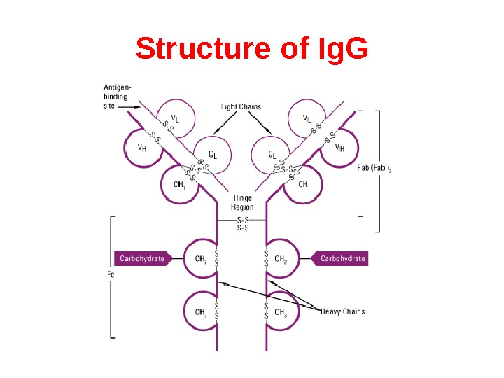
Structure of Ig. G
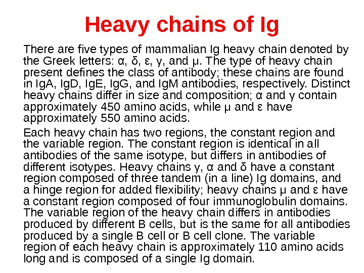
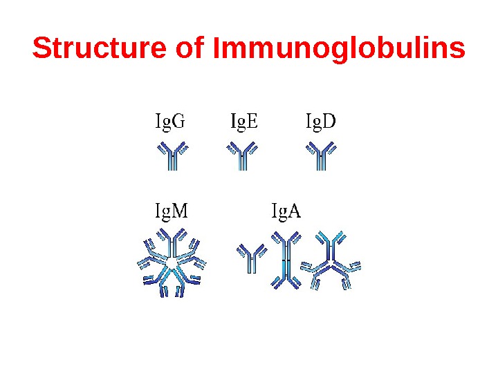
Heavy chains of Ig There are five types of mammalian Ig heavy chain denoted by the Greek letters: α, δ, ε, γ, and μ. The type of heavy chain present defines the class of antibody; these chains are found in Ig. A, Ig. D, Ig. E, Ig. G, and Ig. M antibodies, respectively. Distinct heavy chains differ in size and composition; α and γ contain approximately 450 amino acids, while μ and ε have approximately 550 amino acids. Each heavy chain has two regions, the constant region and the variable region. The constant region is identical in all antibodies of the same isotype, but differs in antibodies of different isotypes. Heavy chains γ, α and δ have a constant region composed of three tandem (in a line) Ig domains, and a hinge region for added flexibility; heavy chains μ and ε have a constant region composed of four immunoglobulin domains. The variable region of the heavy chain differs in antibodies produced by different B cells, but is the same for all antibodies produced by a single B cell or B cell clone. The variable region of each heavy chain is approximately 110 amino acids long and is composed of a single Ig domain.
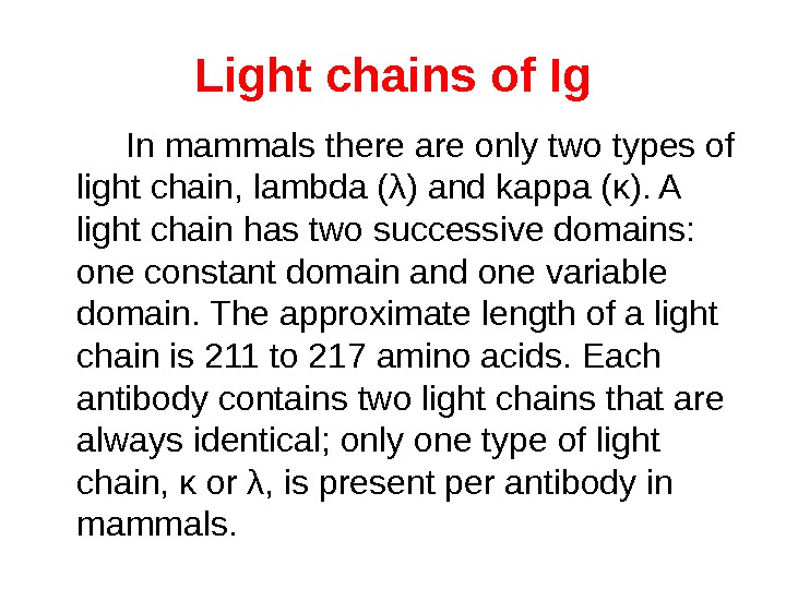
Light chains of Ig In mammals there are only two types of light chain, lambda (λ) and kappa (κ). A light chain has two successive domains: one constant domain and one variable domain. The approximate length of a light chain is 211 to 217 amino acids. Each antibody contains two light chains that are always identical; only one type of light chain, κ or λ, is present per antibody in mammals.
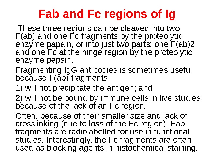
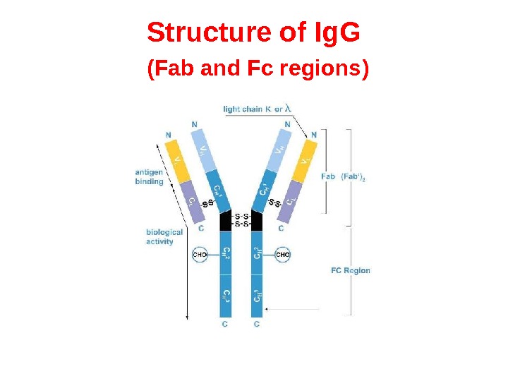
Fab and Fc regions of Ig These three regions can be cleaved into two F(ab) and one Fc fragments by the proteolytic enzyme papain, or into just two parts: one F(ab)2 and one Fc at the hinge region by the proteolytic enzyme pepsin. Fragmenting Ig. G antibodies is sometimes useful because F(ab) fragments 1) will not precipitate the antigen; and 2) will not be bound by immune cells in live studies because of the lack of an Fc region. Often, because of their smaller size and lack of crosslinking (due to loss of the Fc region), Fab fragments are radiolabelled for use in functional studies. Interestingly, the Fc fragments are often used as blocking agents in histochemical staining.
Structure of Ig. G ( Fab and Fc regions )
Structure of Immunoglobulins
Affinity & Avidity Affinity Antibody affinity is the strength of the reaction between a single antigenic determinant and a single combining site on the antibody. It is the sum of the attractive and repulsive forces operating between the antigenic determinant and the combining site of the antibody. Avidity is a measure of the overall strength of binding of an antigen with many antigenic determinants and multivalent antibodies. Avidity is influenced by both the valence of the antibody and the valence of the antigen. Avidity is more than the sum of the individual affinities. . To repeat, affinity refers to the strength of binding between a single antigenic determinant and an individual antibody combining site whereas avidity refers to the overall strength of binding between multivalent antigens and antibodies.
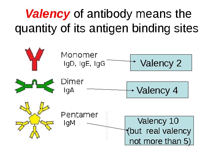
Valency of antibody means the quantity of its antigen binding sites Valency 2 Valency 4 Valency 10 (but real valency not more than 5)
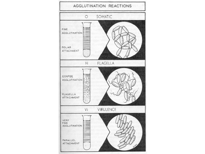
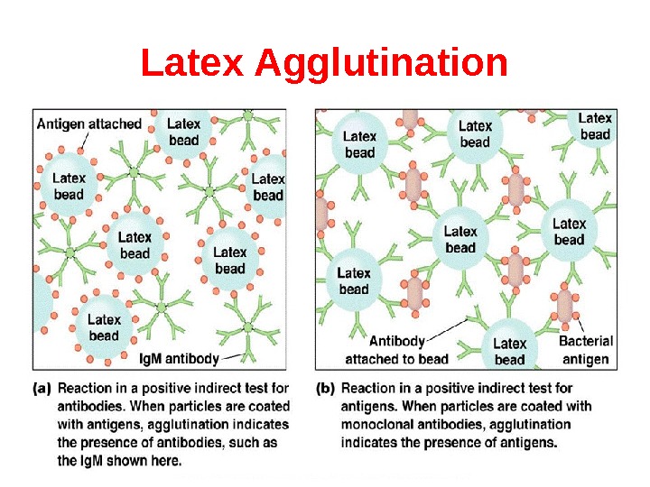
Agglutination reaction When the antigen is particulate (corpuscular, insoluble), the reaction of an antibody with the antigen can be detected by agglutination (clumping) of the antigen. The general term agglutinin is used to describe antibodies that agglutinate particulate (corpuscular, insoluble) antigens. When the antigen is absorbed on erythrocytes the term hemagglutination is used. When the antigen is absorbed on latex beads the term latex-agglutination is used. All antibodies can theoretically agglutinate particulate antigens, but Ig. M, due to its high valence, is particularly good agglutinin and one sometimes infers that an antibody may be of the Ig. M class if it is a good agglutinating antibody.
Mechanism of agglutination
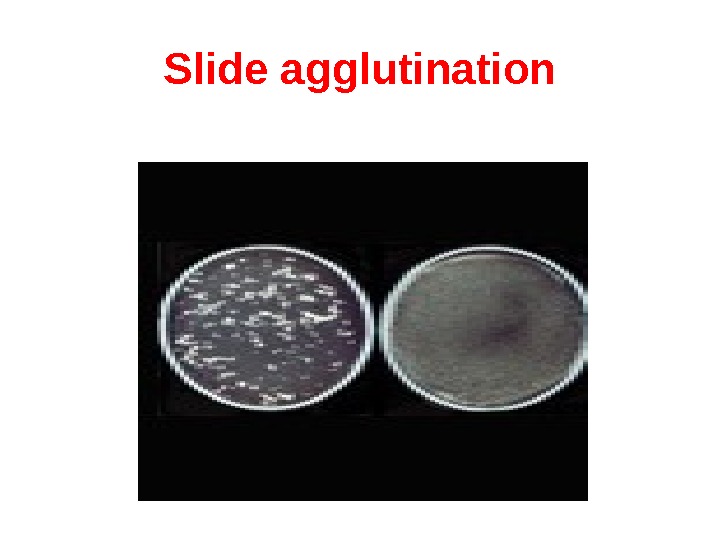
Slide agglutination When a drop of the appropriate antiserum is added to a smooth, uniform suspension of a particulate antigen in a drop of saline on a slide, agglutination takes place. A positive result is indicated by the clumping together of the particles and the clearing of the drop instantly or within minutes. It is essential to have on the same slide a control consisting of the antigen suspension in saline, without the antiserum, to ensure that the antigen is not autoagglutinable. Slide agglutination is a routine procedure for the identification of many bacterial isolates from clinical specimens.
Slide agglutination
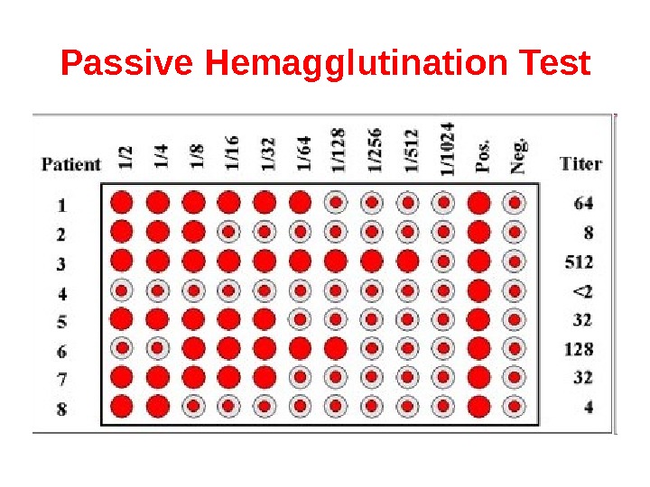
Tube agglutination is a standard quantitative method for the measurement of antibodies. When a fixed volume of a particulate antigen suspension is added to an equal volume of serial dilutions of an antiserum in tubes, the agglutination titer of the serum can be estimated. Agglutination titer of the specimen is compared with diagnostical titer for the particular disease. If the result for agglutination titer is equal or more than diagnostical titer, the sample is positive. If the result for agglutination titer is less than diagnostical titer, the sample is negative.
Tube agglutination Agglutination Control of antigen Control of serum
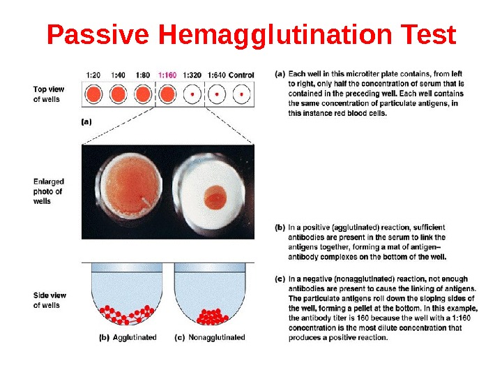
Passive Hemagglutination Test The agglutination test only works with particulate antigens. However, it is possible to coat erythrocytes with a soluble antigen (e. g. viral antigen, a polysaccharide or a hapten) and use the coated red blood cells in an agglutination test for antibody to the soluble antigen. This is called passive hemagglutination. The test is performed just like the agglutination test. Applications include detection of antibodies to soluble antigens and detection of antibodies to viral antigens.
Passive Hemagglutination Test
Passive Hemagglutination Test
Reverse Passive Hemagglutination Test It is possible to coat erythrocytes with specific antibodies and use the coated red blood cells in an agglutination test for detection of antigen (usually toxin) to the specific antibodies. The visual result of reverse passive hemagglutination test is the same as for passive hemagglutination test: “umbrella” means positive result, “button” means negative result.
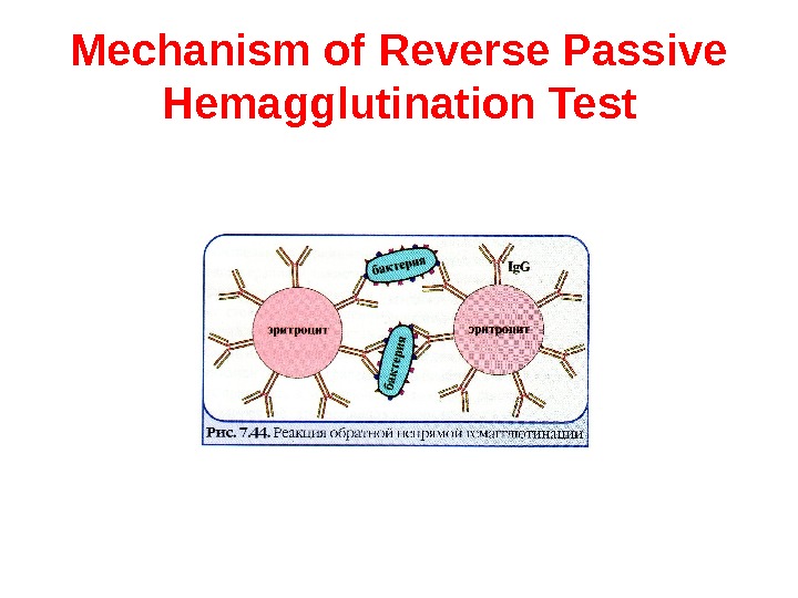
Mechanism of Reverse Passive Hemagglutination Test
Latex Agglutination
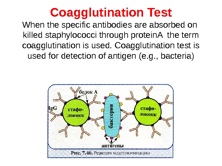
Coagglutination Test When the specific antibodies are absorbed on killed staphylococci through protein. A the term coagglutination is used. Coagglutination test is used for detection of antigen (e. g. , bacteria)
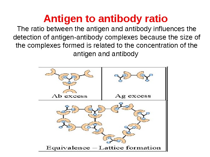
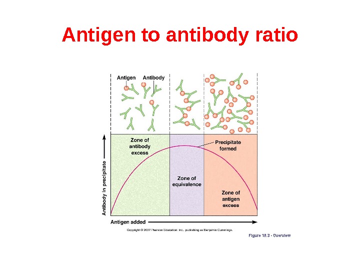
Precipitation reaction is the test when a soluble antigen combines with its antibody in the presence of electrolytes (Na. Cl) at a suitable temperature and p. H. The antigen antibody complex forms an insoluble precipitate. Precipitation can take place in liquid media or in gels. Precipitation reactions depend on the formation of lattices and occur best when antigen and antibody are present in optimal proportions. Excesses of either component decrease lattice formation and subsequent precipitation.
Antigen to antibody ratio The ratio between the antigen and antibody influences the detection of antigen-antibody complexes because the size of the complexes formed is related to the concentration of the antigen and antibody
Antigen to antibody ratio
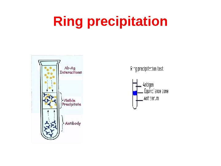
Ring test consists of layering the antigen solution over a column of antiserum in a narrow tube. A precipitate forms at the junction of the two liquids. Ring tests are Ascoli’s thermoprecipitin test and the grouping of streptococci by Lancerfield technique.
Ring precipitation
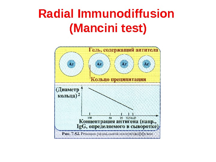
Radial Immunodiffusion (Mancini test ) In radial immunodiffusion antibody is incorporated into the agar gel as it is poured and different dilutions of the antigen are placed in holes punched into the agar. As the antigen diffuses into the gel, it reacts with the antibody and when the equivalence point is reached a ring of precipitation is formed as illustrated. The diameter of the ring is proportional to the log of the concentration of antigen since the amount of antibody is constant. Thus, by running different concentrations of a standard antigen one can generate a standard cure from which one can quantitate the amount of an antigen in an unknown sample. Thus, this is a quantitative test. If more than one ring appears in the test, more than one antigen/antibody reaction has occurred. This could be due to a mixture of antigens or antibodies. This test is commonly used in the clinical laboratory for the determination of immunoglobulin levels in patient samples.
Radial Immunodiffusion (Mancini test )
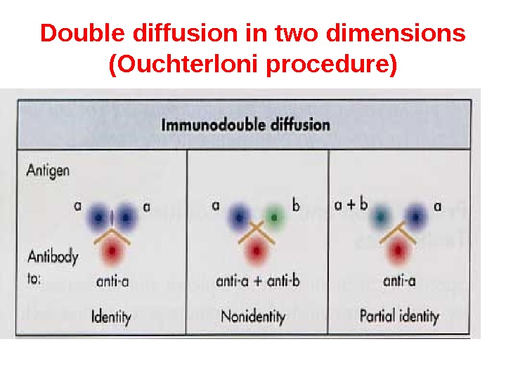
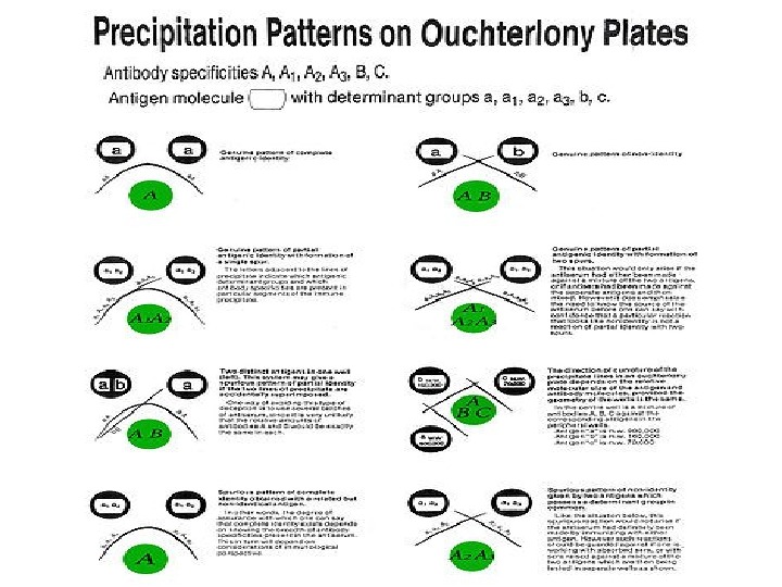
Double diffusion in two dimensions (Ouchterloni procedure) is the immunodiffusion method, which helps to compare different antigens and antisera directly. Agar gel is poured on a slide and wells are cut using a template. The antiserum is placed in the central well and different antigens in the surrounding wells. If two adjacent antigens are identical, the lines of precipitate formed by them will fuse. If they are unrelated, the lines will cross each other. Cross reaction or partial identity is indicated by spur formation.
Double diffusion in two dimensions (Ouchterloni procedure)
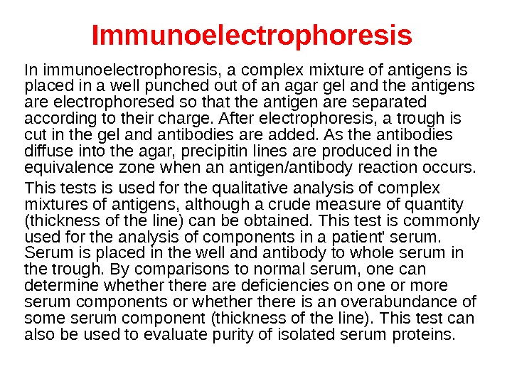
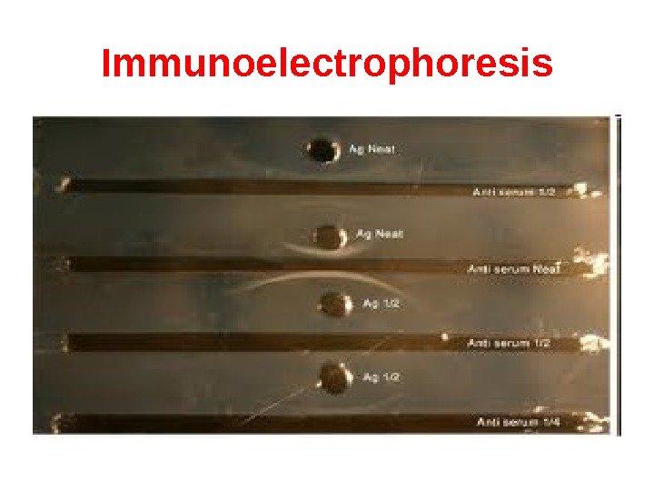
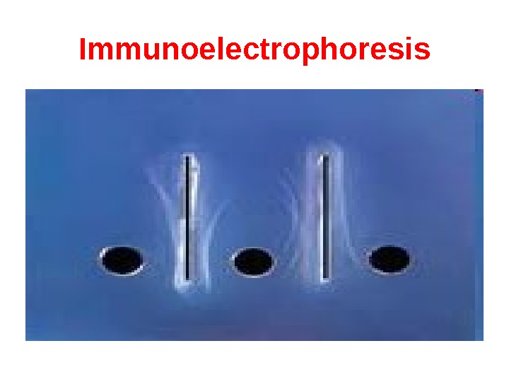
Immunoelectrophoresis In immunoelectrophoresis, a complex mixture of antigens is placed in a well punched out of an agar gel and the antigens are electrophoresed so that the antigen are separated according to their charge. After electrophoresis, a trough is cut in the gel and antibodies are added. As the antibodies diffuse into the agar, precipitin lines are produced in the equivalence zone when an antigen/antibody reaction occurs. This tests is used for the qualitative analysis of complex mixtures of antigens, although a crude measure of quantity (thickness of the line) can be obtained. This test is commonly used for the analysis of components in a patient’ serum. Serum is placed in the well and antibody to whole serum in the trough. By comparisons to normal serum, one can determine whethere are deficiencies on one or more serum components or whethere is an overabundance of some serum component (thickness of the line). This test can also be used to evaluate purity of isolated serum proteins.
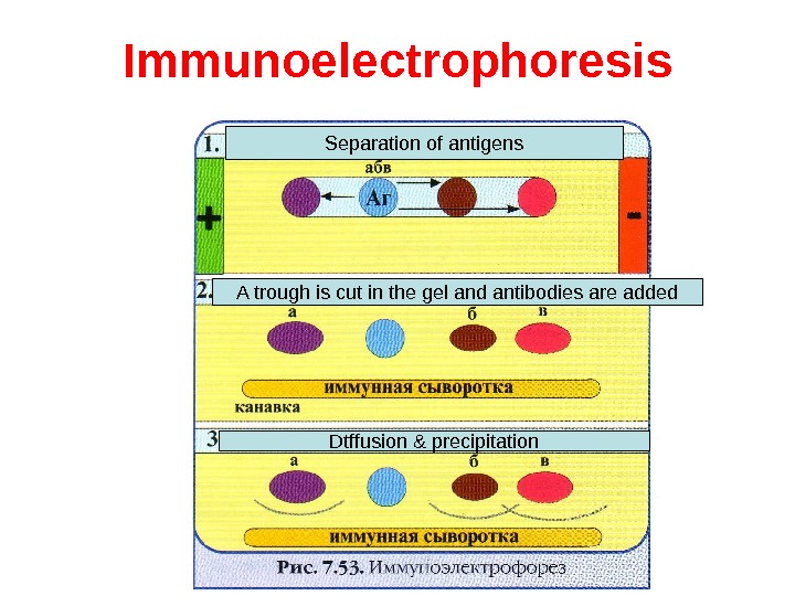
Immunoelectrophoresis Separation of antigens A trough is cut in the gel and antibodies are added Dtffusion & precipitation
Immunoelectrophoresis
Immunoelectrophoresis