

AML_lecture2studfeb06.ppt
- Количество слайдов: 41
 ACUTE MYELOID LEUKEMIA
") What is an Acute Myeloid Leukemia ? Accumulation of early myeloid progenitors (blast cells) in bone marrow and blood Definition requests presence of 20% or more blasts in BM Normally- less than 5%
What is an Acute Myeloid Leukemia ? Accumulation of early myeloid progenitors (blast cells) in bone marrow and blood Definition requests presence of 20% or more blasts in BM Normally- less than 5%
 ETIOLOGY • Environment: irradiation, chemotherapeutic agents, organic solvents – benzene etc. • Genetic diseases: neurofibromatosis, Wiscott-Aldrich synd. , defective DNA repair – Fanconi, Down synd. • Acquired disorders: Aplastic Anemia, PNH • MOST OF THE CASES APPEAR WITH NO APPARENT RISK FACTORS!!!
 AML Aggressive disease with an acute onset Can occur De Novo or following a known leukomogemic trigger (radiation, chemotherapy, diseases): Secondary AML
 Leukemia Malignant Transformation Proliferation and Accumulation Peripheral blood Blasts in BM Visceral organs Cytopenias
") BM - Acute Leukemia (low power)
BM - Acute Leukemia (low power)
 Morphology AML
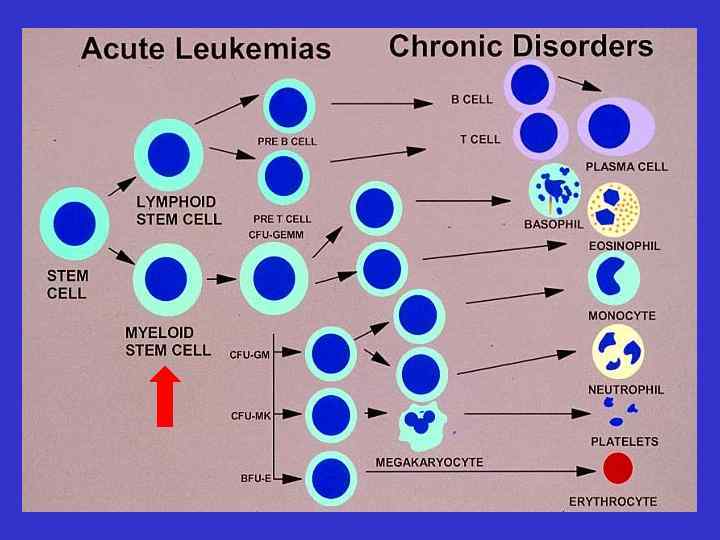
, M 2 t(15;") Myeloid Stem Cell Pathophysiology Radiation Chemotherapy Viruses chromosomal damage t(8; 21), M 2 t(15; 17) M 3 Inv 16; M 4 e protooncogen Inhibition/Enhancements of regulatory genes Inhibition of suppressor genes Enhancements of proliferation Inhibition of apoptosis
Myeloid Stem Cell Pathophysiology Radiation Chemotherapy Viruses chromosomal damage t(8; 21), M 2 t(15; 17) M 3 Inv 16; M 4 e protooncogen Inhibition/Enhancements of regulatory genes Inhibition of suppressor genes Enhancements of proliferation Inhibition of apoptosis
 Epidemiology
 Predisposing factors Environmental Acquired diseases Genetic Benzen, herbicies Chemotherapy : AK ; NU; PRC Radiation Meyloproliferative(CML; PV. . ) Aplastic anemia Congenital abnormality to repair DNA : Down syndrome Ashkenazi Jews >> orientals Relatives(1 st degree x 3)
 Clinical symptoms of Acute Leukemia Bone marrow expansion Bone pain Bone marrow failure Leucopoenia infections Thrombopenia bleeding Leucostasis Anemia >50, 000 blasts Dispnea, CNS
 Skin CNS Gingiva Kidney") Clinical symptoms Extramedullary (Chloroma) Skin CNS Gingiva Kidney
Clinical symptoms Extramedullary (Chloroma) Skin CNS Gingiva Kidney
 Extramedullary: Gingival hypertrophy
 Clinical symptomes DIC Bleeding Thrombosis Metabolic Hyperuricemia Tumor lysis syndrome K, phosphor, Ca Uric Acid
 Normal bone marrow M AML ; blasts") Diagnosis >20% blasts in bone marrow/peripheral blood) Normal bone marrow M AML ; blasts B
Diagnosis >20% blasts in bone marrow/peripheral blood) Normal bone marrow M AML ; blasts B
 Auer Rods Aggregation") Acute leukemia - AUER Rods ( FAB; AML M 3 ) Auer Rods Aggregation of granules
Acute leukemia - AUER Rods ( FAB; AML M 3 ) Auer Rods Aggregation of granules
 Acute promyelocytic leukemia - AML M 3
 Myeloblasts - AML d ro uer A
 AML M 2 blasts
 classification -Based on morphology and staining (cytochemistry) -Divides patients into") French American British (FAB) classification -Based on morphology and staining (cytochemistry) -Divides patients into 7 AML subtypes -A morphological rather than biological classification -Correlation between morphological and biological characteristics may exist , but not always
French American British (FAB) classification -Based on morphology and staining (cytochemistry) -Divides patients into 7 AML subtypes -A morphological rather than biological classification -Correlation between morphological and biological characteristics may exist , but not always
 AML – WHO classification • AML with recurrent cytogenetic translocations – M 2 with t(8; 21), M 3 with t(15; 17) and variants, M 4 eo with (inv 16), AML with 11 q 23 abnormalities • AML with multilineage dysplasia MDS • AML or MDS therapy related (alkylating agents, epydiphylotoxin, other) • FAB subtypes without other features • Acute biphenotypic leukemia
 Cytochemistry Myeloblasts - myeloproxidase positive
 Diagnosis : >20% blasts in BM Cytochemical stains : ALL Td. T +, MPO AML Td. T -, MPO+ 19 FACS 22 B cells 22 20 3 T cells 3 7 15 5 Myeloblast 13 33 13 Classified into subgroups based on cell surface markers and cytogenetics
 Diagnosis : Karyotype, cytogenetics chromosomal abnormalities: M 3
 AML M 2
") Chromosomal abnormalities (cytogenetics)
Chromosomal abnormalities (cytogenetics)
 Prognosis Risk factors Cytogentics Flt-3 mutation Age White blood cell count at presentation FAB classification De-novo /secondary Response to first course of chemotherapy
 Inv(16) t(8; 21)+8 Intermediate normal karyotype MRC ;") Cytogenetic Classification SWOG Favorable t(15; 17) Inv(16) t(8; 21)+8 Intermediate normal karyotype MRC ; As for SWOG, except: _ t(8; 21) –– + other abnormality 11 q 23 del(9 q), del(7 q) –– alone Complex karyotypes (> 3 abn, but < 5 abn) All abnormalities of unknown prognostic significance -5/del(5 q), -7/del(7 q), Unfavorable inv(3 q), 11 q 23, 20 q, 21 q, del(9 q), t(6; 9) t(9; 22), 17 p, Complex (> 3 abn) Unknown All other clonal chromosomal aberrations with less than 3 abn Complex karyotypes (> 5 abn)
Cytogenetic Classification SWOG Favorable t(15; 17) Inv(16) t(8; 21)+8 Intermediate normal karyotype MRC ; As for SWOG, except: _ t(8; 21) –– + other abnormality 11 q 23 del(9 q), del(7 q) –– alone Complex karyotypes (> 3 abn, but < 5 abn) All abnormalities of unknown prognostic significance -5/del(5 q), -7/del(7 q), Unfavorable inv(3 q), 11 q 23, 20 q, 21 q, del(9 q), t(6; 9) t(9; 22), 17 p, Complex (> 3 abn) Unknown All other clonal chromosomal aberrations with less than 3 abn Complex karyotypes (> 5 abn)
 Favorable n=377 75 67% 64% 62% 50") Cytogenetic and prognosis 100 Overall Survival (%) Favorable n=377 75 67% 64% 62% 50 Intermediate n=1, 072 41% 25 Adverse n=163 15% 11% 0 Years 0 1 2 3 4 5 D. Grimwade, et al, Blood, 1998
Cytogenetic and prognosis 100 Overall Survival (%) Favorable n=377 75 67% 64% 62% 50 Intermediate n=1, 072 41% 25 Adverse n=163 15% 11% 0 Years 0 1 2 3 4 5 D. Grimwade, et al, Blood, 1998
 Treatment % Still Alive 50 40 30 20 10 0 1970 -74 1975 -79 1980 -84 1985 -89 Years 1990 -94 1995 -99
 Supportive care : Hydration Allopurinol to prevent hyperuricemia Cytopharesis") Treatment of acute leukemia (I) Supportive care : Hydration Allopurinol to prevent hyperuricemia Cytopharesis Blood products Patient workup: History for occupational exposure or exposure Bone marrow aspiration and biopsy Bone marrow sample for cytogenetic, FACS, PCR
Treatment of acute leukemia (I) Supportive care : Hydration Allopurinol to prevent hyperuricemia Cytopharesis Blood products Patient workup: History for occupational exposure or exposure Bone marrow aspiration and biopsy Bone marrow sample for cytogenetic, FACS, PCR
 Treatment in the Younger AML Patient<60 yrs Course I of chemotherapy INDUCTION Intensive Chemotherapy Allogeneic Stem Cell Transplantation Autologous Stem Cell Transplantation
 Outcome at 5 years Allo Relapse Overall survival TRM Chemotherapy 20 -30% 50% 20 -30% 40 -60% 5%
 So how to choose which therapy to a specific patient? use the prognostic factors to estimate relapse rate and survival
 Unfavorable Cytogenetics 100% Allogeneic BMT Autologous BMT Chemotherapy Survival 80% 60% 44% 40% 20% 0% Years 15% 0 2 4 6 8 Slovak M. , et al, Blood, 2000
 What is the best treatment? Who should have a Patients with poor risk matched related Allo and standard risk younger than SCT ? 35/40 years in CR 1 Patients in CR 2 or beyond Who should have an Favourable/standard risk patients who relapsed, Auto SCT? responded again to chemotherapy and have no matched donor Patients in CR 1 ?
 The majority of the patients are older than 60") AML in Elderly patients(>60 years) The majority of the patients are older than 60 Lower remission rate Higher treatment –related morbidity & mortality Very poor outcome higher frequency of poor risk cytogenetics & resistance to chemotherapy
AML in Elderly patients(>60 years) The majority of the patients are older than 60 Lower remission rate Higher treatment –related morbidity & mortality Very poor outcome higher frequency of poor risk cytogenetics & resistance to chemotherapy
 Future directions Identify new prognostic factors New therapies : Modulation of drug resistance Biological, specific treatments: Monoclonal antibodies ATRA in APL, t (15; 17)
 Summary The majority of patients still die of their disease (significantly poor outcome in elderly patients) Further improvement is needed: Better ability to predict patients outcome Tailoring treatment to patient’s risk factors Improving therapy & supportive care New strategies for elderly patients
 Suggested Reading Hoffbrand Hematology Williams Hematology Harrison’s Text book of Internal Medicine תודה