

3653f60d046a9df2f24476939e61016c.ppt
- Количество слайдов: 153
 Acute Gastrointestinal Hemorrhage Sirikan Yamada, M. D. , F. R. C. S. T Assistant Professor Department of Surgery Faculty of Medicine, CMU Chiang Mai, Thailand
 “Learning without thinking is useless. Thinking without learning is dangerous. ” - Confucius

 : Bleeding upon") Acute Gastrointestinal Hemorrhage Definition and Terminology I> Upper gastrointestinal hemorrhage (UGIH) : Bleeding upon the ligament of treitz • Hematemesis : vomiting for fresh blood shown active/ massive bleeding
Acute Gastrointestinal Hemorrhage Definition and Terminology I> Upper gastrointestinal hemorrhage (UGIH) : Bleeding upon the ligament of treitz • Hematemesis : vomiting for fresh blood shown active/ massive bleeding
 • Coffee ground: blood+gastric secretion shown resent") Acute Gastrointestinal Hemorrhage Definition and Terminology (cont) • Coffee ground: blood+gastric secretion shown resent subside UGIH • Melena: Hb+acid= acid hematin, since 50 cc of blood 1000 cc of blood caused melena persist For 5 -7 days, and occult blood can be positive for 21 days
Acute Gastrointestinal Hemorrhage Definition and Terminology (cont) • Coffee ground: blood+gastric secretion shown resent subside UGIH • Melena: Hb+acid= acid hematin, since 50 cc of blood 1000 cc of blood caused melena persist For 5 -7 days, and occult blood can be positive for 21 days
: bleeding below ligament of Treitz Hematochezia:") Acute Gastrointestinal Hemorrhage II> Lower gastrointestinal hemorrhage (LGIH): bleeding below ligament of Treitz Hematochezia: means fresh blood, clot, or current jelly stool
Acute Gastrointestinal Hemorrhage II> Lower gastrointestinal hemorrhage (LGIH): bleeding below ligament of Treitz Hematochezia: means fresh blood, clot, or current jelly stool
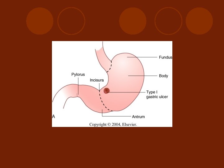
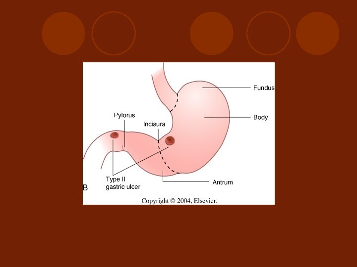
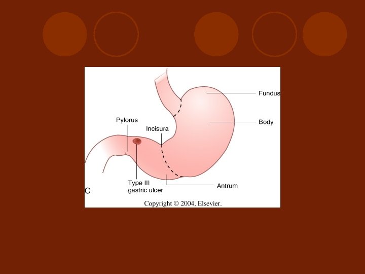
 Divisions of the stomach (From Zuidema G: Shackelford’s Surgery of the Alimentary Tract, 4 th ed. Philadelphia , WB Saunders, 1995 (.
 l ***Guideline for approach and management non-variceal bleeding l -Related Surgical") Upper gastrointestinal hemorrhage(UGIH) l ***Guideline for approach and management non-variceal bleeding l -Related Surgical Anatomy and pathophysiology of Stomach and Duodenum l - Group of diseases caused UGIH and specific consideration l - Endoscopic and surgical management for UGIH
Upper gastrointestinal hemorrhage(UGIH) l ***Guideline for approach and management non-variceal bleeding l -Related Surgical Anatomy and pathophysiology of Stomach and Duodenum l - Group of diseases caused UGIH and specific consideration l - Endoscopic and surgical management for UGIH
 variceal bleeding l -Cirrhosis and portal hypertension l -Endoscopic diagnosis and") Upper gastrointestinal hemorrhage(UGIH) variceal bleeding l -Cirrhosis and portal hypertension l -Endoscopic diagnosis and management l -Surgical management
Upper gastrointestinal hemorrhage(UGIH) variceal bleeding l -Cirrhosis and portal hypertension l -Endoscopic diagnosis and management l -Surgical management
 Lower gastrointestinal hemorrhage ( LGIH( l -Relate Surgical Anatomy of small and large intestine l -Guideline for approach and treatment l -Group of diseases cause LGIH and specific consideration l -Historical background of investigation for localization and surgical management for LGIH
 PERIOD I …
 Why we have to learn …. . l Over all mortality rate is still high in upper GI hemorrhage, about 5 -8% Dudnick R, Martin P, Friedman LS; management of bleeding ulcer. Med Clin North Am 75: 948, 1991
 Blood supply to the stomach and duodenum with anatomical relationships to the spleen and pancreas. The stomach is reflected cephalad. (From Zuidema G: Shackelford’s Surgery of the Alimentary Tract, 4 th ed. Philadelphia, WB Saunders, 1995. )
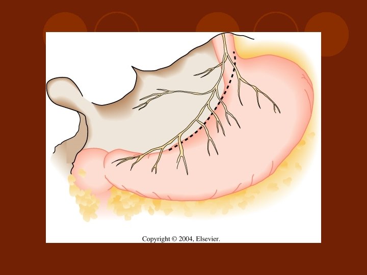
 NERVE SUPPY TO THE STOMACH
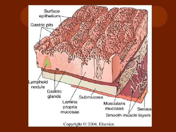
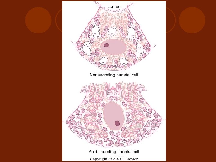
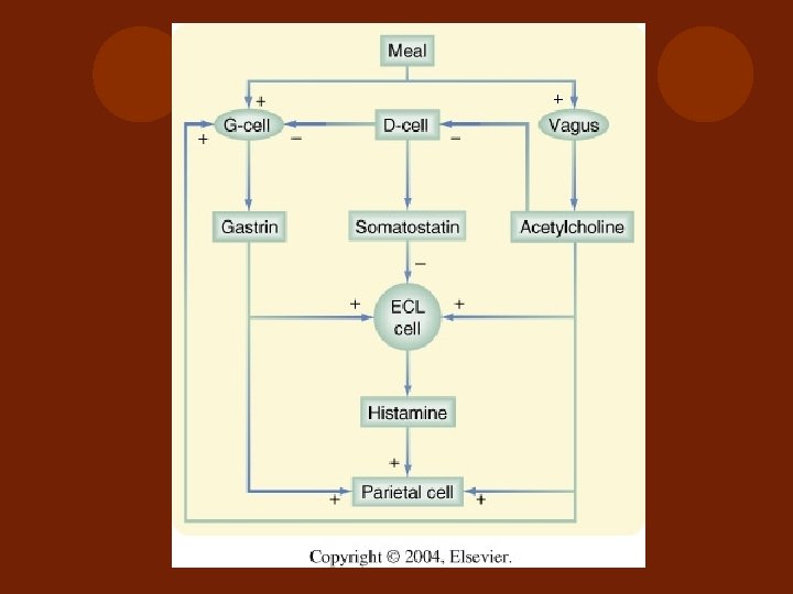
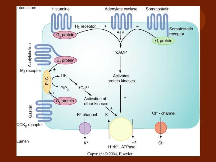
 GASTRIC GLAND
 : since 1899 l Non-steroidal") Risk Factor for Peptic Ulcer Hemorrhage l Aspirin (ASA) : since 1899 l Non-steroidal antiinflammatory drug ( N-SAID) : has both Cyclooxygenase-1 and 2: COX - 1(house keeping enzyme) & COX 2( Coenzyme) l Selective COX- 2 inhibitor: 1999 EX: Clelcoxib, Rofecoxib
Risk Factor for Peptic Ulcer Hemorrhage l Aspirin (ASA) : since 1899 l Non-steroidal antiinflammatory drug ( N-SAID) : has both Cyclooxygenase-1 and 2: COX - 1(house keeping enzyme) & COX 2( Coenzyme) l Selective COX- 2 inhibitor: 1999 EX: Clelcoxib, Rofecoxib
 COX theory l ASA – inhibit COX- 1, decrease Thromboxane& decrease prostaglandin caused of lost of protection for gastric mucosa, and decrease hemostasis l N-SAID- inhibits both COX-1 and COX-2 : results like in ASA user. Increase risk of complication in PU patients =6. 1 (relative risk) and in recent GI bleeding patients= 13. 5 (relative risk) l Selective COX-2 inhibitor: results more protection for gastric mucosal barrier, and hemostasis
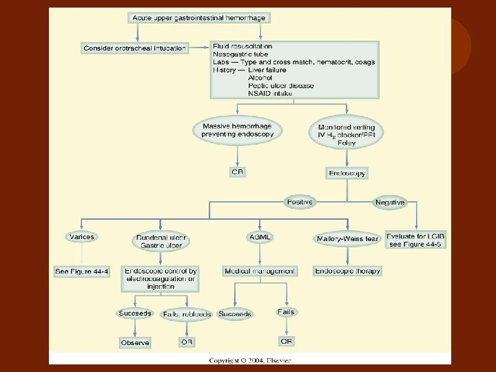
 Acute Gastrointestinal Hemorrhage IDENTIFICATION SOURCE OF BLEEDING AND SPECIFIC THERAPY RESUSCITATION and STABILIZATION INTENSIVE MONITORING INITIAL ASSESSMENT **Initial evaluation and treatment of patients with acute gastrointestinal hemorrhage
 UPPER GI HEMORRHAGE How should surgeons deal with and step up this complicated problem ? l Initial Assessment l Initial Resuscitation l Critical care and monitoring l Definite diagnosis and management Evidence based critical care, Paul Ellis Marick , 2001
 I. Initial Assessment A: General assessment and Scoring to categorize the patients - Active or ongoing/ massive/ continue/ or intermittent bleeding B: Hx and PE (Cirrhotic patient or Non cirrhotic patient ) C: NG tube should be inserted to confirm the level of bleeding
 A. General Assessment Hemodynamic assessment : BP, pulse, postural changes, peripheral perfusion l The presence of co morbid diseases l Estimation of blood loss by nasogastric tube intubation and hemodynamic response to fluid challenge ** remarked that - to use 2 L of crystalloid to stabilize v/s , blood loss is about 15 -30% - If BP raises but fall again, blood loss is about 30 -40% - If BP continues to fall, blood loss is more than 40% l
") Category of Hypovolemic Shock l Class I: Impending (< 10% of blood volumn loss) no symptom, pulse > 90 -100 , BP normal l Class II: mild (10 -20% of blood volumn loss) fainting, pallor, cool skin, BP drop, pulse>120 l Class III: modurate(20 -30% of blood volumn loss): urine output -oliguria l Class IV: severe ( >40% of blood volumn loss) may caused unconcious and cardiac arrest
Category of Hypovolemic Shock l Class I: Impending (< 10% of blood volumn loss) no symptom, pulse > 90 -100 , BP normal l Class II: mild (10 -20% of blood volumn loss) fainting, pallor, cool skin, BP drop, pulse>120 l Class III: modurate(20 -30% of blood volumn loss): urine output -oliguria l Class IV: severe ( >40% of blood volumn loss) may caused unconcious and cardiac arrest
 Category and scoring of patients l To evaluate and predict further ulcer hemorrhage l To select the method of management “ It is dictated by the rate of bleeding”

 of cases") Clinical bleeding 1. Trace heme-positive stools and without severe anemia ( OPD) of cases 2. Visible blood, coffee ground, melena ( IPD/ further evaluation) 1+ 2 = 80 % of cases Fleischer D, et al Gastroenterology, 1983 3. Persistent or massive bleeding / referred due to rebleeding with hemodynamic instability (ICU) ** Massive/ ongoing bleeding is defined as loss of > 30% of estimated blood volume or bleeding required blood transfusion > 6 U/ 24 hours
Clinical bleeding 1. Trace heme-positive stools and without severe anemia ( OPD) of cases 2. Visible blood, coffee ground, melena ( IPD/ further evaluation) 1+ 2 = 80 % of cases Fleischer D, et al Gastroenterology, 1983 3. Persistent or massive bleeding / referred due to rebleeding with hemodynamic instability (ICU) ** Massive/ ongoing bleeding is defined as loss of > 30% of estimated blood volume or bleeding required blood transfusion > 6 U/ 24 hours
 Scoring to categorize the patients l Forrest classification severe, moderate, mild Lancet 1974 l Rockall Risk Scoring Gut 1996 l New Scoring System by Blatchford Lancet 2000 l Modified Rockall Score for both Non-variceal and Variceal bleeding AJG 2002
 Endoscopic diagnosis Stigmata of") Rockall Scoring Age Shock Co morbid disease ( cancer diseases) Endoscopic diagnosis Stigmata of recent hemorrhage Pre-endoscope score 0 -7 Post –op endoscope score 0 -11 * this scoring system is good to predict for the mortality rate much than rebleeding 0 -3 : mortality rate = 0 – 11% 4 -7 : mortality rate = 24 - 27% l > 8 : motality rate = > 40% l l l Rockall TA et al GUT 1996; 38: 316 -21
Rockall Scoring Age Shock Co morbid disease ( cancer diseases) Endoscopic diagnosis Stigmata of recent hemorrhage Pre-endoscope score 0 -7 Post –op endoscope score 0 -11 * this scoring system is good to predict for the mortality rate much than rebleeding 0 -3 : mortality rate = 0 – 11% 4 -7 : mortality rate = 24 - 27% l > 8 : motality rate = > 40% l l l Rockall TA et al GUT 1996; 38: 316 -21
 New Scoring System by Blatchford l Admission Hb l BUN l Pulse l Systolic BP l Fainting or melena as chief complaint l Liver disease or cardiac disease to predict the need for clinical interventions • But it is in only one study •
 High Risk ~Criteria l l l Host Factors Age >60 yr Co-morbid conditions e. g. renal failure, cirrhosis, cardiovascular disease, COPD Hemodynamic instability; mod to severe shock Coagulopathy include drug-related Bleeding character ; Active continue red blood from NG after irriagtion and red blood per rectum Patient course; massive blood transfution> 4 -6 units to maintain Hb in 24 hr , re-bleeding in 72 hr , return to have hemodynamic instability 2004 Concensus for Clinical Practice Guideline for the Management of Upper GI bleeding; สมาคมโรคทางเดนอาหารแหงประเทศไทย
 I. Initial Assessment A: General assessment and Scoring to categorize the patients - Active or ongoing/ massive/ continue/ or intermittent bleeding B: Hx and PE (Cirrhotic patient or Non cirrhotic patient ) C: NG tube should be inserted to confirm the level of bleeding
 - History") B. Take Hx and PE (Cirrhotic patient or Non cirrhotic patient ) - History taking of previous medication and underlying diseases/ anticoagulant usage. - Esophageal varices is more suspicious for 60% - 80% in severe upper GI bleeding with history of advanced liver disease or a history of previous variceal bleeding.
B. Take Hx and PE (Cirrhotic patient or Non cirrhotic patient ) - History taking of previous medication and underlying diseases/ anticoagulant usage. - Esophageal varices is more suspicious for 60% - 80% in severe upper GI bleeding with history of advanced liver disease or a history of previous variceal bleeding.
 Duodenal ulcer 24.") Prediction of UGI bleeding etiology l l l l l Incidence(%) Duodenal ulcer 24. 3 Gastric erosions 23. 4 Gastric ulcer 21. 3 Esophageal varices 10. 3 -----20% (in cirrhosis) Malorry-Weiss tear 7. 2 Esophagitis 6. 3 Duodenitis 5. 8 Neoplasm 2. 9 Marginal( stomal) ulcer 1. 8 Esophageal ulcer 1. 7 Miscellaneous 6. 8 l Silverstein FE, Gilbert DA, Tadeseo FJ, et al, The national ASGE Survey on upper gastrointestinal bleeding Gastrointestinal Endoscopy, 1981
Prediction of UGI bleeding etiology l l l l l Incidence(%) Duodenal ulcer 24. 3 Gastric erosions 23. 4 Gastric ulcer 21. 3 Esophageal varices 10. 3 -----20% (in cirrhosis) Malorry-Weiss tear 7. 2 Esophagitis 6. 3 Duodenitis 5. 8 Neoplasm 2. 9 Marginal( stomal) ulcer 1. 8 Esophageal ulcer 1. 7 Miscellaneous 6. 8 l Silverstein FE, Gilbert DA, Tadeseo FJ, et al, The national ASGE Survey on upper gastrointestinal bleeding Gastrointestinal Endoscopy, 1981
 I. Initial Assessment A: General assessment and Scoring to categorize the patients - Active or ongoing/ massive/ continue/ or intermittent bleeding B: Hx and PE (Cirrhotic patient or Non cirrhotic patient ) C: NG tube should be inserted to confirm the level of bleeding
 C. NG tube placement v. Should perform in all UGI hemorrhage vto confirmation that it is the upper GI bleeding , monitoring of bleeding and , decompressed the stomach v. No report that it may potentiate bleeding in case of esophageal varices, just careful in patients who had severe coagulopathy.
 UGI LGI Melena l Hematemasis or coffee ground l Maroon stool * l Red stool ** * l Guaiac test ( can positive more 2 weeks after bleeding stopped) l * Bile was seen via NG tube ** Massive bleeding
 Necessary Laboratories l CBC, plt l BS, BUN, Cr, electrolyte l PT, PTT, bleeding time l LFT l G/M l EKG l Cx. R
 II. Initial Resuscitation l How to do for good resuscitation? l When will we give blood transfusion? l Which medication will be used?
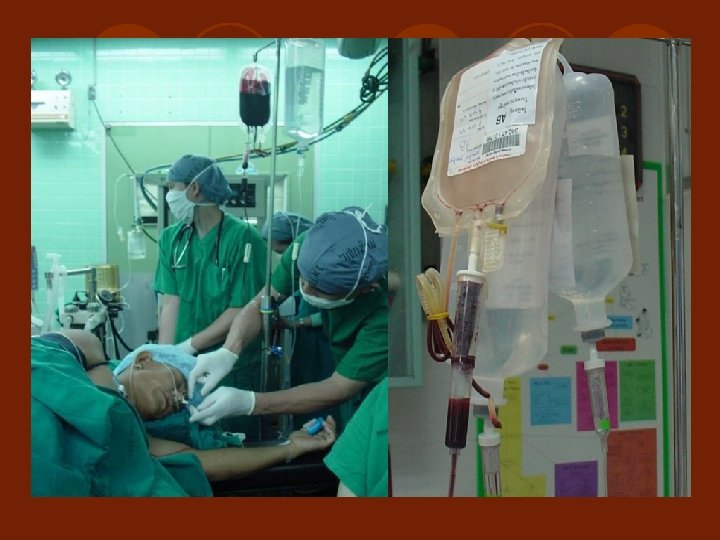
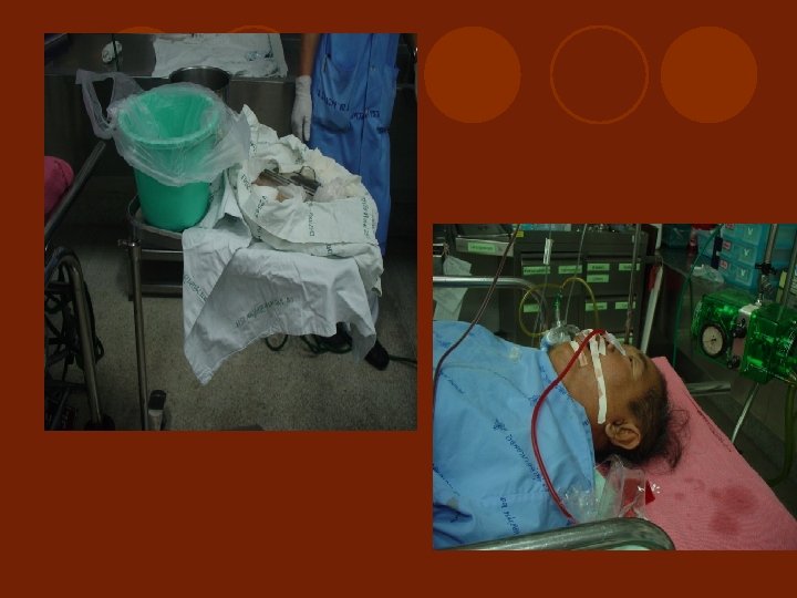
 to decompress") Large- bore intravenous lines or central lines NG tube aspiration (by hand) to decompress clot in stomach l Volume expansion with colloid or crystalloid l Transfusion of blood immediately if patient has hemodynamically unstable l l * Blood products are the most efficient volume expanders ** It take about 72 hours for Hct to reach its nadir; therefore, a normal or moderate low Hct does not exclude significant bleeding *** Conversely, minimally falling of Hct also represent fluid disequilibrium much rather than continued bleeding
Large- bore intravenous lines or central lines NG tube aspiration (by hand) to decompress clot in stomach l Volume expansion with colloid or crystalloid l Transfusion of blood immediately if patient has hemodynamically unstable l l * Blood products are the most efficient volume expanders ** It take about 72 hours for Hct to reach its nadir; therefore, a normal or moderate low Hct does not exclude significant bleeding *** Conversely, minimally falling of Hct also represent fluid disequilibrium much rather than continued bleeding
 If patients have coagulopathy, they should be corrected. - PTT prolong > 1. 5 times - Platelet < 50, 000/ mm 3 - FFP should be given after 6 unit of PRC and plt should add after 10 unit of PRC l Monitoring V/S, urine out put /hour l Airway protection in those who have alteration of consciousness or endotracheal intubations may facilitate to investigate and give treatment in these patients l
 Recommendation for empiric Acid- suppression therapy l Traditionally treated, even before the cause is determined, with acid suppression therapy. Medications are extremely safe, although the efficacy of this practice has not been proven conclusively. Kupfer, et al Gastroenterol Clin of North Amer, 2000 I. V. Proton pump inhibitor is more effective than I. V. H 2 blocker in increasing intragastric p. H Vasopressin should not be used due to its systemic side effect
 High dose omeprazole significantly reduces the frequency of further bleeding and of surgery in patients with bleeding ulcer. dosage 40 mg i. v. every 12 hrs. for 5 days Saltzman JR, N Engl J Med, 1997 NEW GENERATION PPI Lanzoplazole - Pantoprazole - Rabeprazole - Esomeprazole -
 SOMATOSTATIN Somatostatin / Octreotide infusion - In massive UGIH with Hx of advance liver disease is recommended PROSTAGLANDIN ANALOQUE - Cytoprotective agent
 Somatostatin causes - Splanchnic vasocostriction - Reduces Azygos venous blood flow - Reduces portal colatteral circulation and decreases portal pressure Octreotide (Somatostatin analoque) 50 microgram i. v. bolus then 50 microgram/ hr for infusion rate for 5 days it can be discontinued without tapering.
 III. Critical care and monitoring l ICU is needed, when? - Massive/ continue or on going bleeding with or without coagulopathy - High Rockall scoring patients ( high risk of morbidity & mortality due to continue or rebleeding - Severe co-morbid disease
 l") IV. Definite diagnosis and management l Esophagogastroduodenoscope ( EGD for Dx and Rx) l Technique of operative intervention
IV. Definite diagnosis and management l Esophagogastroduodenoscope ( EGD for Dx and Rx) l Technique of operative intervention
 Indication and Timing - In high score") Endoscogastroduodenoscope ( EGD for Dx and Rx) Indication and Timing - In high score patients ( > 3) - Shock Category II, III - Promptly as a double set up in active /massive bleeding - Under specialist to perform endoscopic therapy for hemostasis or localized potential angiographic or surgical therapy * Initial diagnostic procedure of choice should be performed in first 6 - 24 hour after onset of bleeding
Endoscogastroduodenoscope ( EGD for Dx and Rx) Indication and Timing - In high score patients ( > 3) - Shock Category II, III - Promptly as a double set up in active /massive bleeding - Under specialist to perform endoscopic therapy for hemostasis or localized potential angiographic or surgical therapy * Initial diagnostic procedure of choice should be performed in first 6 - 24 hour after onset of bleeding
 Precaution and contraindication Absolute contraindication - GI perforation - Acute uncontrolled unstable angina - Severe untreated coagulopathy - uncontrolled respiratory decompensation - unexperience endoscopist and patient agitation and uncooperation * Intraoperative endoscopy ( on ET-tube and G/A ) in selected cases or shift the intervention to surgery or conservative only l
 General Complications of EGD l GI perforation l Sepsis l Pulmonary aspiration l Respiratory failure l Induce bleeding l Ventricular tachycardia l Myocardial infarction l Death
 Prediction of further ulcer hemorrhage The most important endoscopic predictor of persistent or recurrent bleeding is active bleeding( arterial spurting or oozing) at the time of endoscopy l The rate of rebleeding is approximately 3 % in the low risk group 25% in the high risk group l Number of blood transfusion units > 5 units = 57% needing Surgery, mortality = 43% l
 Adverse Prognostic Factors in UGIH Endoscopic criteria for endoscopic intervention because of high rate of continue or re-bleeding Stigmata of recent hemorrhage: Forrest classification I, II L Active bleeding lesion, oozing L Visible vessel, Adherent clot Ulcer location L Posterior duodenal bulb L Higher lessor gastric curvature, High lying ulcer Ulcer size and character L Large and hard edge
 Forrest’s Endscopic finding Classification l IA : l IB : l IIA: l IIB: l III : Active or Spurting Oozing ulcer Non-bleeding visible vessel Adherent clot other unspecsified
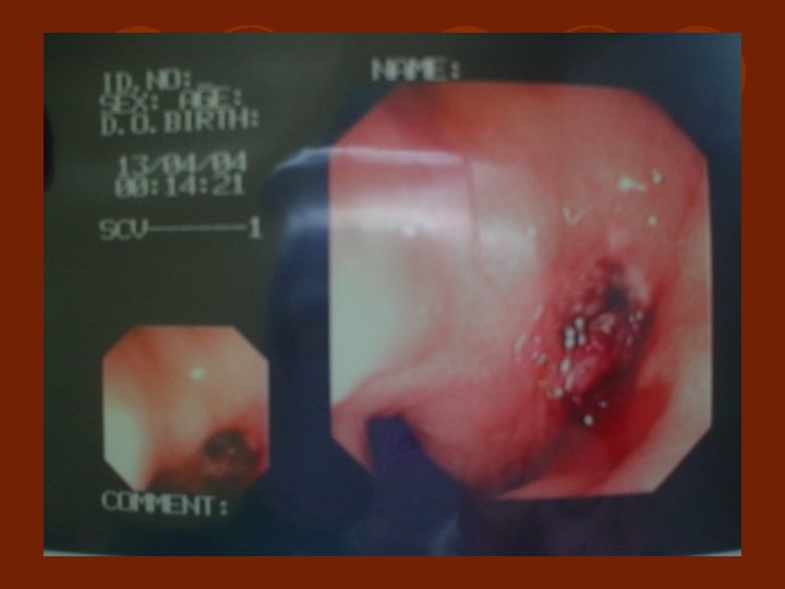
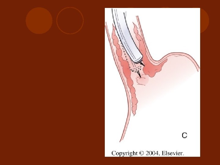
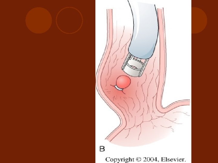
 Injection") Endoscopic Intervention For PU bleeding Thermal Techniques ; monopolar/bipolar/heater probe/laser photo coagulation 2) Injection Methods; 3. 6% hypertonic saline+1: 20, 000 adrenalin 9 -12 cc or 1: 10, 000 10 cc via 23 -25 gauge needles. 0. 5 cc each point 3) Topical Agents; cyanoacrylate tissue glues/ microcrystalline collagen hemostat : * good for diffuse gastric mucosal lesions or adjunct to other modalities 4) Mechanical methods; Hemoclips (1. 5 mm)/ balloon tamponad 1) Sukawa, et al, Surg Clin of North Amer, 1992
Endoscopic Intervention For PU bleeding Thermal Techniques ; monopolar/bipolar/heater probe/laser photo coagulation 2) Injection Methods; 3. 6% hypertonic saline+1: 20, 000 adrenalin 9 -12 cc or 1: 10, 000 10 cc via 23 -25 gauge needles. 0. 5 cc each point 3) Topical Agents; cyanoacrylate tissue glues/ microcrystalline collagen hemostat : * good for diffuse gastric mucosal lesions or adjunct to other modalities 4) Mechanical methods; Hemoclips (1. 5 mm)/ balloon tamponad 1) Sukawa, et al, Surg Clin of North Amer, 1992
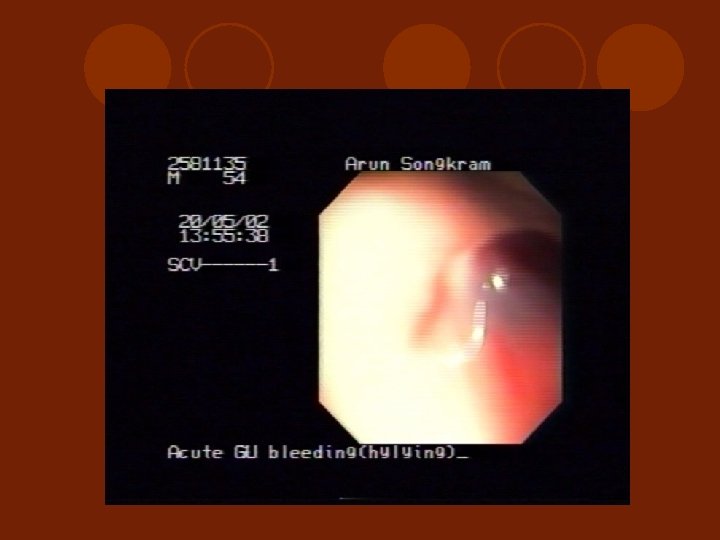
 Post combind adrenalin injection And Heat probe coagulation in acute GU bleeding
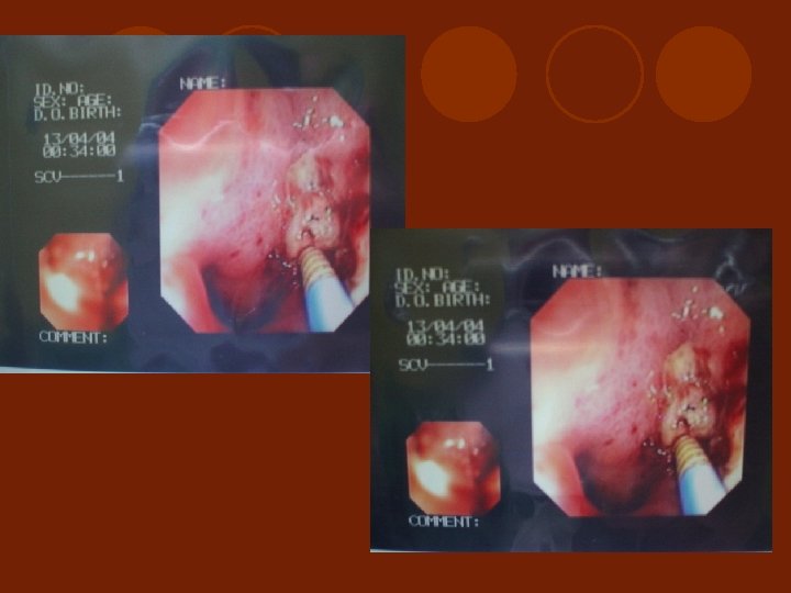
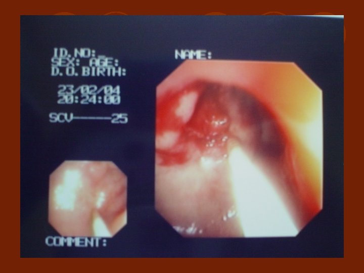
 Post injection + Heater probe coagulation in active DU bleeding
 Follow up EGD of DU bleeding 1 month
 Dieulafoy’s lesion : Therapeutic Hemoclip via EGD
 Complication l Perforation ; 1 -3 % l Necrosis on high dose epinephrine l Induce acute and delayed hemorrhage 5 -30% in visible vv. those treated by thermal therapy or injection therapy * most common is cardiopulmonary in nature or related to sedation given ** Prophylaxis antibiotic should be applied
 Endoscopic band ligation combind 2) Endoscopic sclerosing") Endoscopic Intervention For Esophageal varices : 1) Endoscopic band ligation combind 2) Endoscopic sclerosing therapy: 1% Ethoxyscleral solution 0. 5 -1 cc /point 3) Combined Ballon Tamponad for temporary control after fail endoscopic intervetion control ( Senstaken Blakemore tube preparation)
Endoscopic Intervention For Esophageal varices : 1) Endoscopic band ligation combind 2) Endoscopic sclerosing therapy: 1% Ethoxyscleral solution 0. 5 -1 cc /point 3) Combined Ballon Tamponad for temporary control after fail endoscopic intervetion control ( Senstaken Blakemore tube preparation)
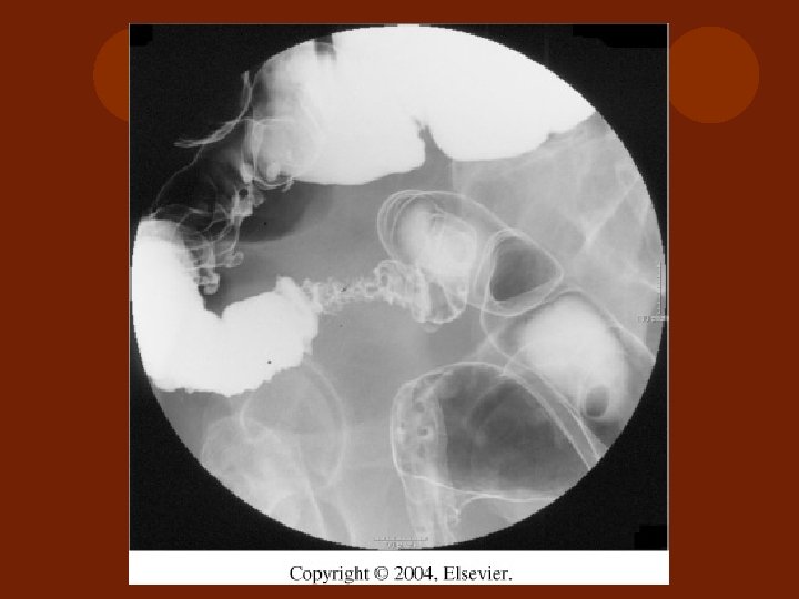
") INTRAVARICEAL INJECTION ( underfluoroscope and venogram)
INTRAVARICEAL INJECTION ( underfluoroscope and venogram)
 PARAVARICEAL INJECTION
 ENDOSCOPIC MUCOSAL VARICEAL BAND LIGATION
 SB- tube
 Complication Prophylaxis antibiotics cover gram negative bacteria such as ciprofoxacin, levofloxacin, ceftacidime, amoxicillin-culvulanic acid, and aztreonam are appropriate choices
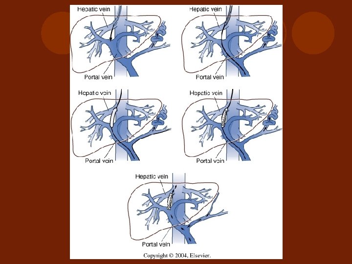
 l Non-operative shunt l Use in stage of cirrhosis") TIPS( Transcutaneous-jugular intrahepatic portosystemic shunts) l Non-operative shunt l Use in stage of cirrhosis with liver failure l In non-randomize trial : Less effective to stop GI bleeding than operative shunt, but less invasive. l Technique need radiointervention ( described by Zemel G, Katzen B T, Becker G J, et al TIPS, JAMA, 266: 390, 1991 )
TIPS( Transcutaneous-jugular intrahepatic portosystemic shunts) l Non-operative shunt l Use in stage of cirrhosis with liver failure l In non-randomize trial : Less effective to stop GI bleeding than operative shunt, but less invasive. l Technique need radiointervention ( described by Zemel G, Katzen B T, Becker G J, et al TIPS, JAMA, 266: 390, 1991 )
 Topic of interest. . … l Video capsule Endoscopy l Intraoperative endoscopy l Rare causes of upper gastrointestinal hemorrhage from an obscure source; small intestine above ligament of treiz that EGD could not exam, new scope was developed
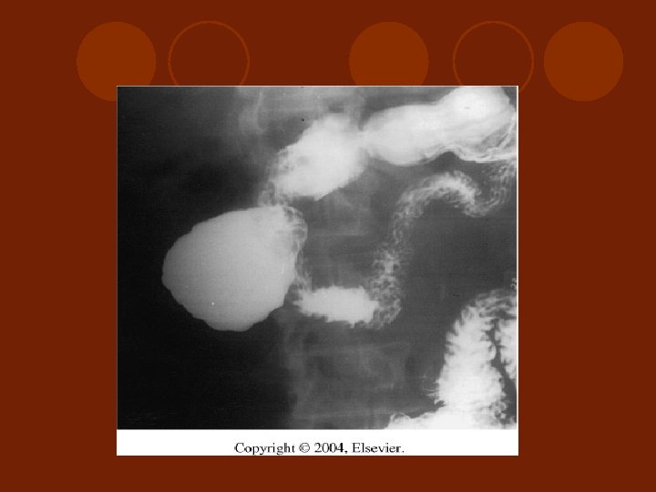
 Gastric Diverticulum
 Post operative EGD to follow up l For non-definite surgical procedure cases ; after 2 week post operation (in Japan) l To check for malignant potential and adjunct medical treatment including H. pylori eradication in some case * There is a report of unnecessary management to eradicate H. pylori at the time of hemorrhage occurrence.
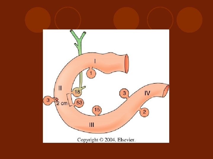
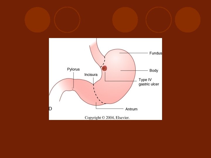
 l Acute or Chronic l Type") Type of GASTRIC ULCER GU ( Johnston’s criteria) l Acute or Chronic l Type I: at Lessor curvature l Type II: GU anywhere with DU l Type III: at Prepylorus( prepyloric ulcer) l Type IV: in 5 CM below EG junction (high lying GU) Type V - ?
Type of GASTRIC ULCER GU ( Johnston’s criteria) l Acute or Chronic l Type I: at Lessor curvature l Type II: GU anywhere with DU l Type III: at Prepylorus( prepyloric ulcer) l Type IV: in 5 CM below EG junction (high lying GU) Type V - ?
 DUODENAL ULCER (DU( l Acute or Chronic l Post bulbar l Kissing ulcer l Aortoenteric fistula
 Operative intervention l To stop bleeding by suture in emergency situation or failure bleeding control by endoscopic intervention 1 -2 times. l To definite treatment for the cause of bleeding l High risk patient, massive and no time, no blood( rare blood group AB, Rh negative , no skillful endoscopist (
 Operative Finding Do not forget to palpate and look for the scar at stomach and duodenum both anterior and posterior wall.
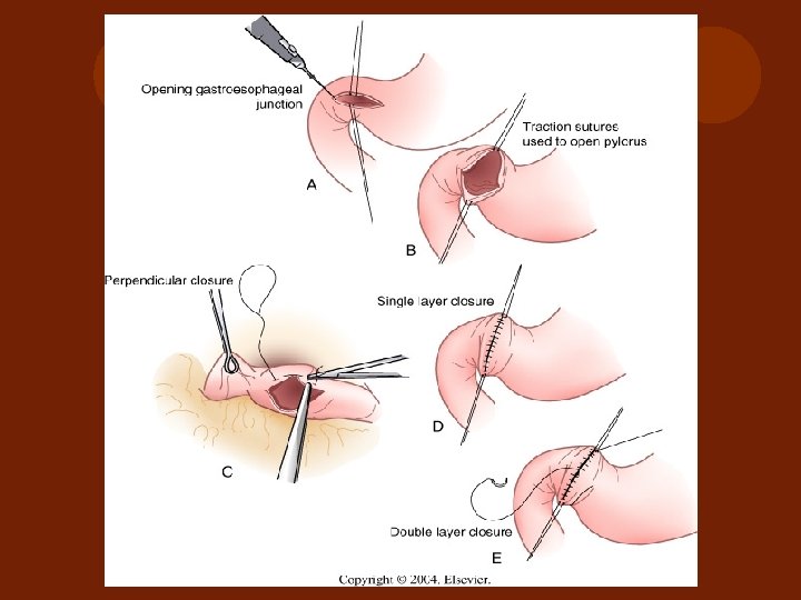
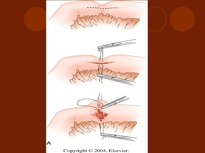
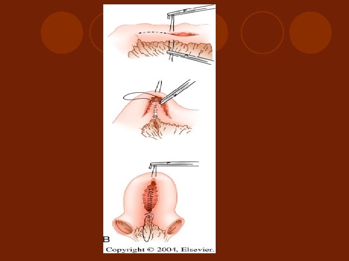
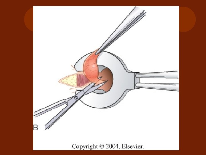
 Technicques for suture and tissue handling l Do not forget to pack the spleen before mobilization of stomach l Hanging suture should be used l Atraumatic non-absorable suture should be used l Babcock preferred to use for temporary handling stomach incision edge l Incision should start at anterior stomach wall longitudinally or at place that EGD suspected the cause of bleeding.
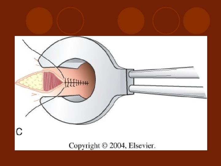
 Technicques for suture and tissue handling Suture bleeding point by Transfixing U stitch l it should be performed in acute massive DU hemorrhage l For the difficult large duodenal ulcer and chronic ulcer, alternative surgical procedures may be added depend on condition of patient - Ulcerectomy - TV with drainage procedure ( various type of pyloroplasty, gastrojejunostomy) ** selection for complete diverticulization l
 Technicques for suture and tissue handling l Difficult chronic GU ; need tissue ? > 10% of GU > 1 cm are malignant ulcer Chua CL and Jeyaraj PR, Am J Surg; 1992 Difficult type - posterior wall ulcer; ulcerectomy with or without leaving the ulcer to capsule of pancrease - high lying GU type IV ( Johnston’s type)
 Acid Reducing and Drainage Precedure VAGOTOMY : reduce acid secretion in cephalic phase - TV ; resect vagus n. both ant. and post. trunk And need to do drainage procedure, always - SV ; resect ant. Vagus n. and post. N. of Latajet ( vagus n. after separation of celiac and hepatic branch). Need to do drainage procedue - HSV ; resect only branch of Latajet and preserve branch at Craw foot to preserve function of pylorus l ANTRACTOMY : reduce G-cell ( Billroth I, or Billroth II) l
 TV
TV
 HSV
HSV
 ANTRECTOMY

 BILLROTH I
 BILLROTH I WITH TV
 BILLROTH II
 Operative Procedure for chronic GU and DU ; Antrectomy include ulcer or ulcer excision l GU Type II ; Ulcer excision& TV & pyloroplasty or highly selective vagotomy( HSV) l GU Type III and DU ; 3 options 1) Suture bleeding point & TV/SV with pyloroplasty 2) Suture bleeding point & HSV 3) Antrectomy with TV or SV l GU Type I In GI hemorrhage – To stop bleeding is the main aim ****
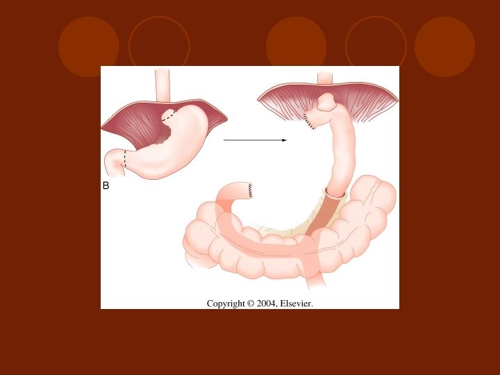
 Most aggressive distal gastrectomy including portion of") Surgical tecniques for highlying GU( type IV) Most aggressive distal gastrectomy including portion of ulcer at esophageal wall with roux-en –Y esophagogastrojejunostomy; “ Csendes procedure” l Am J Surg; 1978 Antrectomy only with leaving the ulcer in place due to it close to EG junction; “ Kelling- Made- lener procedure” , Maingot ; 1997 l Greenfield; 1992 l For 2 -5 cm ulcer at lessor curvature from EG junction; distal gastric resection with a vertical extension( tonque) to include the lesser curvature with end-to-end gastroduodenostomy. “ Pauchet procedure” Shackelford’s; 1991 l Wedge of anterior and posterior gastric wall at lessor curvature to include the ulcer, with ligation of left gastric vessels close to stomach wall ; “ Shoe- maker”
Surgical tecniques for highlying GU( type IV) Most aggressive distal gastrectomy including portion of ulcer at esophageal wall with roux-en –Y esophagogastrojejunostomy; “ Csendes procedure” l Am J Surg; 1978 Antrectomy only with leaving the ulcer in place due to it close to EG junction; “ Kelling- Made- lener procedure” , Maingot ; 1997 l Greenfield; 1992 l For 2 -5 cm ulcer at lessor curvature from EG junction; distal gastric resection with a vertical extension( tonque) to include the lesser curvature with end-to-end gastroduodenostomy. “ Pauchet procedure” Shackelford’s; 1991 l Wedge of anterior and posterior gastric wall at lessor curvature to include the ulcer, with ligation of left gastric vessels close to stomach wall ; “ Shoe- maker”
 LOCAL Post. Op COMPLICATION q Re-bleeding q Mediastinitis q Leakage q Post gastrectomy syndrome
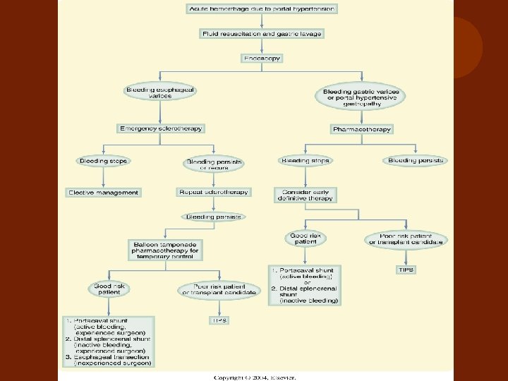
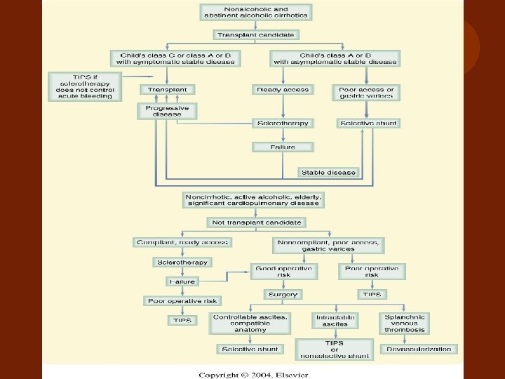
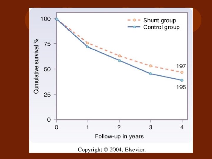
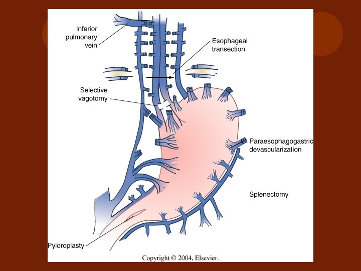
 Emergency Surgery for EV Bleeding PURPOSE l Indirect control of bleeding site l Do not change the mortality after bleeding EV in cirrhotic patient 1) Emergency Total or nonselective- Shunt Operation: Portocaval shunt is the procedure of choice 2) Non-Shunt Operation: Sugiura’s( Esophageal transection with devascularizatoion) , Hassab’s operation(Total devascularization)
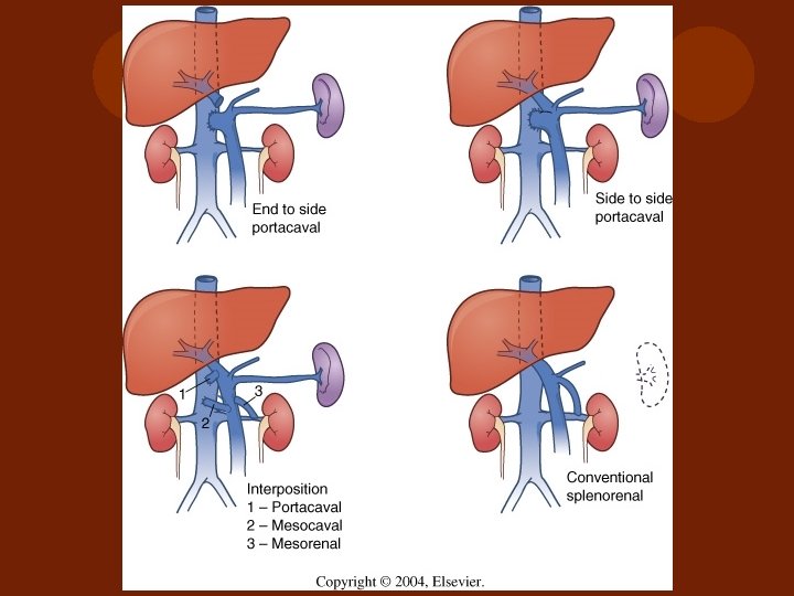
") Selective shunt ( distal splenorenal)
Selective shunt ( distal splenorenal)
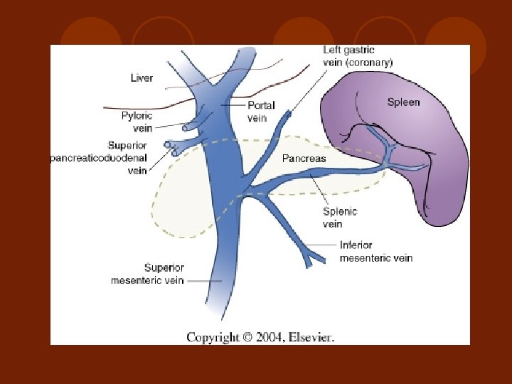
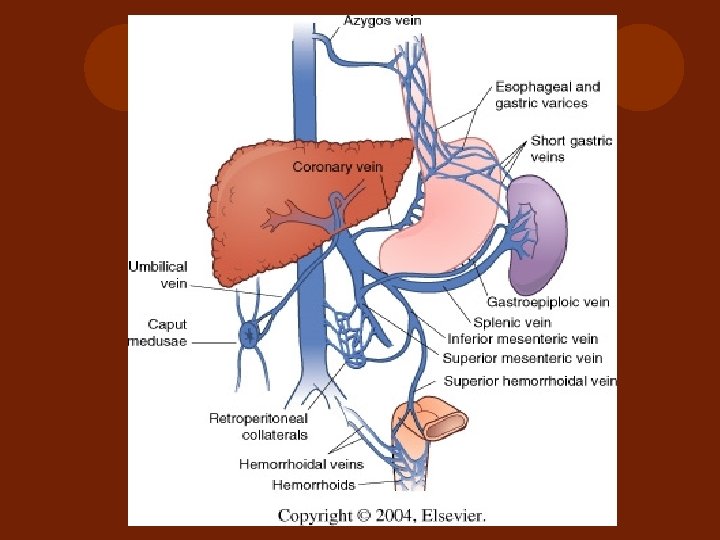
 Gastric Varices Primary l Lessor curvature : common- resolute after Endoscopic intervention for RX of EV l Greater curvature: Less common Secondary Isolated Gastric Varices: Fundus, due to splenic vein thrombosis treated by SPLENECTOMY
 PERIOD II
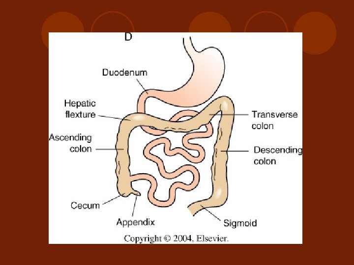
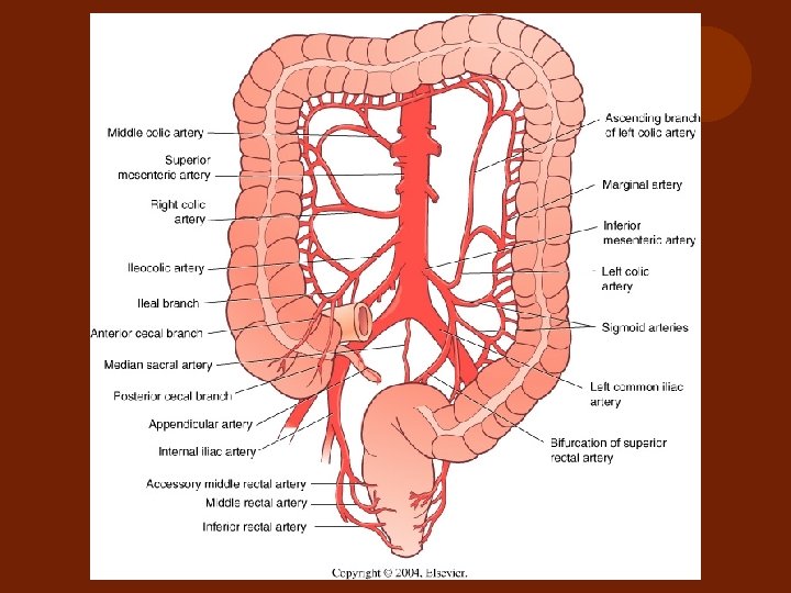
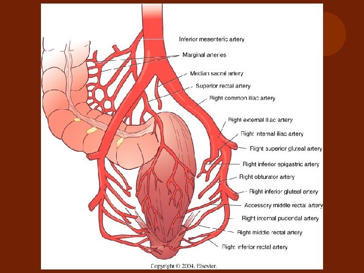
 Lower gastrointestinal hemorrhage ( LGIH( l -Relate Surgical Anatomy of small and large intestine l -Guideline for approach and treatment l -Group of diseases cause LGIH and specific consideration l -Historical background of investigation for localization and surgical management for LGIH
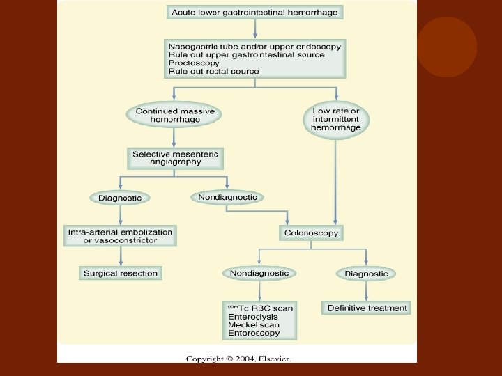
 Approach to Hematochezia Massive Ongoing Bleeding 10 -20% of patients with massive bleeding l Major Self Limited Bleeding 80 -90% of patients with massive bleeding l Minor Self Limited Bleeding l
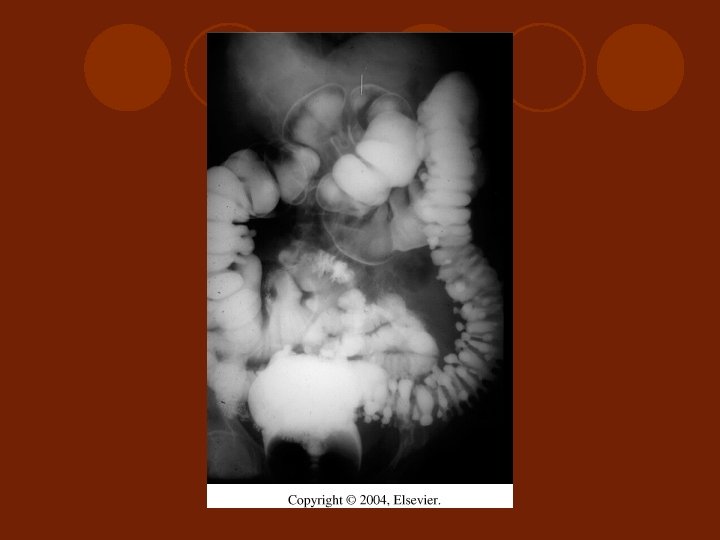
 Diagnosis investigation for LGIH Nuclear Scintigraphy - Sulfur colloid scan - Technetium 99 -m labeled RBC scan l Selective visceral Angiography l Colonoscope l Barium Enema l Ix for small intestine bleeding l
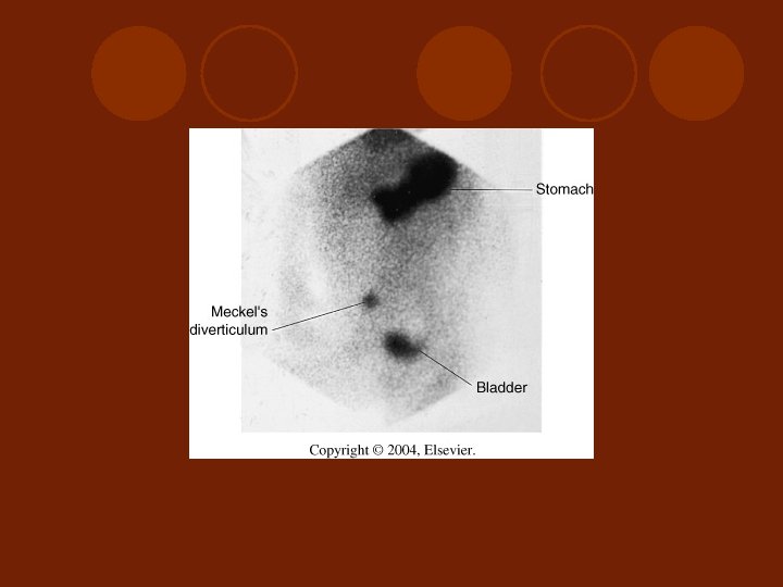
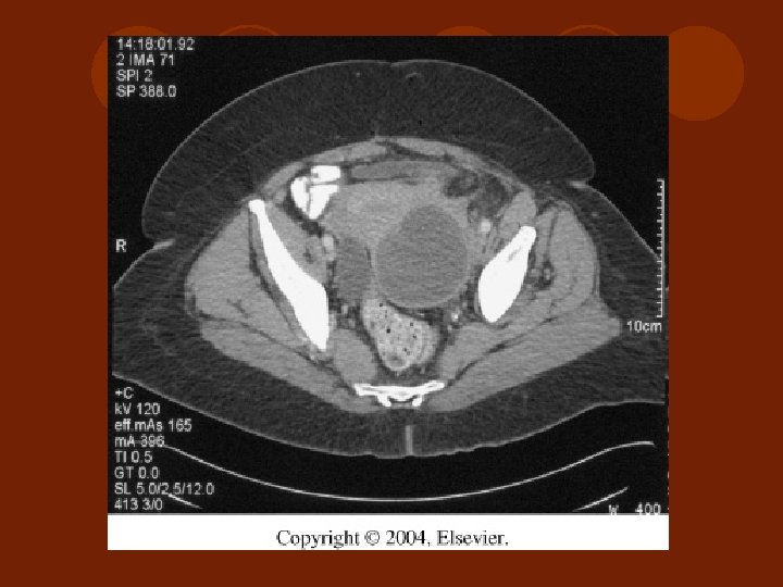
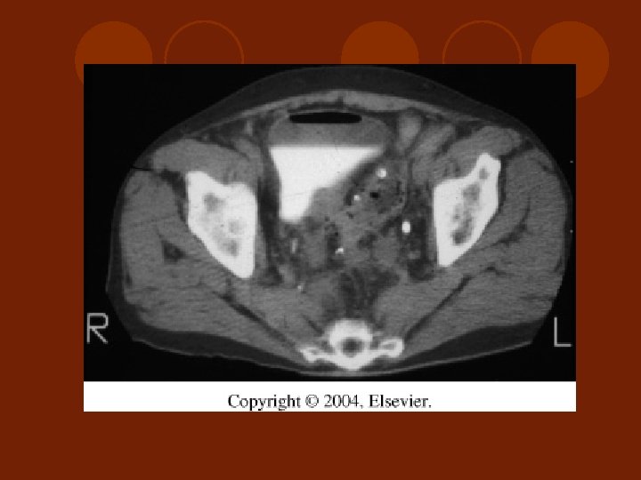
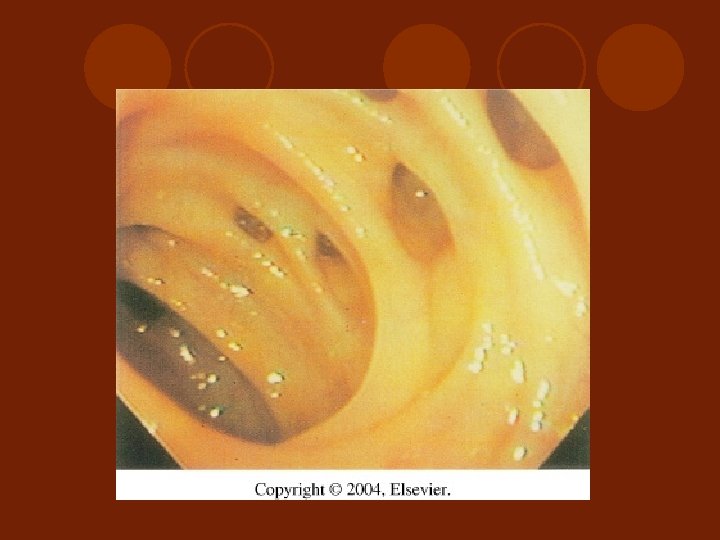
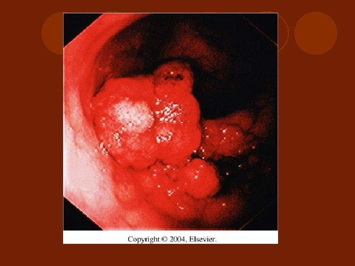
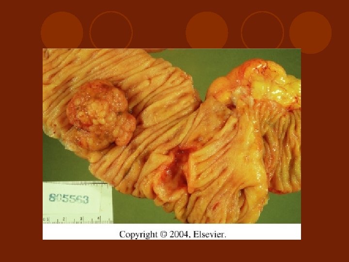
 Causes and Treatment of Lower GI Hemorrhage l l l l l Colonic Diverticular Disease Arteriovenous Malformation (AVM) Inflammatory Bowel Disease Radiation Injury to Small and Large Bowel Tumor of Colon and Rectum Intussusception Ishemic Colitis Colon and Anorectal Varices Meckel’s and other small intestinal diverticula
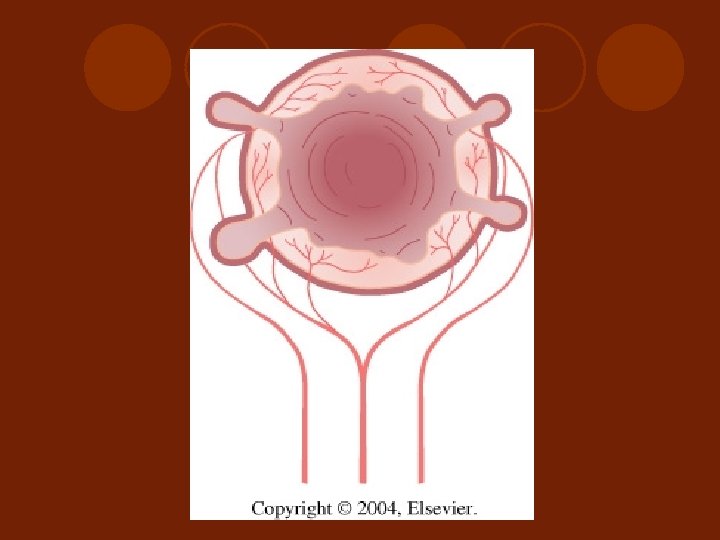
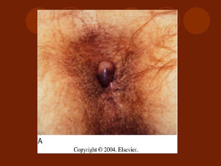
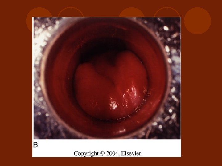
 Baron ligation
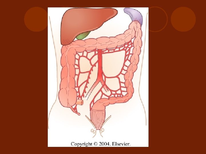
 Operative Intervention l Exploratory Laparotomy l Looking for the cause of bleeding as the information of localization preoperatively l Perform small bowel resection or colonic resection if localization l Incase of none localization and negative intraoperative localization, right hemicolectomy may be performed