28-1 Human Anatomy, First Edition McKinley & O’Loughlin

9844-ch28_reproductive_system.ppt
- Количество слайдов: 74
 28-1 Human Anatomy, First Edition McKinley & O'Loughlin Chapter 28 : The Reproductive System
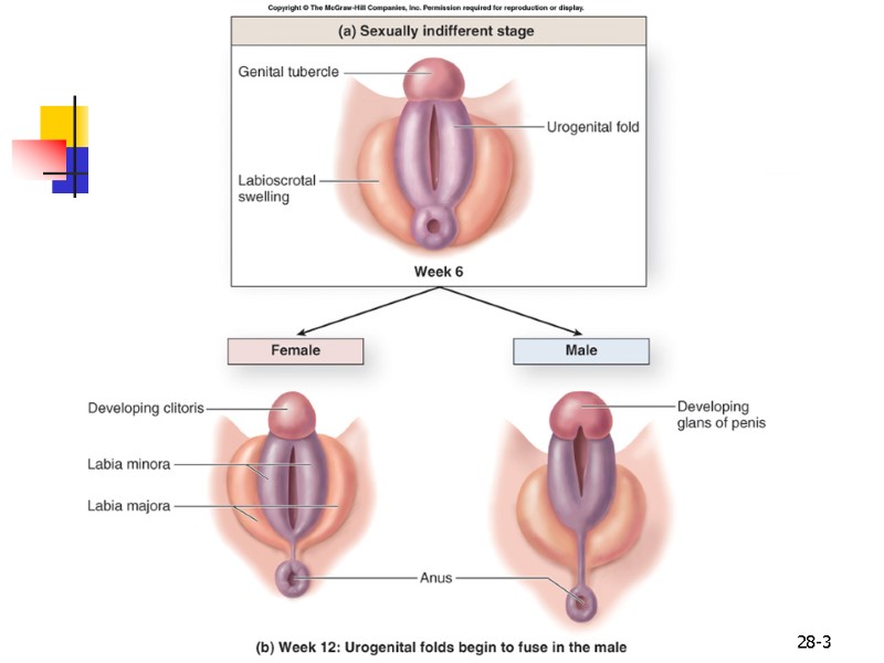
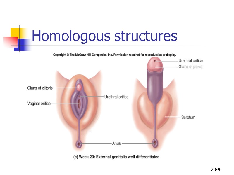
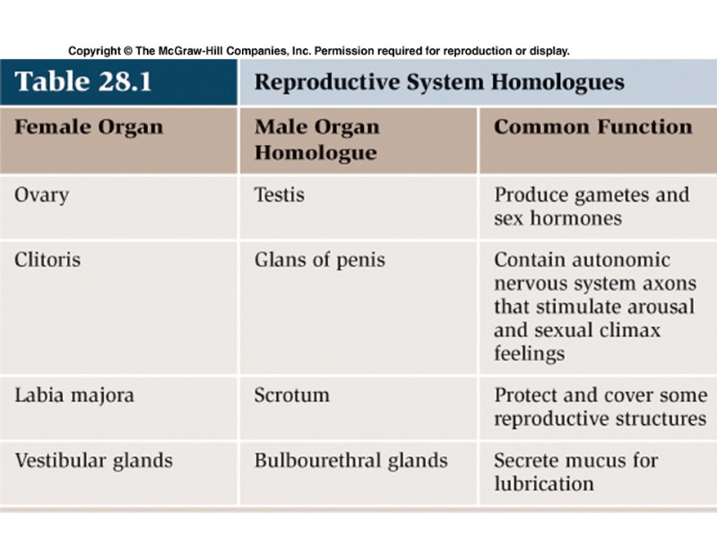
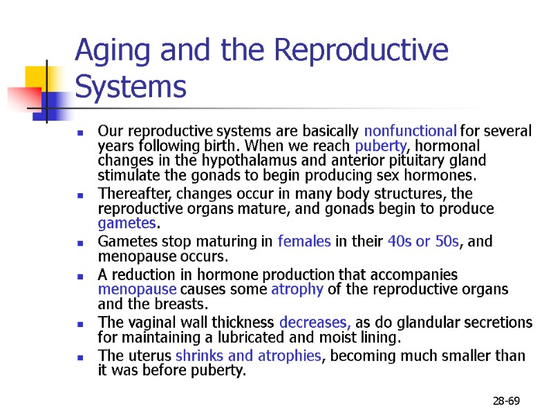
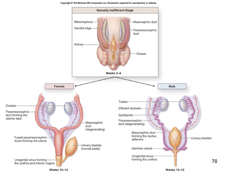
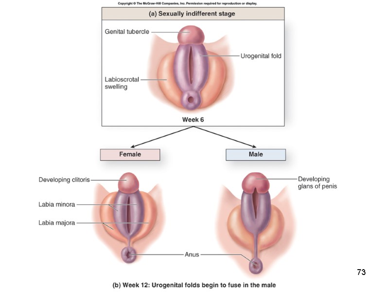
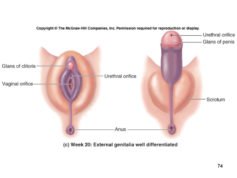
 28-2 Reproductive Systems Main function: propagation of the species To achieve this goal: Must ensure sexual maturation Produce gametes (n). Male and female structures are homologues: derived from common developmental tissues
 28-3
28-3
 28-4 Homologous structures
 28-5 Comparison of the Female and Male Reproductive Systems Primary sex organs called gonads. ovaries in females testes in males Produce gametes which unite to form a new individual. oocytes sperm Gonads produce large amounts of sex hormones which affect maturation, development, and changes in the activity of the reproductive system organs. estrogen and progesterone in the female androgens (esp. testosterone) in the male
 28-6 Comparison of the Female and Male Reproductive Systems Both have accessory reproductive organs duct systems carry gametes away from the gonads toward the site of fertilization in females to the outside of the body in males Fertilization occurs when male and female gametes meet: copulation, coitus, sexual intercourse Restores the diploid number (2n)
 28-7 Comparison of the Female and Male Reproductive Systems Primarily nonfunctional and “dormant” until puberty. At puberty, external sex characteristics become more prominent. breast enlargement in females fat distribution patterns in both sexes pubic hair in both sexes reproductive organs become fully functional gametes mature gonads secrete sex hormones Both reproductive systems produce gametes.
 28-8 Comparison of the Female and Male Reproductive Systems Puberty: Initiated by hypothalamus Secretes GnRH (gonadotropin-releasing hormone Stimulates release of FSH and LH Prior to puberty, not present Stimulate gonads to produce: Sex hormones gametes
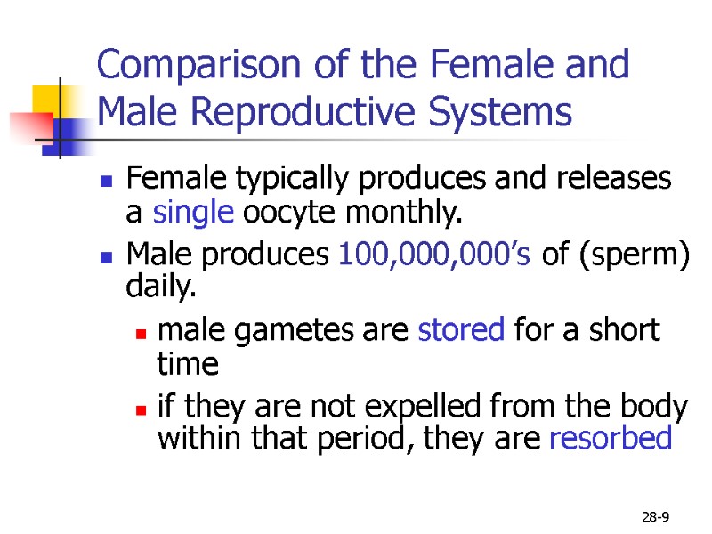
 28-9 Comparison of the Female and Male Reproductive Systems Female typically produces and releases a single oocyte monthly. Male produces 100,000,000’s of (sperm) daily. male gametes are stored for a short time if they are not expelled from the body within that period, they are resorbed
 10
10
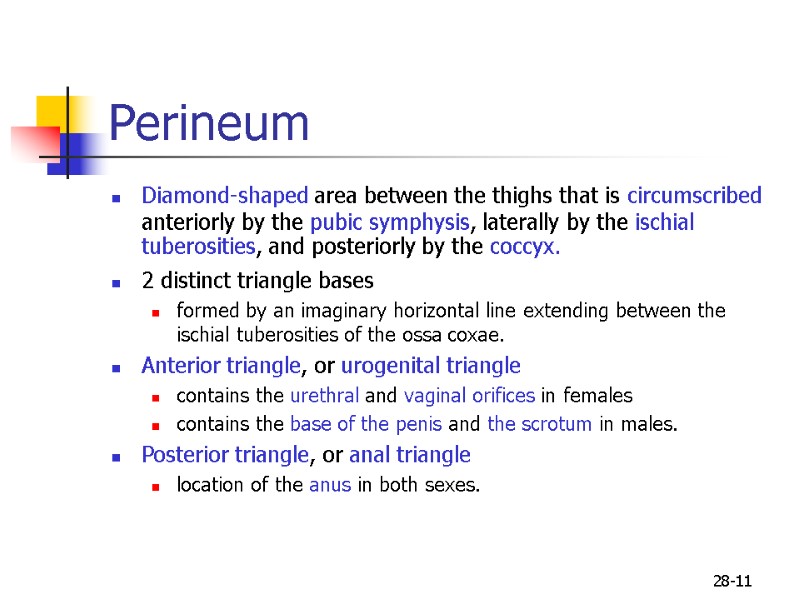
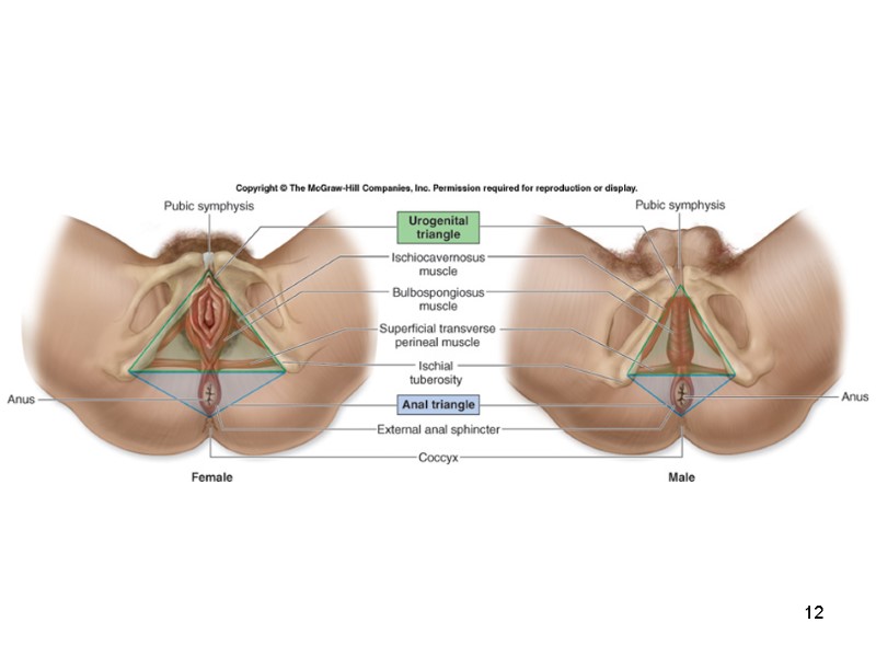
 28-11 Perineum Diamond-shaped area between the thighs that is circumscribed anteriorly by the pubic symphysis, laterally by the ischial tuberosities, and posteriorly by the coccyx. 2 distinct triangle bases formed by an imaginary horizontal line extending between the ischial tuberosities of the ossa coxae. Anterior triangle, or urogenital triangle contains the urethral and vaginal orifices in females contains the base of the penis and the scrotum in males. Posterior triangle, or anal triangle location of the anus in both sexes.
 12
12
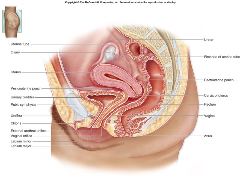
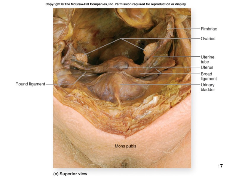
 28-13 Anatomy of the Female Reproductive System Peritoneum folds around the various pelvic organs and creates two major dead-end recesses, or pouches. anterior vesicouterine pouch forms the space between the uterus and the urinary bladder posterior rectouterine pouch forms the space between the uterus anteriorly and the rectum posteriorly Primary sex organs of the female are the ovaries. Accessory sex organs include uterine tubes uterus, vagina, clitoris mammary glands.
 14
14
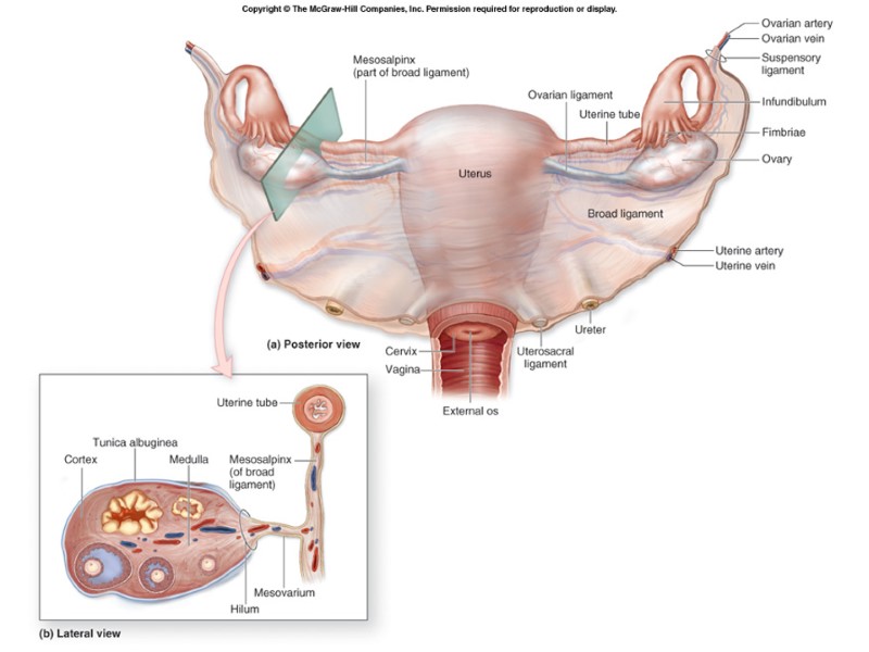
 28-15 Mesovarium: Double folds of peritoneum Attaches ovaries to broad ligament Broad ligament Peritonium Drapes over the uterus Ovarian ligament Ovary to uterus Suspensory ligament Ovary to pelvic wall
 16
16
 17
17
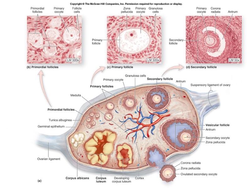
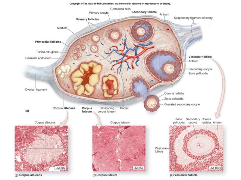
 28-18 Ovarian Follicles Within the cortex are thousands of ovarian follicles. Consist of Follicle cells granulosa cells nurse cells that support the oocyte a type of oocyte. Several different kinds of ovarian follicles, each representing a different stage of development. Oogenesis: maturation of a primary oocyte to a secondary oocyte.
 19
19
 20
20
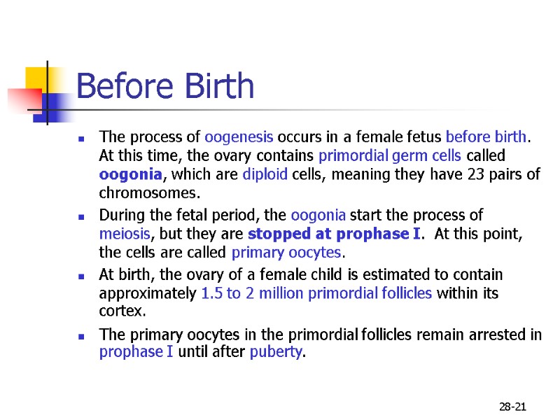
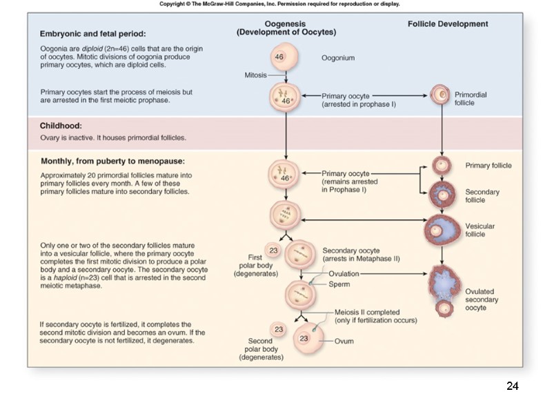
 28-21 Before Birth The process of oogenesis occurs in a female fetus before birth. At this time, the ovary contains primordial germ cells called oogonia, which are diploid cells, meaning they have 23 pairs of chromosomes. During the fetal period, the oogonia start the process of meiosis, but they are stopped at prophase I. At this point, the cells are called primary oocytes. At birth, the ovary of a female child is estimated to contain approximately 1.5 to 2 million primordial follicles within its cortex. The primary oocytes in the primordial follicles remain arrested in prophase I until after puberty.
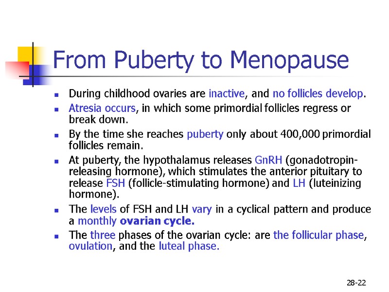
 28-22 From Puberty to Menopause During childhood ovaries are inactive, and no follicles develop. Atresia occurs, in which some primordial follicles regress or break down. By the time she reaches puberty only about 400,000 primordial follicles remain. At puberty, the hypothalamus releases GnRH (gonadotropin-releasing hormone), which stimulates the anterior pituitary to release FSH (follicle-stimulating hormone) and LH (luteinizing hormone). The levels of FSH and LH vary in a cyclical pattern and produce a monthly ovarian cycle. The three phases of the ovarian cycle: are the follicular phase, ovulation, and the luteal phase.
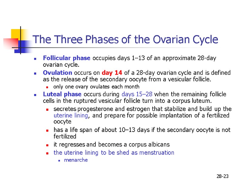
 28-23 The Three Phases of the Ovarian Cycle Follicular phase occupies days 1–13 of an approximate 28-day ovarian cycle. Ovulation occurs on day 14 of a 28-day ovarian cycle and is defined as the release of the secondary oocyte from a vesicular follicle. only one ovary ovulates each month Luteal phase occurs during days 15–28 when the remaining follicle cells in the ruptured vesicular follicle turn into a corpus luteum. secretes progesterone and estrogen that stabilize and build up the uterine lining, and prepare for possible implantation of a fertilized oocyte has a life span of about 10–13 days if the secondary oocyte is not fertilized it regresses and becomes a corpus albicans the uterine lining to be shed as menstruation menarche
 24
24
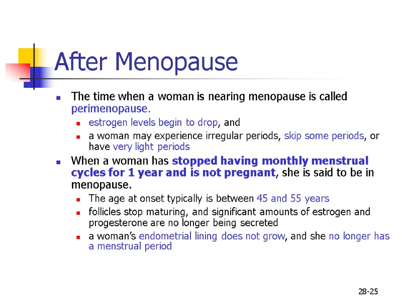
 28-25 After Menopause The time when a woman is nearing menopause is called perimenopause. estrogen levels begin to drop, and a woman may experience irregular periods, skip some periods, or have very light periods When a woman has stopped having monthly menstrual cycles for 1 year and is not pregnant, she is said to be in menopause. The age at onset typically is between 45 and 55 years follicles stop maturing, and significant amounts of estrogen and progesterone are no longer being secreted a woman’s endometrial lining does not grow, and she no longer has a menstrual period
 26
26
 27
27
 28
28
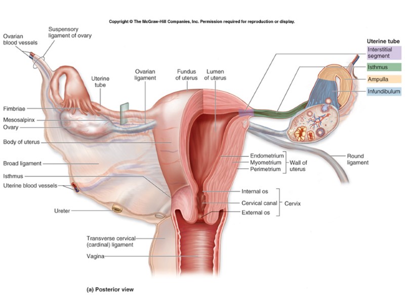
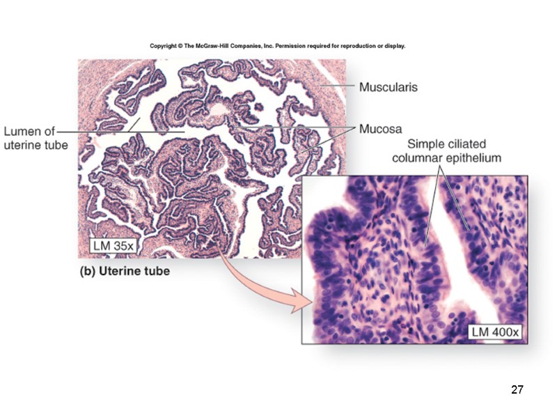
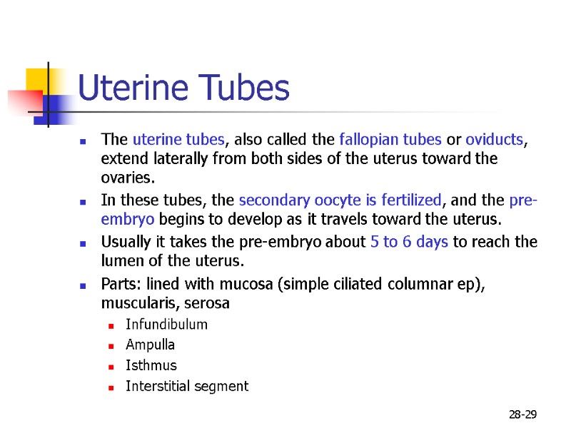
 28-29 Uterine Tubes The uterine tubes, also called the fallopian tubes or oviducts, extend laterally from both sides of the uterus toward the ovaries. In these tubes, the secondary oocyte is fertilized, and the pre-embryo begins to develop as it travels toward the uterus. Usually it takes the pre-embryo about 5 to 6 days to reach the lumen of the uterus. Parts: lined with mucosa (simple ciliated columnar ep), muscularis, serosa Infundibulum Ampulla Isthmus Interstitial segment
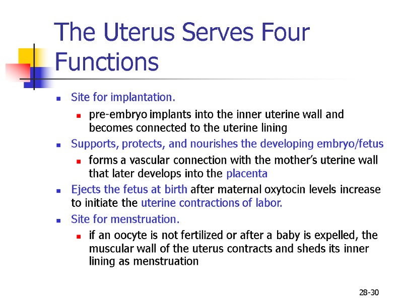
 28-30 The Uterus Serves Four Functions Site for implantation. pre-embryo implants into the inner uterine wall and becomes connected to the uterine lining Supports, protects, and nourishes the developing embryo/fetus forms a vascular connection with the mother’s uterine wall that later develops into the placenta Ejects the fetus at birth after maternal oxytocin levels increase to initiate the uterine contractions of labor. Site for menstruation. if an oocyte is not fertilized or after a baby is expelled, the muscular wall of the uterus contracts and sheds its inner lining as menstruation
 28-31 Regions of the Uterus Fundus Body Isthmus Cervix Cervical canal Internal os External os
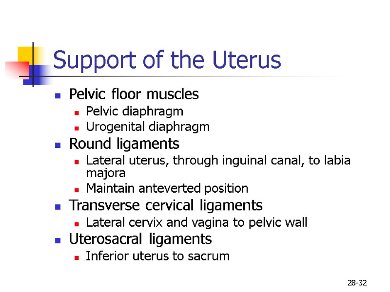
 28-32 Support of the Uterus Pelvic floor muscles Pelvic diaphragm Urogenital diaphragm Round ligaments Lateral uterus, through inguinal canal, to labia majora Maintain anteverted position Transverse cervical ligaments Lateral cervix and vagina to pelvic wall Uterosacral ligaments Inferior uterus to sacrum
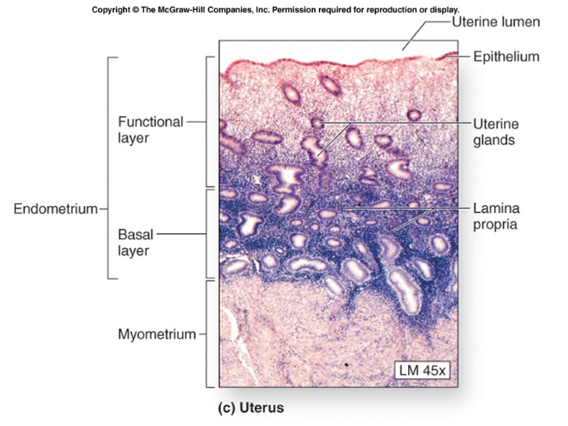
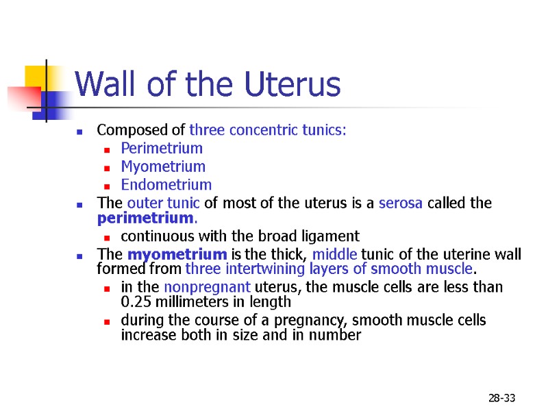
 28-33 Wall of the Uterus Composed of three concentric tunics: Perimetrium Myometrium Endometrium The outer tunic of most of the uterus is a serosa called the perimetrium. continuous with the broad ligament The myometrium is the thick, middle tunic of the uterine wall formed from three intertwining layers of smooth muscle. in the nonpregnant uterus, the muscle cells are less than 0.25 millimeters in length during the course of a pregnancy, smooth muscle cells increase both in size and in number
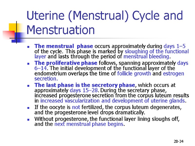
 28-34 Uterine (Menstrual) Cycle and Menstruation The menstrual phase occurs approximately during days 1–5 of the cycle. This phase is marked by sloughing of the functional layer and lasts through the period of menstrual bleeding. The proliferative phase follows, spanning approximately days 6–14. The initial development of the functional layer of the endometrium overlaps the time of follicle growth and estrogen secretion. The last phase is the secretory phase, which occurs at approximately days 15–28. During the secretary phase, increased progesterone secretion from the corpus luteum results in increased vascularization and development of uterine glands. If the oocyte is not fertilized, the corpus luteum degenerates, and the progesterone level drops dramatically. Without progesterone, the functional layer lining sloughs off, and the next menstrual phase begins.
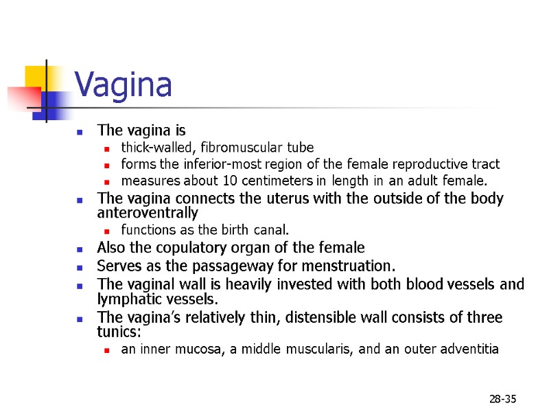
 28-35 Vagina The vagina is thick-walled, fibromuscular tube forms the inferior-most region of the female reproductive tract measures about 10 centimeters in length in an adult female. The vagina connects the uterus with the outside of the body anteroventrally functions as the birth canal. Also the copulatory organ of the female Serves as the passageway for menstruation. The vaginal wall is heavily invested with both blood vessels and lymphatic vessels. The vagina’s relatively thin, distensible wall consists of three tunics: an inner mucosa, a middle muscularis, and an outer adventitia
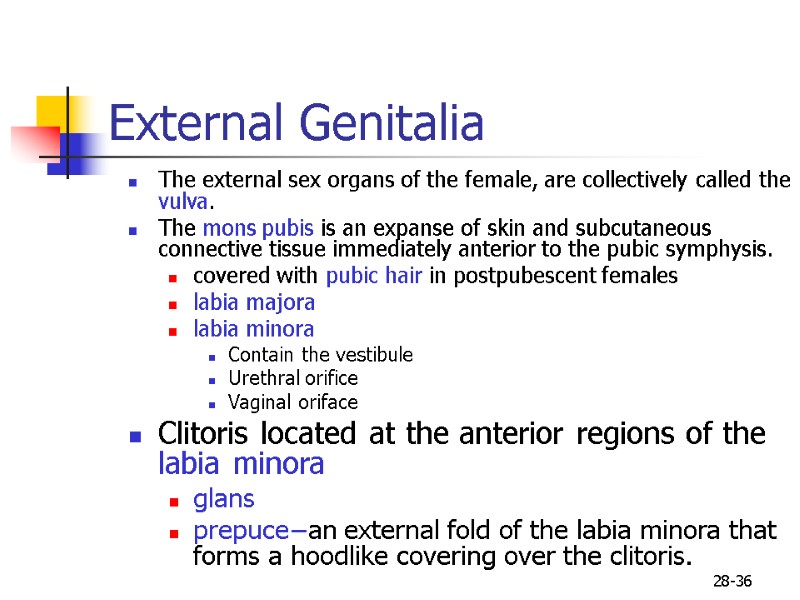
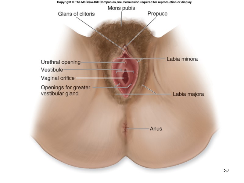
 28-36 External Genitalia The external sex organs of the female, are collectively called the vulva. The mons pubis is an expanse of skin and subcutaneous connective tissue immediately anterior to the pubic symphysis. covered with pubic hair in postpubescent females labia majora labia minora Contain the vestibule Urethral orifice Vaginal oriface Clitoris located at the anterior regions of the labia minora glans prepuce−an external fold of the labia minora that forms a hoodlike covering over the clitoris.
 37
37
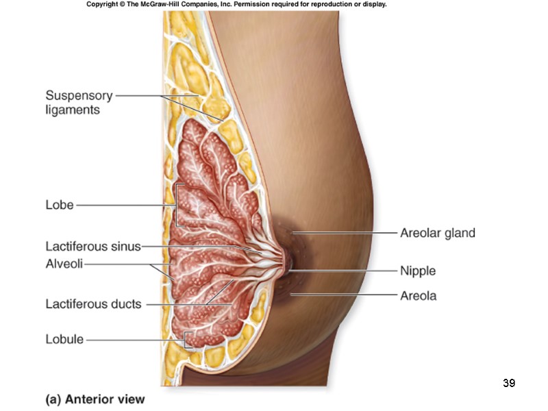
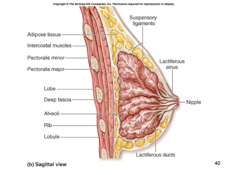
 28-38 Mammary Glands Each mammary gland, or breast, is located within the anterior thoracic wall and is composed of a compound tubuloalveolar exocrine gland. Breast milk contains proteins, fats, and a sugar to provide nutrition to infants. The nipple is a cylindrical projection on the center of the breast. It contains multiple tiny openings of the excretory ducts that produce breast milk. The areola is the pigmented rosy or brownish ring of skin around the nipple. Its surface often appears uneven and grainy due to the numerous sebaceous glands immediately internal to the surface. The color of the areola may vary, depending upon whether or not a woman has given birth. In a nulliparous woman (a woman who has never given birth), the areola is rosy or light brown in color. In a parous woman (a woman who has given birth), the areola may change to a darker rose or brown color.
 39
39
 40
40
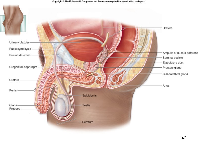
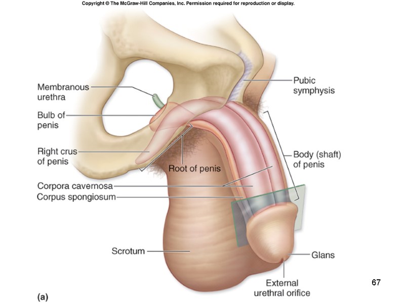
 28-41 Anatomy of the Male Reproductive System Primary organs: gonads are the testes Accessory sex organs include: a complex set of ducts and tubules leading from the testes to the penis a group of male accessory glands the penis, which is the organ of copulation
 42
42
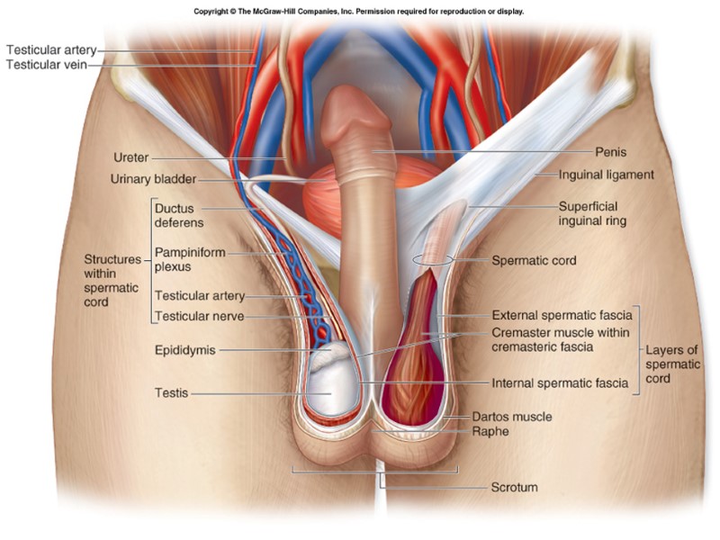
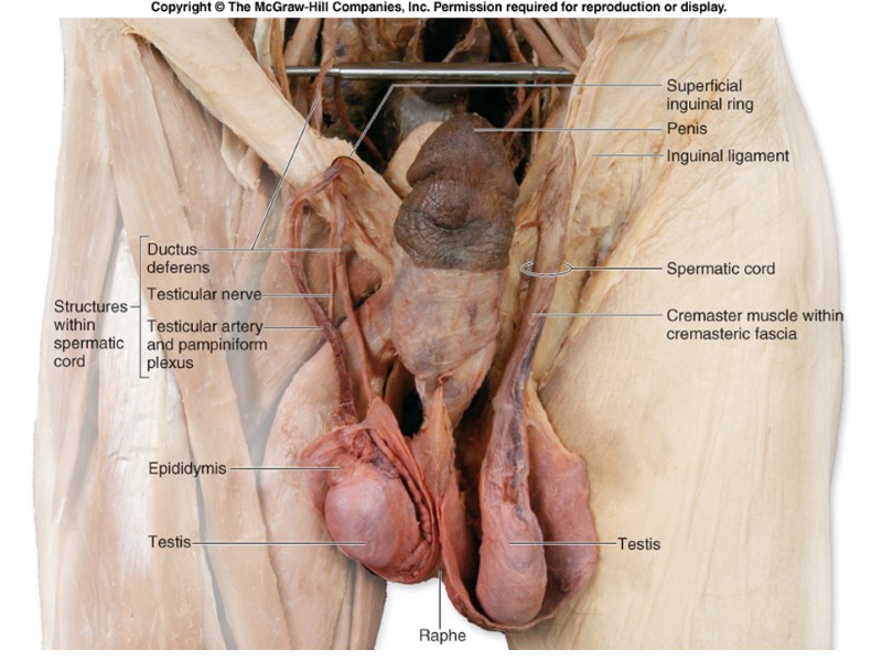
 28-43 Scrotum Skin covered sac Raphe: external midline seam Continues on inferior surface of the penis, and to anus. Components of scrotal wall. Skin Fascia Dartos muscle External spermatic fascia Cremaster muscle Internal spermatic fascia Tunica vaginalis.
 28-44 Scrotum Male gametes are sensitive to elevated temperatures often exhibit abnormal or completely curtailed development Gamete development occurs outside the body Scrotum: a skin-covered sac that houses: male gonads first portion of the duct system site of early sperm maturation and development, reside outside the body proper. Testes exposed to elevated temperatures Skin of the scrotal sac becomes thin result of dartos muscle relaxation. The cremaster muscle relaxes allows the testes to move inferiorly away from the body The testes temperature becomes less than normal body temperature. The opposite occurs if the testes are exposed to cold.
 45
45
 46
46
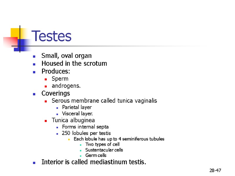
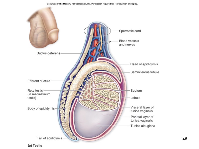
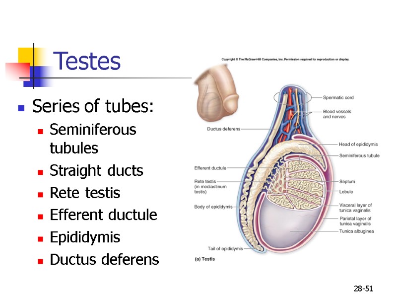
 28-47 Testes Small, oval organ Housed in the scrotum Produces: Sperm androgens. Coverings Serous membrane called tunica vaginalis Parietal layer Visceral layer. Tunica albuginea Forms internal septa 250 lobules per testis Each lobule has up to 4 seminiferous tubules Two types of cell Sustentacular cells Germ cells Interior is called mediastinum testis.
 48
48
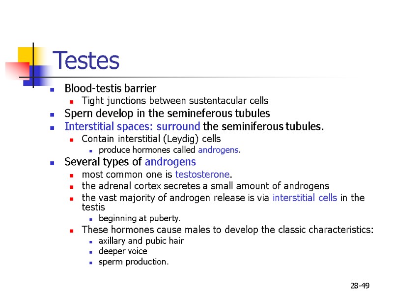
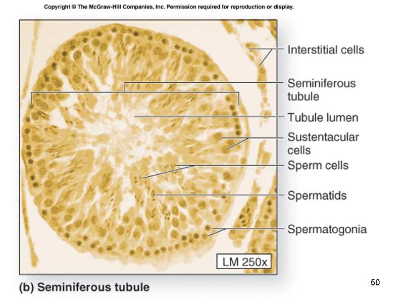
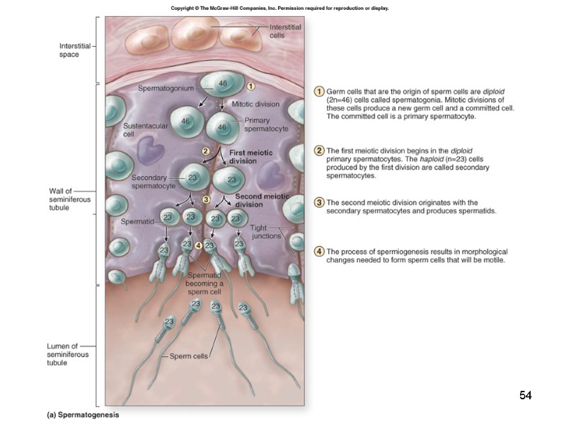
 28-49 Testes Blood-testis barrier Tight junctions between sustentacular cells Spern develop in the semineferous tubules Interstitial spaces: surround the seminiferous tubules. Contain interstitial (Leydig) cells produce hormones called androgens. Several types of androgens most common one is testosterone. the adrenal cortex secretes a small amount of androgens the vast majority of androgen release is via interstitial cells in the testis beginning at puberty. These hormones cause males to develop the classic characteristics: axillary and pubic hair deeper voice sperm production.
 50
50
 28-51 Testes Series of tubes: Seminiferous tubules Straight ducts Rete testis Efferent ductule Epididymis Ductus deferens
 28-52 Spermatic Cord The blood vessels and nerves to the testis travel from within the abdomen to the scrotum in a multilayered structure called the spermatic cord. Layers Contain Testicular artery Pampiniform plexus Autonomic nerves
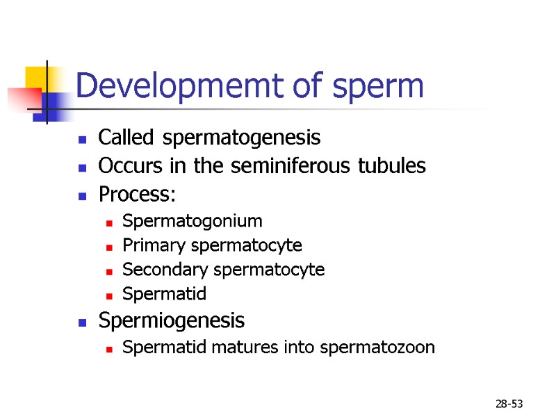
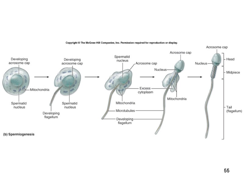
 28-53 Developmemt of sperm Called spermatogenesis Occurs in the seminiferous tubules Process: Spermatogonium Primary spermatocyte Secondary spermatocyte Spermatid Spermiogenesis Spermatid matures into spermatozoon
 54
54
 55
55
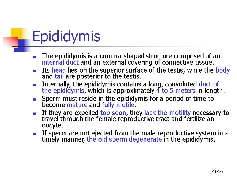
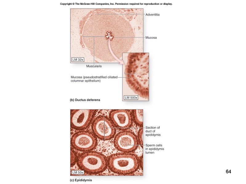
 28-56 Epididymis The epididymis is a comma-shaped structure composed of an internal duct and an external covering of connective tissue. Its head lies on the superior surface of the testis, while the body and tail are posterior to the testis. Internally, the epididymis contains a long, convoluted duct of the epididymis, which is approximately 4 to 5 meters in length. Sperm must reside in the epididymis for a period of time to become mature and fully motile. If they are expelled too soon, they lack the motility necessary to travel through the female reproductive tract and fertilize an oocyte. If sperm are not ejected from the male reproductive system in a timely manner, the old sperm degenerate in the epididymis.
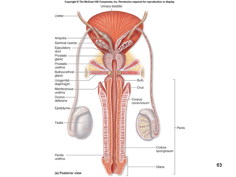
 28-57 Ductus Deferens When sperm leave the epididymis, they enter the ductus deferens, also called the vas deferens. The ductus deferens is a thick-walled tube that travels within the spermatic cord, through the inguinal canal, and within the pelvic cavity before it reaches the prostate gland. The ampulla of the ductus deferens unites with the proximal region of the seminal vesicle to form the terminal portion of the reproductive duct system, called the ejaculatory duct.
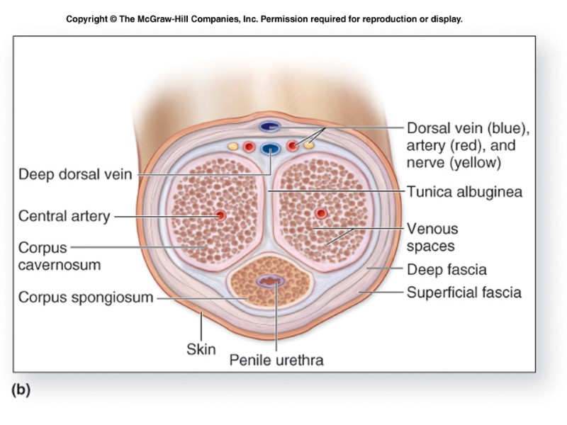
 28-58 Urethra Transports semen from the ejaculatory duct to the outside of the body. Subdivided into: prostatic urethra that extends through the prostate gland membranous urethra that travels through the urogenital diaphragm penile urethra that ends through the penis Sperm leave the body through the urethra.
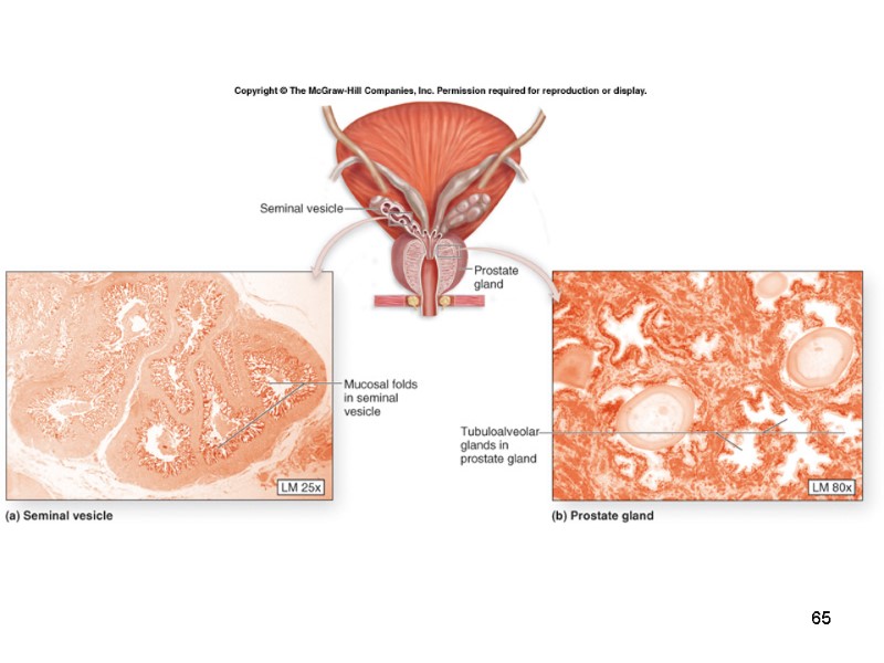
 28-59 Accessory Glands The vagina has a highly acidic environment to prevent bacterial growth. Sperm cannot survive in this type of environment, so an alkaline secretion called seminal fluid is needed to lessen the acidity of the vagina and bring pH values closer to neutral. As the sperm travel through the reproductive tract (a process that can take several days), they are nourished by nutrients within the seminal fluid. The components of seminal fluid are produced by accessory glands: seminal vesicles prostate gland bulbourethral glands
 28-60 Seminal Vesicles The paired seminal vesicles are located on the posterior surface of the urinary bladder adjacent to the ampulla of the ductus deferens. Each seminal vesicle is an elongated, pouchlike hollow organ approximately 5–8 centimeters long. It is the proximal portion of each seminal vesicle that merges with a ductus deferens to form the ejaculatory duct. The seminal vesicles secrete a viscous, whitish-yellow alkaline fluid containing both fructose and prostaglandins. The fructose is a sugar that nourishes the sperm as they travel through the female reproductive tract, while the prostaglandins promote the widening and slight dilation of the external os of the cervix.
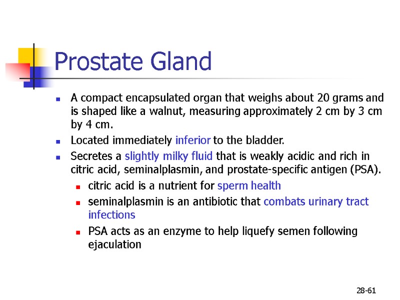
 28-61 Prostate Gland A compact encapsulated organ that weighs about 20 grams and is shaped like a walnut, measuring approximately 2 cm by 3 cm by 4 cm. Located immediately inferior to the bladder. Secretes a slightly milky fluid that is weakly acidic and rich in citric acid, seminalplasmin, and prostate-specific antigen (PSA). citric acid is a nutrient for sperm health seminalplasmin is an antibiotic that combats urinary tract infections PSA acts as an enzyme to help liquefy semen following ejaculation
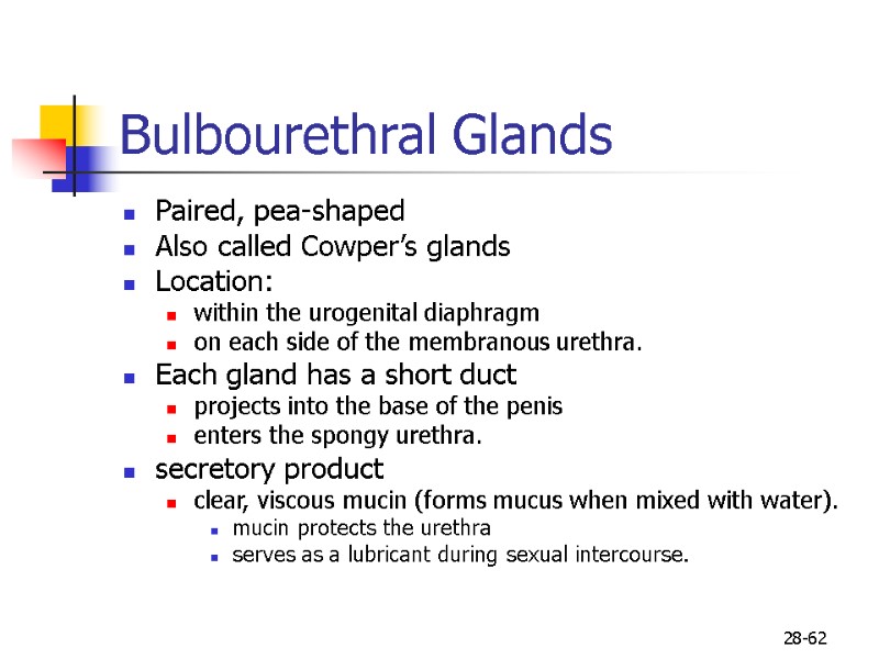
 28-62 Bulbourethral Glands Paired, pea-shaped Also called Cowper’s glands Location: within the urogenital diaphragm on each side of the membranous urethra. Each gland has a short duct projects into the base of the penis enters the spongy urethra. secretory product clear, viscous mucin (forms mucus when mixed with water). mucin protects the urethra serves as a lubricant during sexual intercourse.
 63
63
 64
64
 65
65
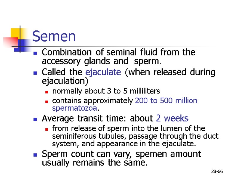
 28-66 Semen Combination of seminal fluid from the accessory glands and sperm. Called the ejaculate (when released during ejaculation) normally about 3 to 5 milliliters contains approximately 200 to 500 million spermatozoa. Average transit time: about 2 weeks from release of sperm into the lumen of the seminiferous tubules, passage through the duct system, and appearance in the ejaculate. Sperm count can vary, spemen amount usually remains the same.
 67
67
 68
68
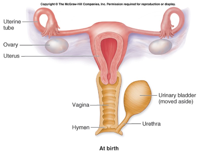
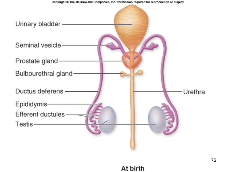
 28-69 Aging and the Reproductive Systems Our reproductive systems are basically nonfunctional for several years following birth. When we reach puberty, hormonal changes in the hypothalamus and anterior pituitary gland stimulate the gonads to begin producing sex hormones. Thereafter, changes occur in many body structures, the reproductive organs mature, and gonads begin to produce gametes. Gametes stop maturing in females in their 40s or 50s, and menopause occurs. A reduction in hormone production that accompanies menopause causes some atrophy of the reproductive organs and the breasts. The vaginal wall thickness decreases, as do glandular secretions for maintaining a lubricated and moist lining. The uterus shrinks and atrophies, becoming much smaller than it was before puberty.
 70
70
 71
71
 72
72
 73
73
 74
74

