1 Modes of Ventilation Dr. Eugenia Mahamid Rambam

1 Modes of Ventilation Dr. Eugenia Mahamid Rambam Medical Center
2 Modes of Ventilation The main indication for ventilatory support is Respiratory Failure
3 Categories of Respiratory Failure HYPOXEMIC ARDS PULMONARY EDEMA PULMONARY HEMORRHAGE PNEUMONIA Low compliance lung disease: Low PO2, Low SaO2
4 HYPERCARBIC OBSTRUCTION TO AIRFLOW NEUROMUSCULAR DISORDERS DRUG OVERDOSE ENDOCRINOPATHIES Increase in PCO2 Respiratory acidosis Decrease in pH Categories of Respiratory Failure
5 ECE CENTRAL DECREASED LEVEL OF CONSCIOUSNESS ACUTE MEDICAL AND SURGICAL CONDITIONS MECHANICAL VENTILATION IS USED TO DECREASE WORK OF BREATHING Categories of Respiratory Failure
6 A MECHANICAL VENTILATOR is a pump providing an external source of energy to push gases into the lungs and allow for passive exhalation (CO2 elimination). Ventilator’s Changeable parameters Vt = Tidal Volume FIO2 = Fraction of Inspired Oxygen RR = Respiratory Rate I:E = Inspiratory to Expiratory ratio EEP = End Expiratory Pressure PIP = Peak Inspiratory Pressure Inspiratory Flow Rate
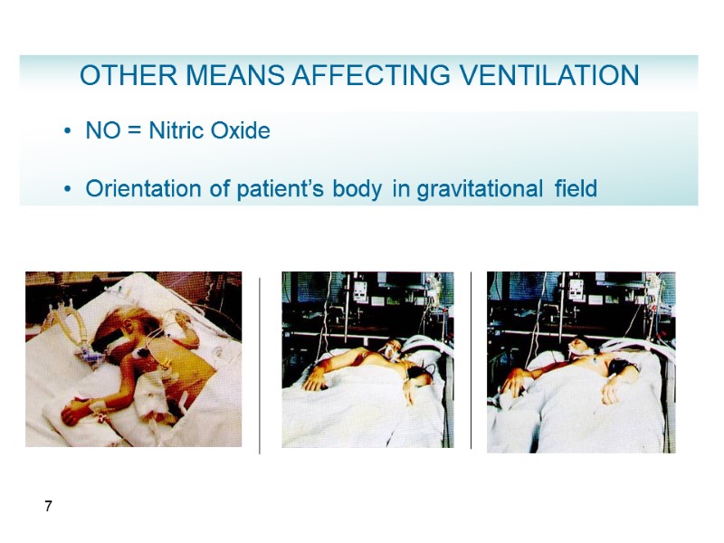
7 NO = Nitric Oxide Orientation of patient’s body in gravitational field OTHER MEANS AFFECTING VENTILATION
8 GENERAL CLASSES OF VENTILATORS Negative pressure application of negative pressure at the chest wall and upper abdomen Positive pressure application of positive pressure at airway opening
9 Negative Pressure Ventilators - Perithoracic pump for replacement failing patients’ muscles, wide-spread use for polio epidemics Manually operated ventilator (Woillez, 1876) Tank respirator “iron lung”, cuirass, body suits (1930 - 1950) Patient care problems: airway obstruction, low efficacy in interstitial lung diseases, patient’s discomfort
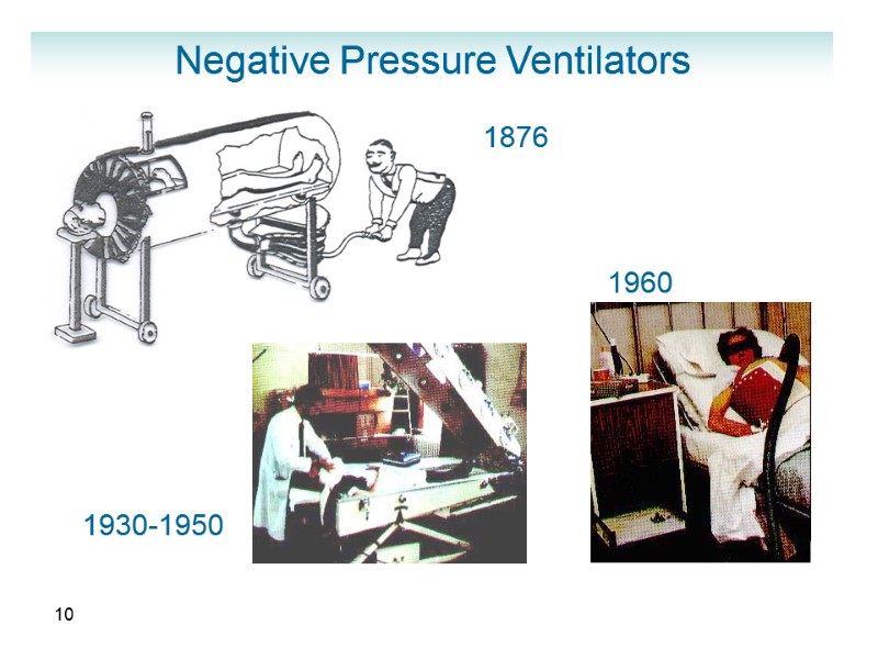
10 Negative Pressure Ventilators 1876 1930-1950 1960
11 Positive pressure ventilators Volume-cycled Delivers set Vt at specified RR and terminates respiration when Vt is delivered. Airway pressures are determined by respiratory system impedance (risk of barotrauma). Pressure-cycled Limits flow, when set pressure is delivered (may decrease minute ventilation)
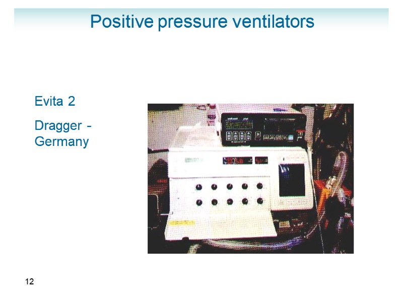
12 Positive pressure ventilators Evita 2 Dragger - Germany
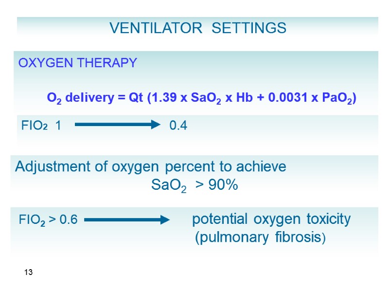
13 VENTILATOR SETTINGS OXYGEN THERAPY O2 delivery = Qt (1.39 x SaO2 x Hb + 0.0031 x PaO2) FIO2 1 0.4 Adjustment of oxygen percent to achieve SaO2 > 90% FIO2 > 0.6 potential oxygen toxicity (pulmonary fibrosis)
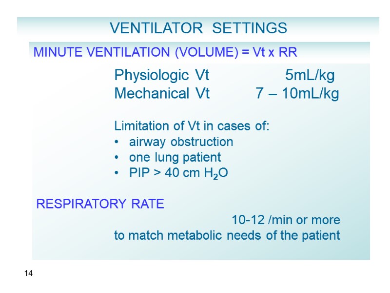
14 MINUTE VENTILATION (VOLUME) = Vt x RR Physiologic Vt 5mL/kg Mechanical Vt 7 – 10mL/kg Limitation of Vt in cases of: airway obstruction one lung patient PIP > 40 cm H2O RESPIRATORY RATE 10-12 /min or more to match metabolic needs of the patient VENTILATOR SETTINGS
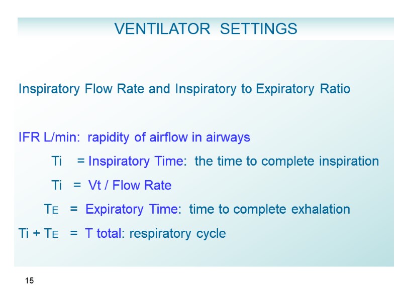
15 Inspiratory Flow Rate and Inspiratory to Expiratory Ratio IFR L/min: rapidity of airflow in airways Ti = Inspiratory Time: the time to complete inspiration Ti = Vt / Flow Rate TE = Expiratory Time: time to complete exhalation Ti + TE = T total: respiratory cycle VENTILATOR SETTINGS
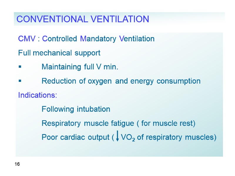
16 CMV : Controlled Mandatory Ventilation Full mechanical support Maintaining full V min. Reduction of oxygen and energy consumption Indications: Following intubation Respiratory muscle fatigue ( for muscle rest) Poor cardiac output ( VO2 of respiratory muscles) CONVENTIONAL VENTILATION
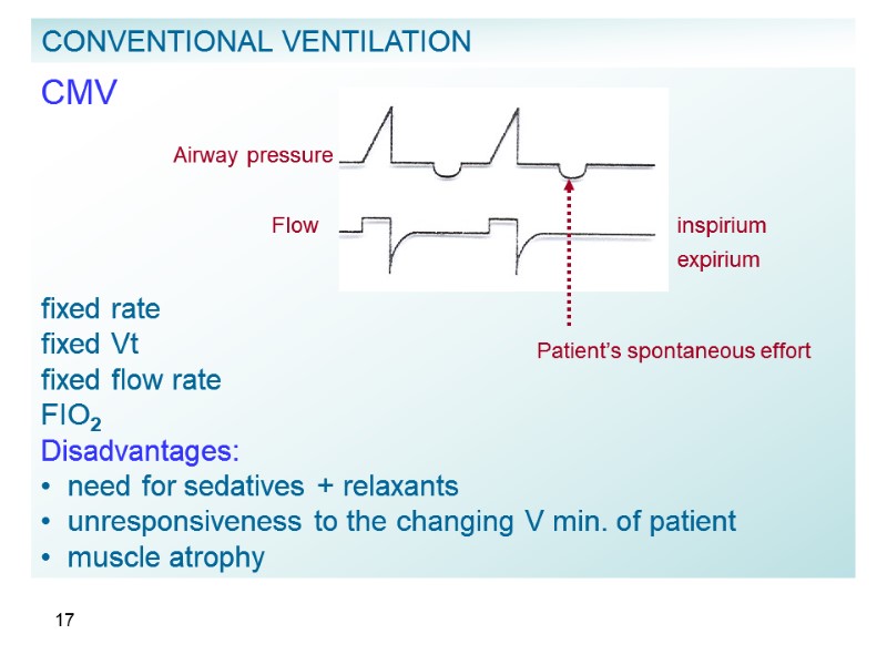
17 CMV fixed rate fixed Vt fixed flow rate FIO2 Disadvantages: need for sedatives + relaxants unresponsiveness to the changing V min. of patient muscle atrophy CONVENTIONAL VENTILATION Airway pressure Flow inspirium expirium Patient’s spontaneous effort
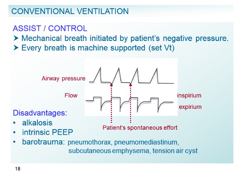
18 ASSIST / CONTROL Mechanical breath initiated by patient’s negative pressure. Every breath is machine supported (set Vt) Disadvantages: alkalosis intrinsic PEEP barotrauma: pneumothorax, pneumomediastinum, subcutaneous emphysema, tension air cyst CONVENTIONAL VENTILATION Airway pressure Flow inspirium expirium Patient’s spontaneous effort
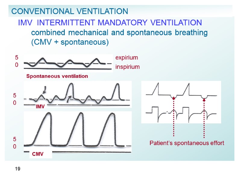
19 IMV INTERMITTENT MANDATORY VENTILATION combined mechanical and spontaneous breathing (CMV + spontaneous) CONVENTIONAL VENTILATION Spontaneous ventilation 5 0 inspirium expirium IMV 5 0 5 0 CMV Patient’s spontaneous effort
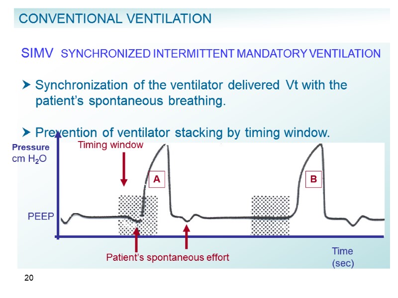
20 SIMV SYNCHRONIZED INTERMITTENT MANDATORY VENTILATION Synchronization of the ventilator delivered Vt with the patient’s spontaneous breathing. Prevention of ventilator stacking by timing window. CONVENTIONAL VENTILATION A B Timing window Patient’s spontaneous effort Time (sec) Pressure cm H2O PEEP
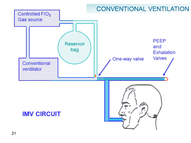
21 Controlled FIO2 Gas source Reservoir bag Conventional ventilator One-way valve PEEP and Exhalation Valves IMV CIRCUIT CONVENTIONAL VENTILATION
22 IMV / SIMV Advantages decreased need in sedatives prevention of muscle atrophy lower airway pressure and intrathoracic pressure hemodynamic stability reduction in alkalosis patient’s ability to regulate his rate and Vt according to metabolic requirements CONVENTIONAL VENTILATION
23 IMV / SIMV Disadvantages respiratory muscle fatigue increased work of breathing due to highly resistant respiratory circuit, small diameter (ETT) possibility of respiratory acidosis risk of cardiac decompensation in patient with heart disease CONVENTIONAL VENTILATION
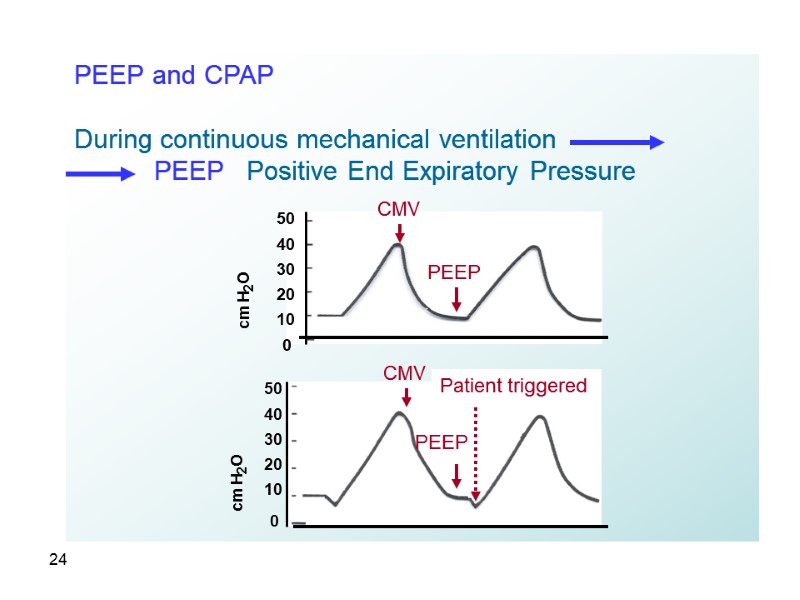
24 PEEP and CPAP During continuous mechanical ventilation PEEP Positive End Expiratory Pressure 50 40 30 20 10 0 cm H2O CMV PEEP 50 40 30 20 10 0 CMV PEEP Patient triggered cm H2O
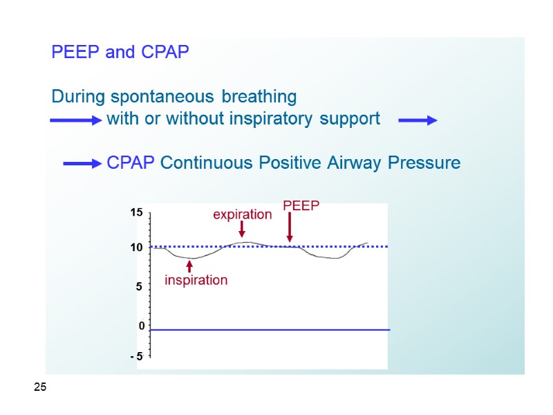
25 PEEP and CPAP During spontaneous breathing with or without inspiratory support CPAP Continuous Positive Airway Pressure 15 10 5 0 - 5 inspiration expiration PEEP
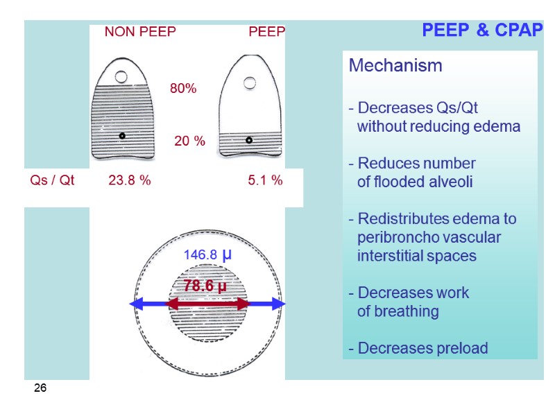
26 Mechanism - Decreases Qs/Qt without reducing edema - Reduces number of flooded alveoli - Redistributes edema to peribroncho vascular interstitial spaces - Decreases work of breathing - Decreases preload NON PEEP PEEP 78.6 μ 146.8 μ 80% 20 % Qs / Qt 23.8 % 5.1 % PEEP & CPAP
27 Goals Reduction of shunt recruitment of previously collapsed alveoli ventilation of non-ventilated zones continuous gas exchange (during expiration) Prevention of atelectasis prevention of brisk alveolar inflation and deflation > protection of surfactant and pulmonary parenchyma PEEP and CPAP
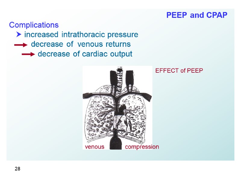
28 Complications increased intrathoracic pressure decrease of venous returns decrease of cardiac output EFFECT of PEEP venous compression PEEP and CPAP
29 Complications Increased ADH secretion, decrease of renal artery perfusion pressure decrease of urinary output and creatinine clearance decreased venous return from brain increased ICP decrease of CPP barotrauma – induced by PEEP ≈ 20% PEEP and CPAP
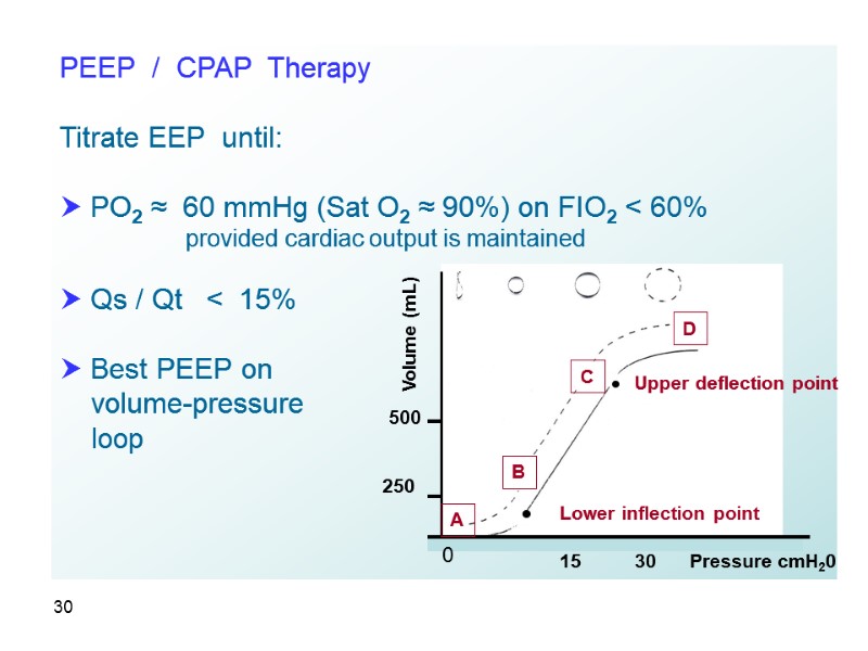
30 PEEP / CPAP Therapy Titrate EEP until: PO2 ≈ 60 mmHg (Sat O2 ≈ 90%) on FIO2 < 60% provided cardiac output is maintained Qs / Qt < 15% Best PEEP on volume-pressure loop 15 30 B D C A Lower inflection point Upper deflection point 500 250 Volume (mL) Pressure cmH20 0
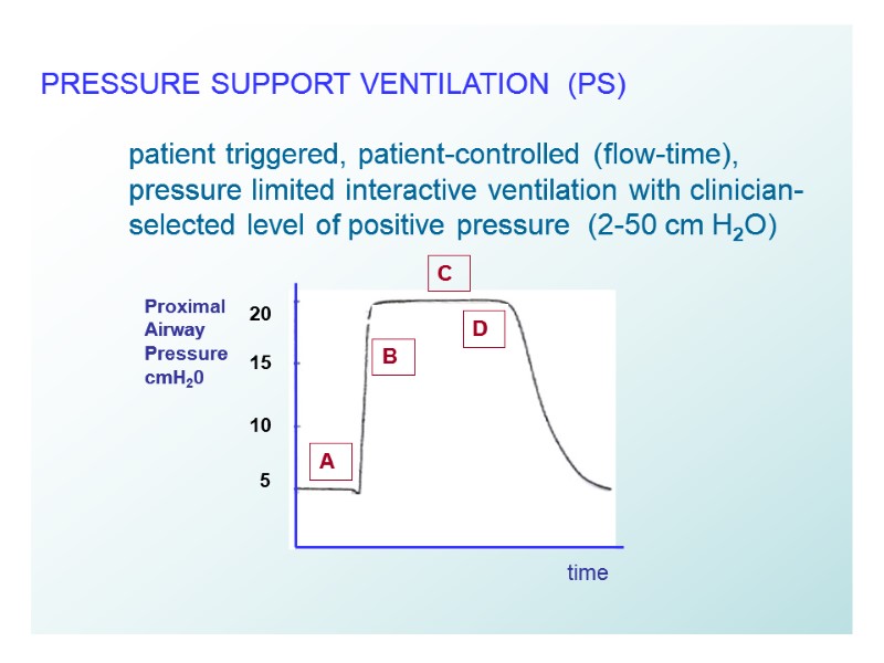
31 PRESSURE SUPPORT VENTILATION (PS) patient triggered, patient-controlled (flow-time), pressure limited interactive ventilation with clinician- selected level of positive pressure (2-50 cm H2O) 20 15 10 5 B C A D time Proximal Airway Pressure cmH20
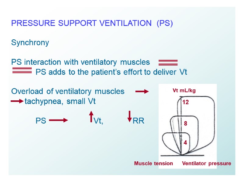
32 PRESSURE SUPPORT VENTILATION (PS) Synchrony PS interaction with ventilatory muscles PS adds to the patient’s effort to deliver Vt Overload of ventilatory muscles tachypnea, small Vt PS Vt, RR Vt mL/kg Ventilator pressure Muscle tension 12 8 4
33 PRESSURE SUPPORT VENTILATION (PS) Synchrony Patient interaction with ventilator: Trigger (prompt breath initiation, ventilator sensitivity and responsiveness) Flow adjustment of the gas delivery to the patient’s effort Cycling ventilator breath termination with the end of patient’s effort 25-30% of peak flow
34 PRESSURE SUPPORT VENTILATION (PS) Titration of PS to overcome endotracheal tube resistance (6-10 cmH2O) to achieve effective Vt and V min without causing respiratory overload non-invasive application BIPAP* = CPAP + Pressure Support *Bi-level Positive Airway Pressure
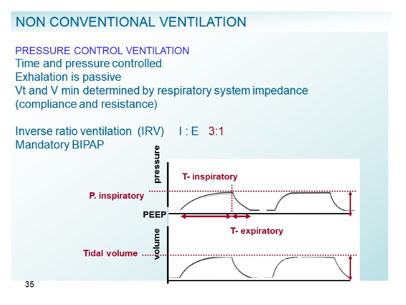
35 PRESSURE CONTROL VENTILATION Time and pressure controlled Exhalation is passive Vt and V min determined by respiratory system impedance (compliance and resistance) Inverse ratio ventilation (IRV) I : E 3:1 Mandatory BIPAP PEEP pressure T- inspiratory T- expiratory P. inspiratory Tidal volume volume NON CONVENTIONAL VENTILATION
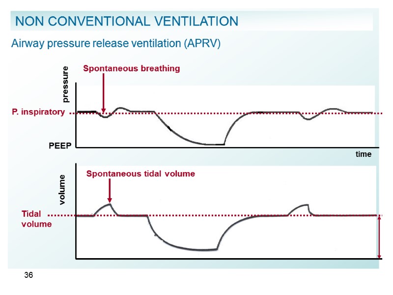
36 Airway pressure release ventilation (APRV) pressure PEEP Spontaneous breathing time volume Spontaneous tidal volume Tidal volume P. inspiratory NON CONVENTIONAL VENTILATION
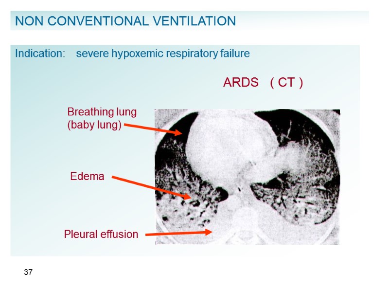
37 NON CONVENTIONAL VENTILATION Indication: severe hypoxemic respiratory failure Breathing lung (baby lung) Edema ARDS ( CT ) Pleural effusion
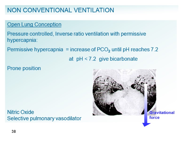
38 NON CONVENTIONAL VENTILATION Open Lung Conception Pressure controlled, Inverse ratio ventilation with permissive hypercapnia: Permissive hypercapnia = increase of PCO2 until pH reaches 7.2 at pH < 7.2 give bicarbonate Prone position Nitric Oxide Selective pulmonary vasodilator Gravitational force
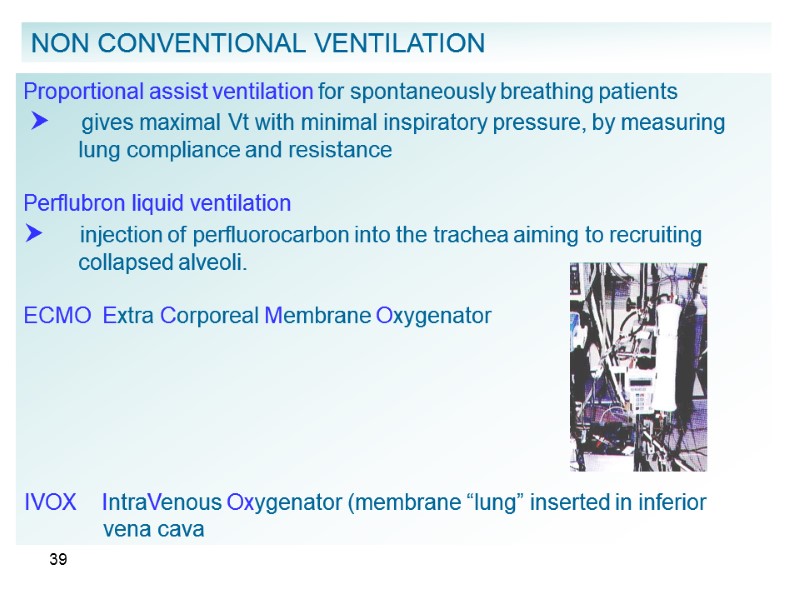
39 NON CONVENTIONAL VENTILATION Proportional assist ventilation for spontaneously breathing patients gives maximal Vt with minimal inspiratory pressure, by measuring lung compliance and resistance Perflubron liquid ventilation injection of perfluorocarbon into the trachea aiming to recruiting collapsed alveoli. ECMO Extra Corporeal Membrane Oxygenator IVOX IntraVenous Oxygenator (membrane “lung” inserted in inferior vena cava
40 HIGH FREQUENCY VENTILATION RR 60 – 3600 / min CONVECTION DIFFUSION TYPES I. HFPPV high frequency positive pressure ventilation II. HFO high frequency oscillation III. HFJT high frequency jet ventilation INDICATIONS I. Broncho-pleural fistula II. Hypoxemic respiratory failure NON CONVENTIONAL VENTILATION
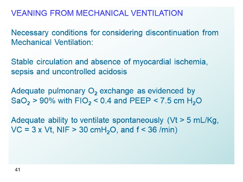
41 VEANING FROM MECHANICAL VENTILATION Necessary conditions for considering discontinuation from Mechanical Ventilation: Stable circulation and absence of myocardial ischemia, sepsis and uncontrolled acidosis Adequate pulmonary O2 exchange as evidenced by SaO2 > 90% with FIO2 < 0.4 and PEEP < 7.5 cm H2O Adequate ability to ventilate spontaneously (Vt > 5 mL/Kg, VC = 3 x Vt, NIF > 30 cmH2O, and f < 36 /min)
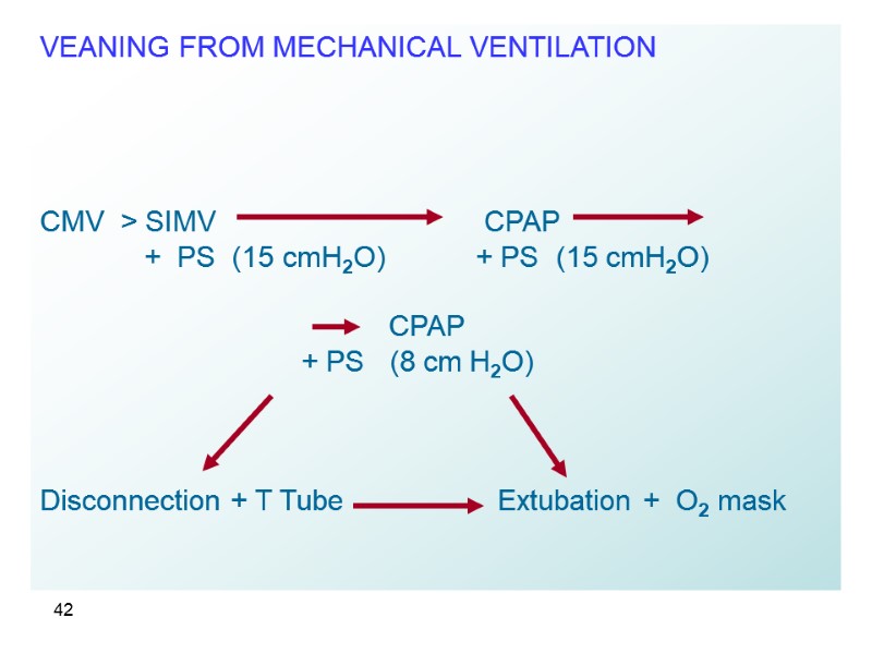
42 VEANING FROM MECHANICAL VENTILATION CMV > SIMV CPAP + PS (15 cmH2O) + PS (15 cmH2O) CPAP + PS (8 cm H2O) Disconnection + T Tube Extubation + O2 mask
18644-modes_of_mechanical_ventilation.ppt
- Количество слайдов: 42

