
1 Human Anatomy, First Edition Mc. Kinley

- Размер: 6.2 Mегабайта
- Количество слайдов: 57
Описание презентации 1 Human Anatomy, First Edition Mc. Kinley по слайдам
1 Human Anatomy, First Edition Mc. Kinley & O’Loughlin Chapter 15 Lecture Outline: Brain and Cranial Nerves
15 — 2 Brain and Cranial Nerves An adult brain weighs between 1. 35 and 1. 4 kilograms (kg) ( around 3 pounds ) and has a volume of about 1200 cubic centimeters (cc). Brain size is not directly correlated with intelligence It is not the physical size of the brain that determines intelligence—it is the number of active synapses.
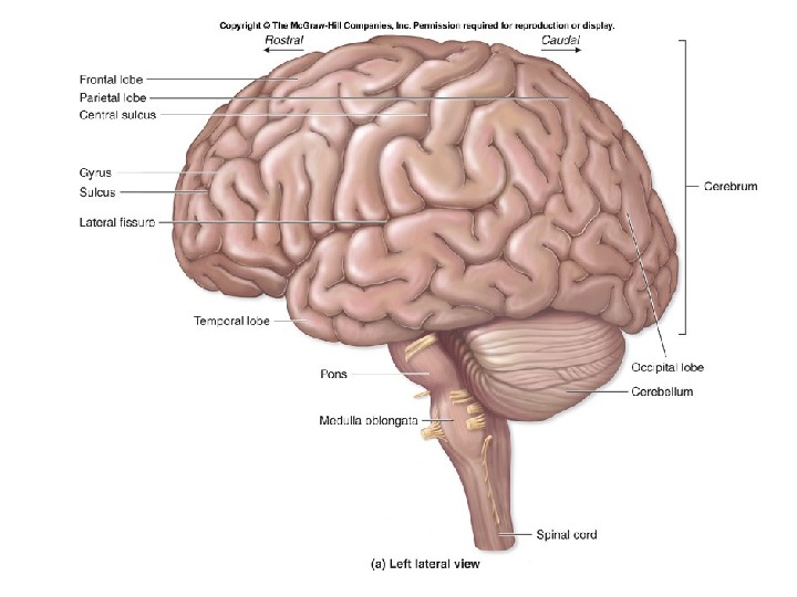
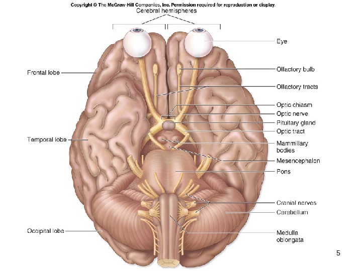
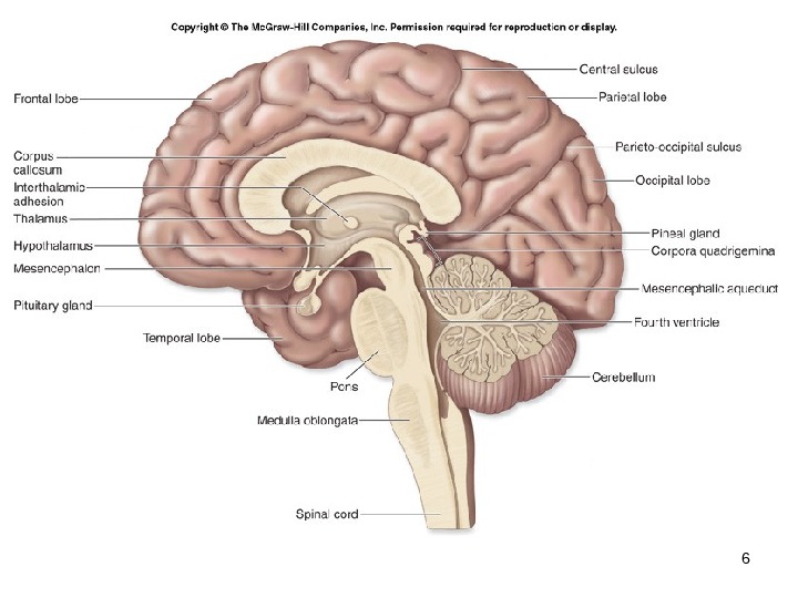
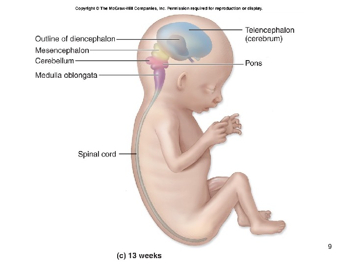
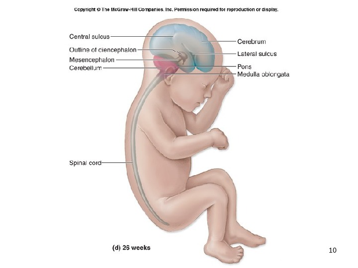
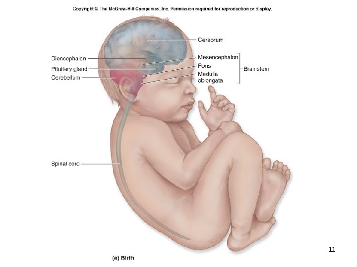
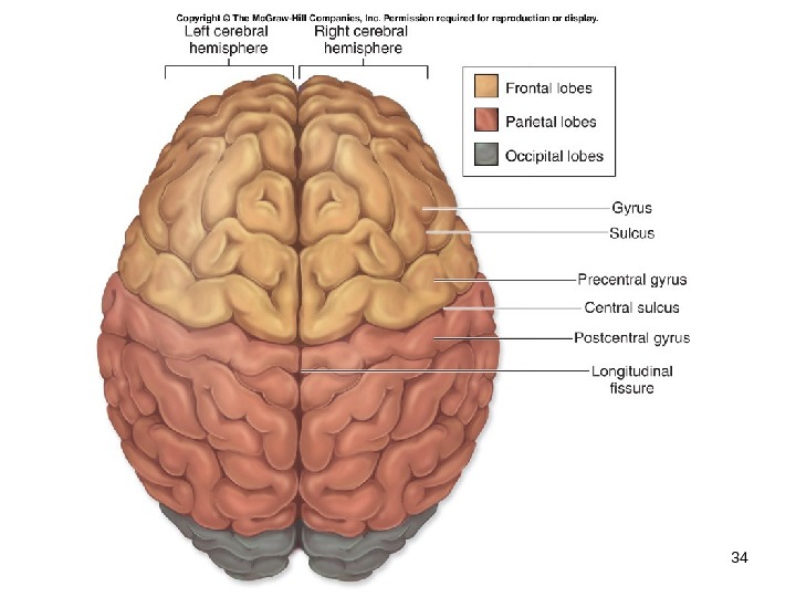
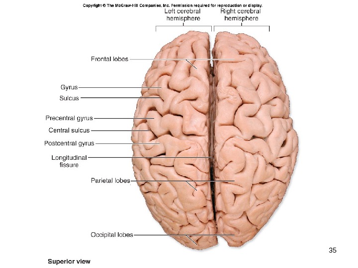
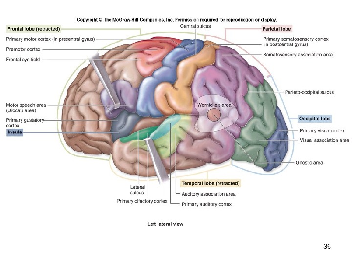
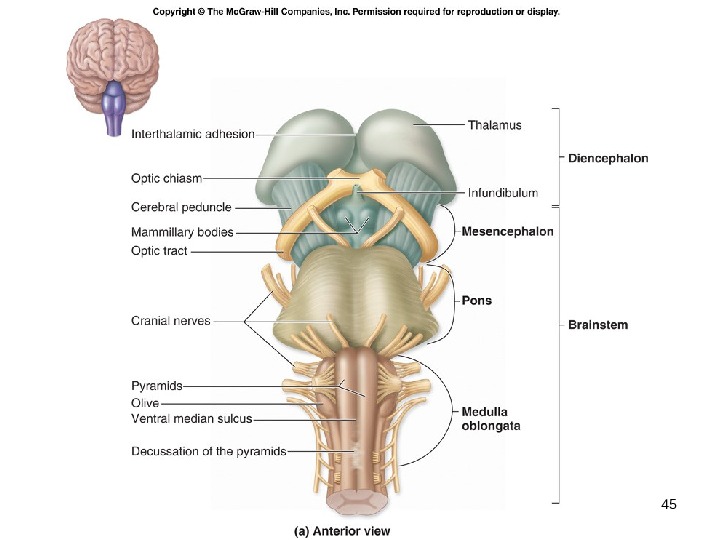
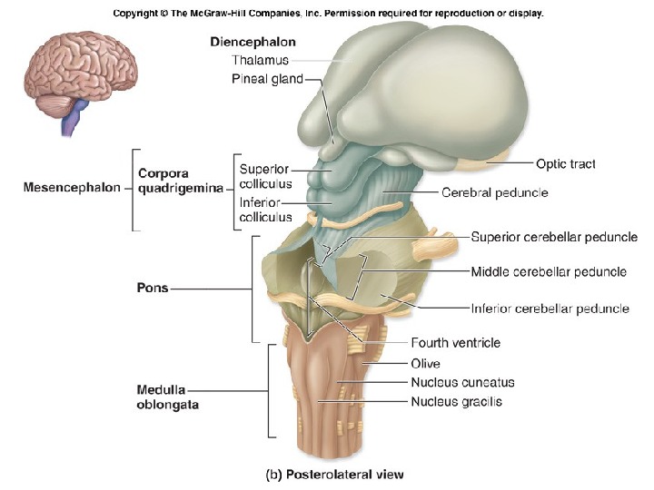
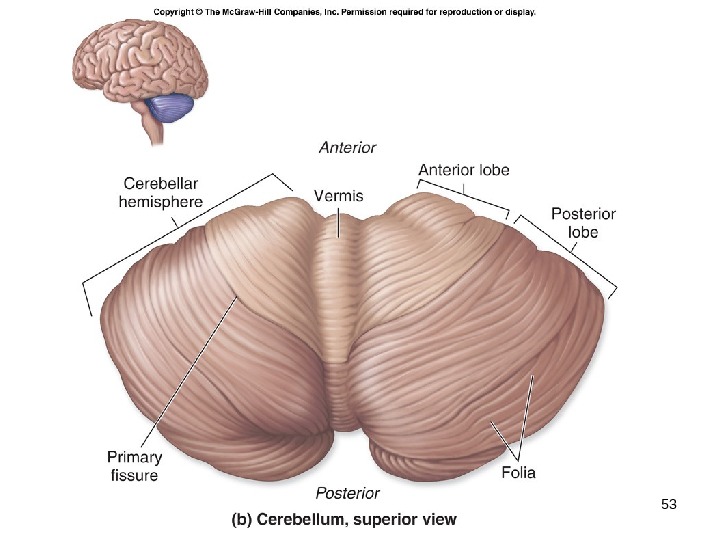
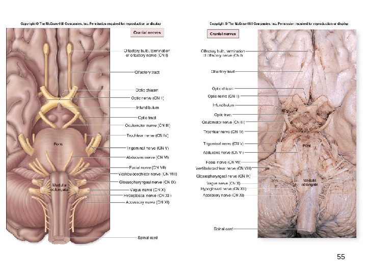
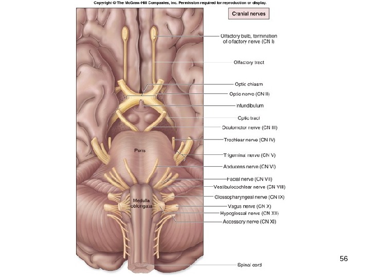
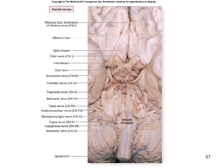
15 — 3 The Brain’s 4 Major Regions Cerebrum , the diencephalon , the brainstem , and the cerebellum. The cerebrum is divided into two halves, called the left and right cerebral hemispheres. Each hemisphere is subdivided into five functional areas called lobes. Outer surface of an adult brain exhibits folds called gyri (gyrus) and shallow depressions between those folds called sulci (sulcus). The brain is associated with 12 pairs of cranial nerves.
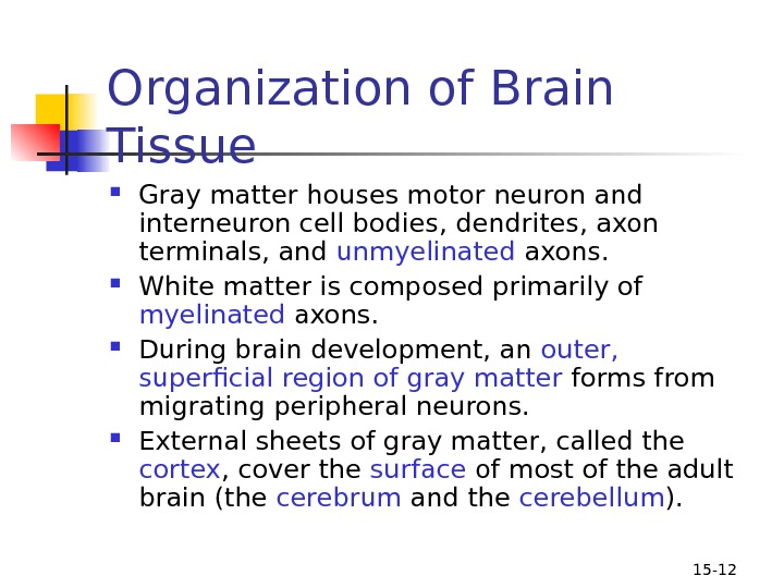
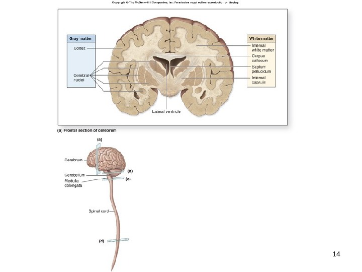
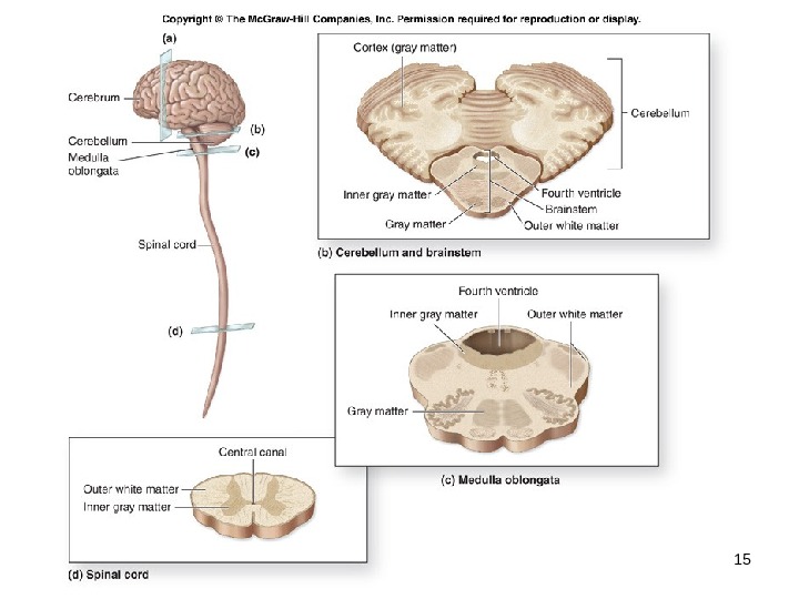
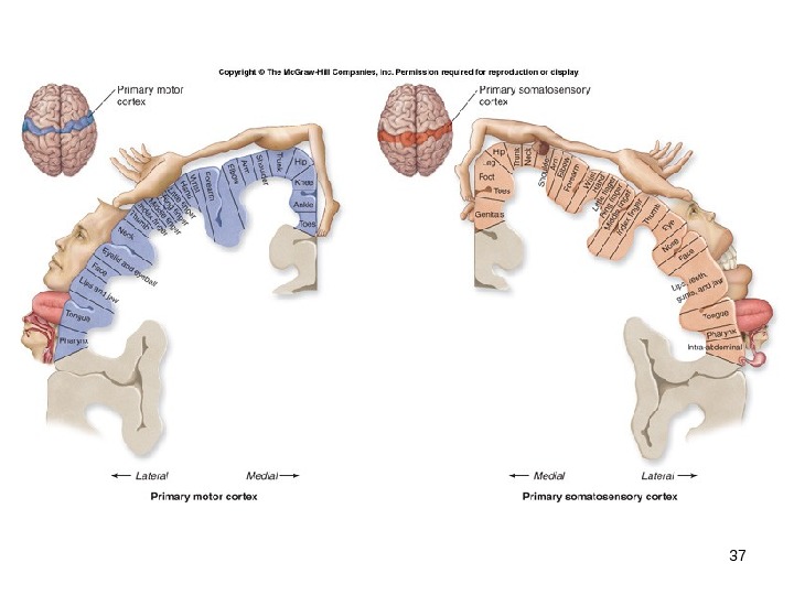
15 — 12 Organization of Brain Tissue Gray matter houses motor neuron and interneuron cell bodies, dendrites, axon terminals, and unmyelinated axons. White matter is composed primarily of myelinated axons. During brain development, an outer, superficial region of gray matter forms from migrating peripheral neurons. External sheets of gray matter, called the cortex , cover the surface of most of the adult brain (the cerebrum and the cerebellum ).
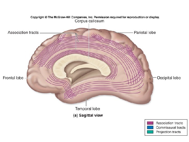
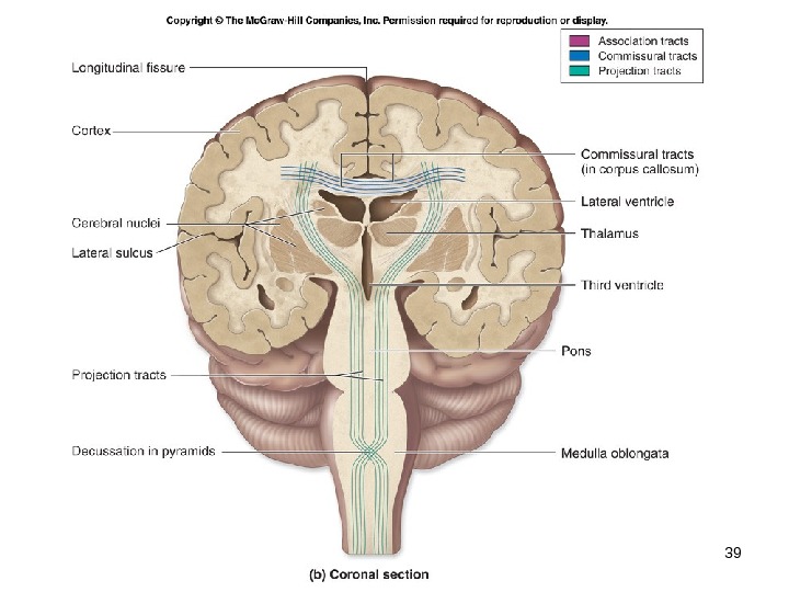
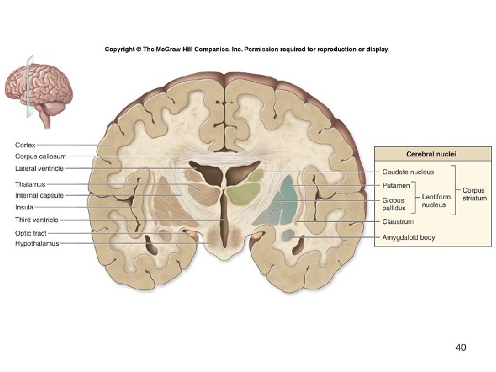
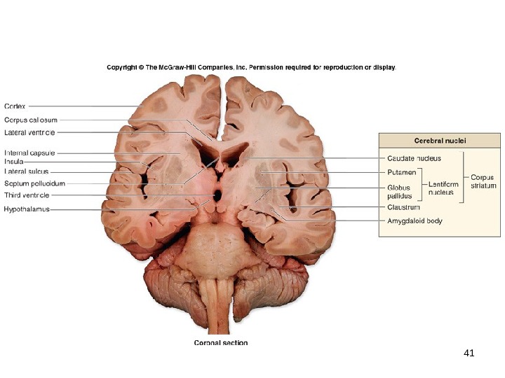
15 — 13 Organization of Brain Tissue White matter lies deep to the gray matter of the cortex. Within the masses of white matter , the brain also contains discrete innermost clusters of gray matter called cerebral nuclei , which are oval, spherical, or sometimes irregularly shaped clusters of neuron cell bodies.
15 — 16 Support and Protection of the Brain The brain is protected and isolated by multiple structures. The bony cranium provides rigid support. Protective connective tissue membranes called meninges surround and partition portions of the brain. Cerebrospinal fluid (CSF) acts as a cushioning fluid. The brain has a blood-brain barrier to prevent entry of harmful materials from the bloodstream.
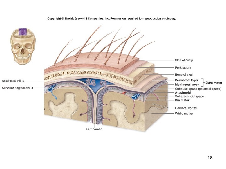
15 — 17 Cranial Meninges Three dense regular connective tissue layers that separate the soft tissue of the brain from the bones of the cranium. Enclose and protect blood vessels that supply the brain. Contain and circulate cerebrospinal fluid. Parts of the cranial meninges form some of the veins that drain blood from the brain. From superficial to deep, the cranial meninges are the dura mater , the arachnoid , and the pia mater.
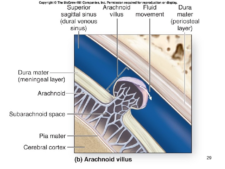
15 — 19 Dura Mater Tough membrane composed of two fibrous layers. Strongest of the meninges. Dura mater is composed of two layers. periosteal layer , the more superficial layer, attaches to the periosteum of the cranial bones meningeal layer lies deep to the periosteal layer The meningeal layer is usually fused to the periosteal layer, except in specific areas where the two layers separate to form large, blood-filled spaces called dural venous sinuses.
15 — 20 Arachnoid Also called the arachnoid mater or the arachnoid membrane. Lies immediately internal to the dura mater. Partially composed of a delicate web of collagen and elastic fibers, termed the arachnoid trabeculae. Between the arachnoid and the overlying dura mater is the subdural space. Immediately deep to the arachnoid is the subarachnoid space.
15 — 21 Pia Mater The innermost of the cranial meninges. Thin layer of delicate connective tissue that tightly adheres to the brain and follows every contour of the brain surface.
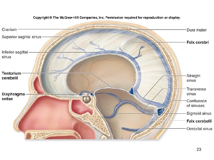
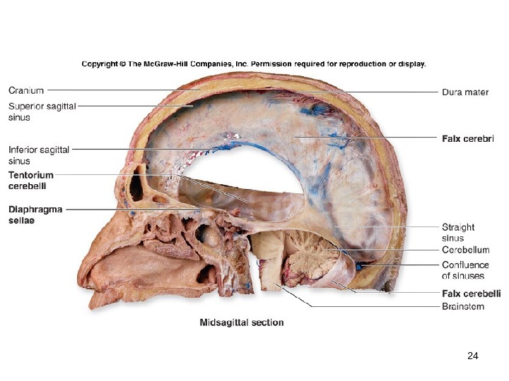
15 — 22 Cranial Dural Septa The meningeal layer of the dura mater extends as flat partitions (septa) deep into the cranial cavity at four locations called cranial dural septa. Membranous partitions separate specific parts of the brain and provide additional stabilization and support to the entire brain. falx cerebri tentorium cerebelli falx cerebelli diaphragma sellae
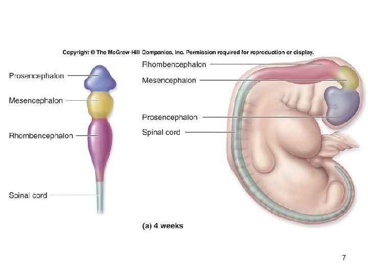
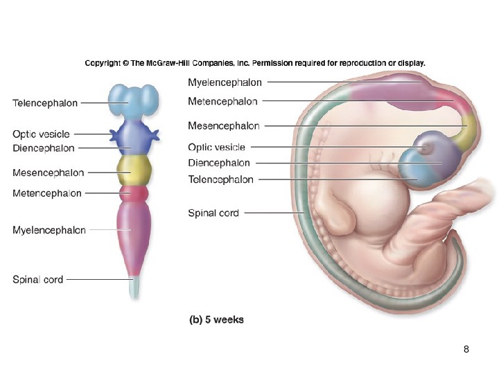
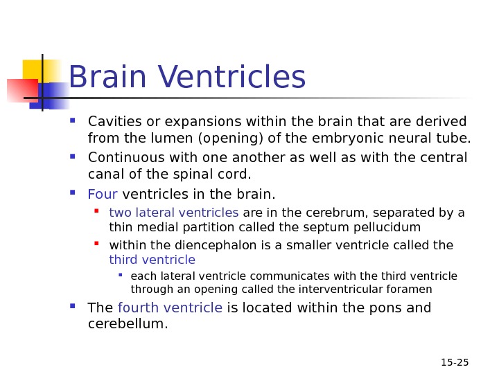
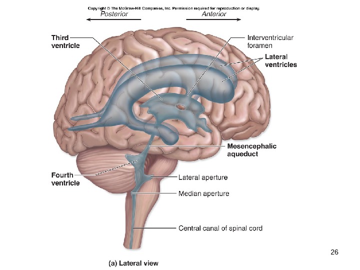
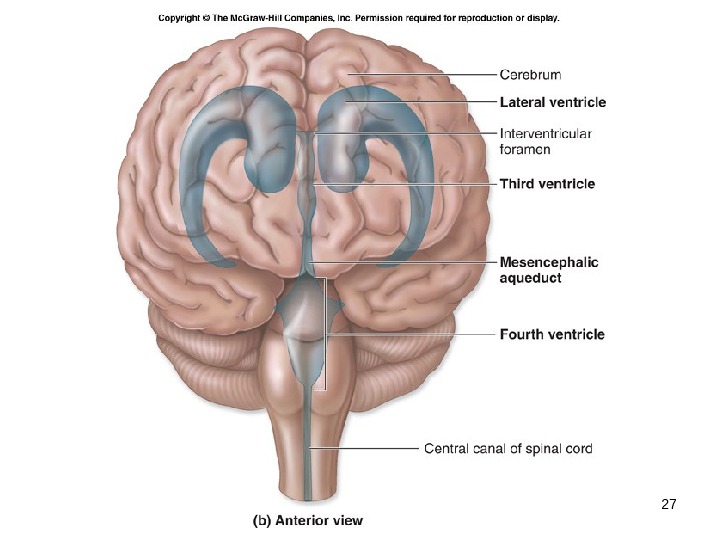
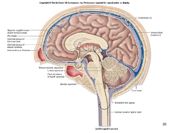
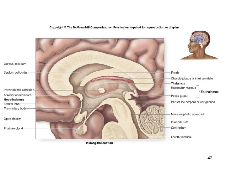
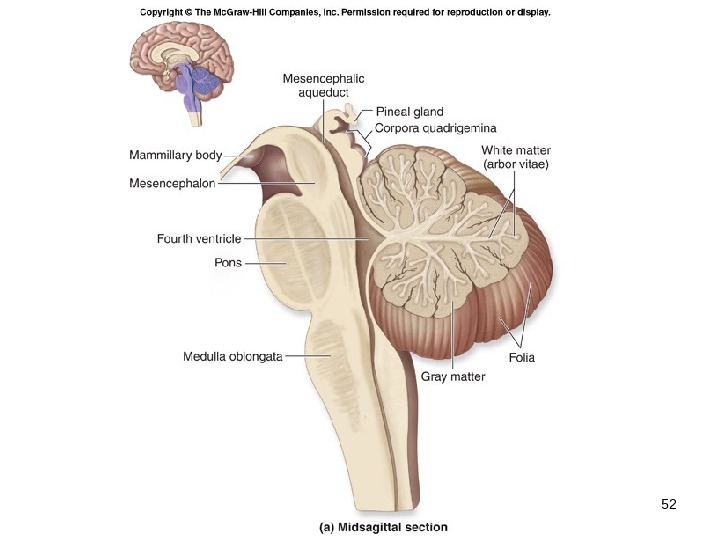
15 — 25 Brain Ventricles Cavities or expansions within the brain that are derived from the lumen (opening) of the embryonic neural tube. Continuous with one another as well as with the central canal of the spinal cord. Four ventricles in the brain. two lateral ventricles are in the cerebrum, separated by a thin medial partition called the septum pellucidum within the diencephalon is a smaller ventricle called the third ventricle each lateral ventricle communicates with the third ventricle through an opening called the interventricular foramen The fourth ventricle is located within the pons and cerebellum.
15 — 28 Cerebrospinal Fluid A clear, colorless liquid that circulates in the ventricles and subarachnoid space. Bathes the exposed surfaces of the central nervous system and completely surrounds it. Performs several important functions. buoyancy protection environmental stability Formed by the choroid plexus in each ventricle. Produced by secretion of a fluid from the ependymal cells that originate from the blood plasma. Is similar to blood plasma.
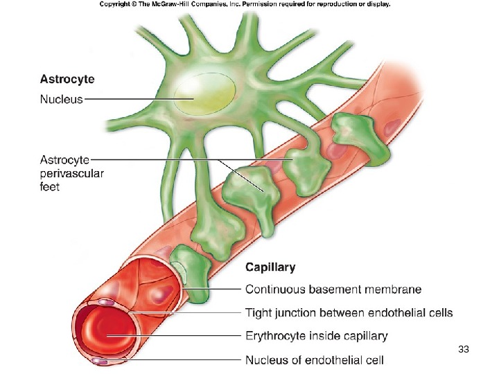
15 — 31 Blood-Brain Barrier Nervous tissue is protected from the general circulation by the blood-brain barrier. Strictly regulates what substances can enter the interstitial fluid of the brain. Prevents exposure of neurons in the brain to drugs, waste products in the blood, and variations in levels of normal substances (ions, hormones) that could adversely affect brain function.
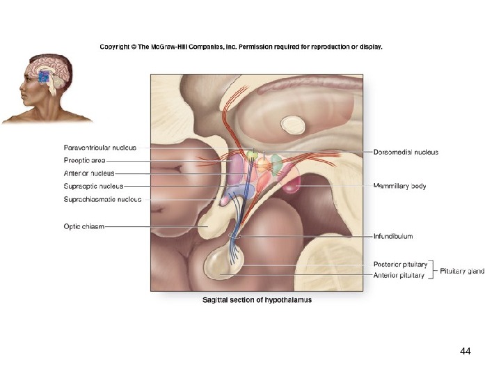
15 — 32 Blood-Brain Barrier Tight junctions prevent materials from diffusing across the capillary wall. Astrocytes act as “gatekeepers” that permit materials to pass to the neurons after leaving the capillaries. Is markedly reduced or missing in three distinct locations in the CNS: the choroid plexus, hypothalamus, and pineal gland.