
1 Gene Expression Systems in Prokaryotes and Eukaryotes


4_expression_systems.ppt
- Размер: 9.5 Mегабайта
- Количество слайдов: 96
Описание презентации 1 Gene Expression Systems in Prokaryotes and Eukaryotes по слайдам
1 Gene Expression Systems in Prokaryotes and Eukaryotes • Expression studies • Expression in Prokaryotes (Bacteria) • Expression in Eukaryotes
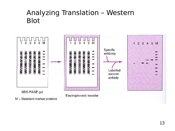
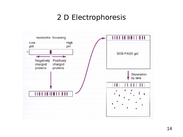
2 Gene Expression Systems in Prokaryotes and Eukaryotes Expression studies: 1. Analyzing Transcription — Northern blot — Micro array — real-time PCR — Primer extension 2. In vivo Expresion studies Use of report genes to study regulatory elements 3. Analyzing Translation — Western blot — immuno assays — 2 D electrophoresis — proteomics
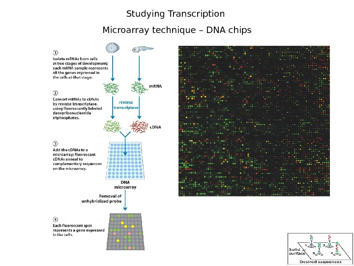
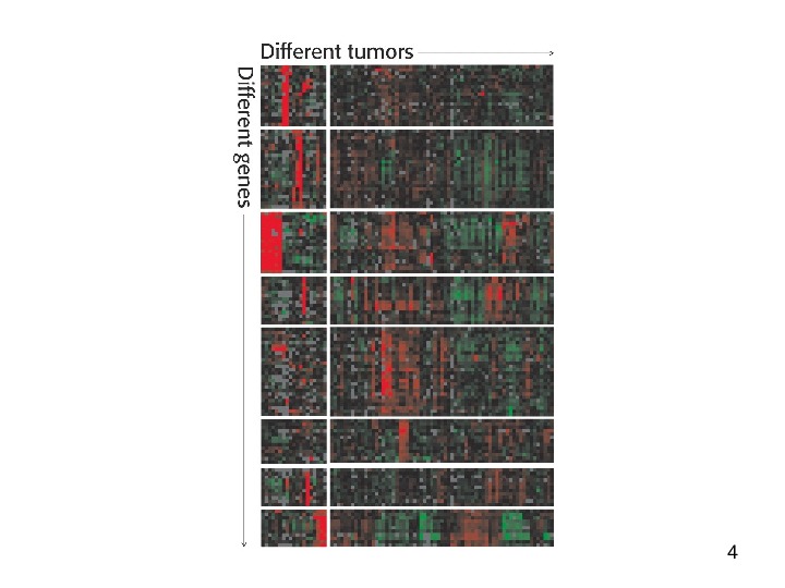
3 Studying Transcription Microarray technique – DNA chips
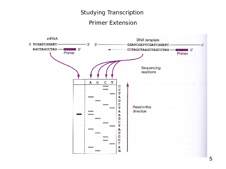
5 Studying Transcription Primer Extension
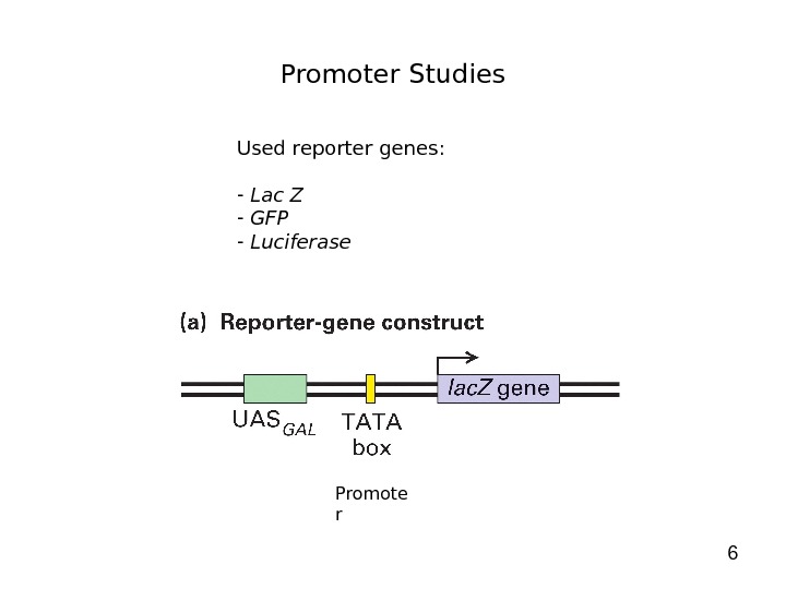
6 Promoter Studies Used reporter genes: — Lac Z — GFP — Luciferase Promote r
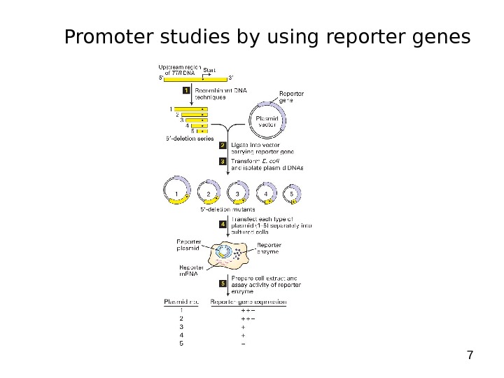
7 Promoter studies by using reporter genes
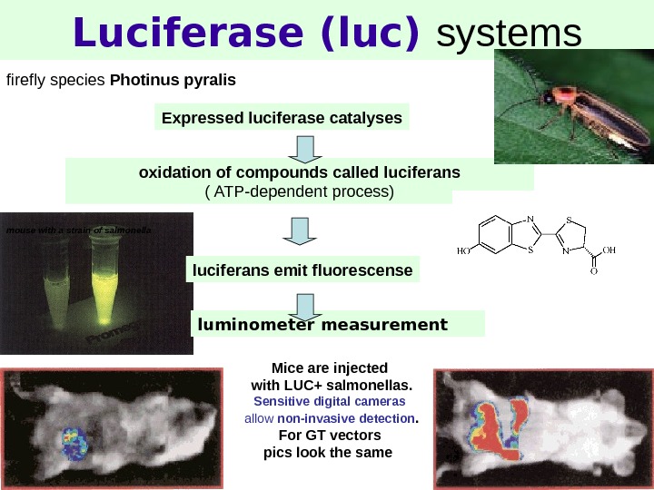
8 Luciferase (luc) systems firefly species Photinus pyralis oxidation of compounds called luciferans ( ATP-dependent process) luciferans emit fluorescense. Expressed luciferase catalyses mouse with a strain of salmonella Mice are injected with LUC+ salmonellas. Sensitive digital cameras allow non-invasive detection. For GT vectors pics look the same luminometer measurement
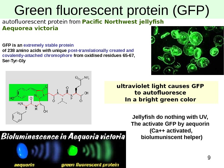
9 Green fluorescent protein (GFP) autofluorescent protein from Pacific Northwest jellyfish Aequorea victoria GFP is an extremely stable protein of 238 amino acids with unique post-translationally created and covalently-attached chromophore from oxidised residues 65 -67, Ser-Tyr-Gly ultraviolet light causes GFP to autofluoresce In a bright green color Jellyfish do nothing with UV, The activate GFP by aequorin (Ca++ activated, biolumuniscent helper)
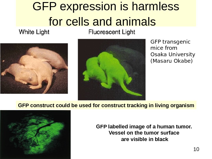
10 GFP expression is harmless for cells and animals GFP transgenic mice from Osaka University (Masaru Okabe) GFP construct could be used for construct tracking in living organism GFP labelled image of a human tumor. Vessel on the tumor surface are visible in black
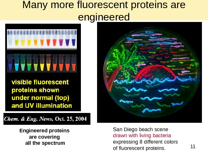
11 Engineered proteins are covering all the spectrum San Diego beach scene drawn with living bacteria expressing 8 different colors of fluorescent proteins. Many more fluorescent proteins are engineered
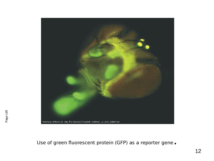
12 Use of green fluorescent protein (GFP) as a reporter gene. Page
13 Analyzing Translation – Western Blot
142 D Electrophoresis
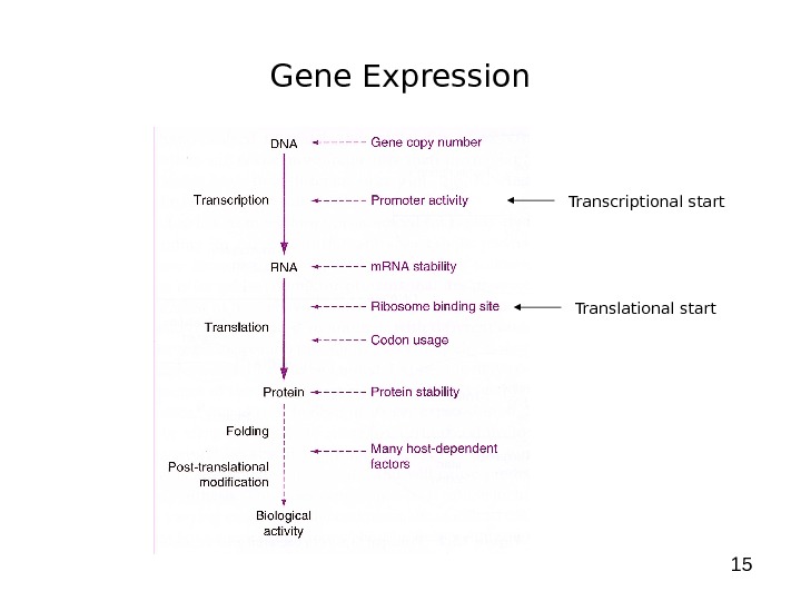
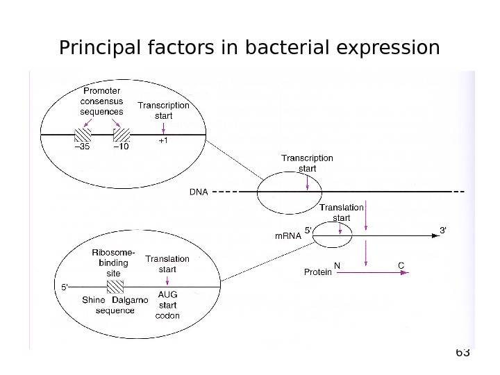
15 Gene Expression Transcriptional start Translational start
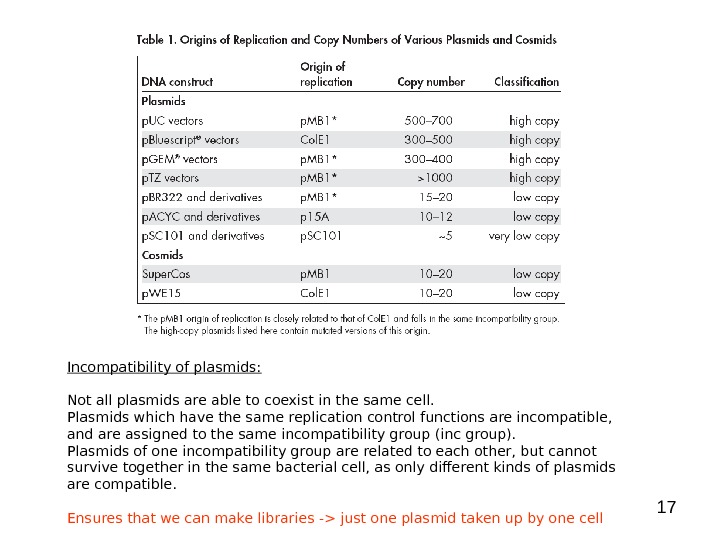
16 Gene Expression Gene copy number: 1. Plasmid copy number: The copy-number of a plasmid in the cell is determined by regulating the initiation of plasmid replication. The initiation of plasmid replication may be controlled by: — the amount of available primer (RNA) — the amount of essential replication proteins — the function of essential replication proteins. 2. Gene dosage -> number of genes integrated into chromosome — prokaryotic systems -> i. e. Transposons, phages, recombinantion — mainly eukaryotic systems
17 Incompatibility of plasmids: Not all plasmids are able to coexist in the same cell. Plasmids which have the same replication control functions are incompatible, and are assigned to the same incompatibility group (inc group). Plasmids of one incompatibility group are related to each other, but cannot survive together in the same bacterial cell, as only different kinds of plasmids are compatible. Ensures that we can make libraries -> just one plasmid taken up by one cell
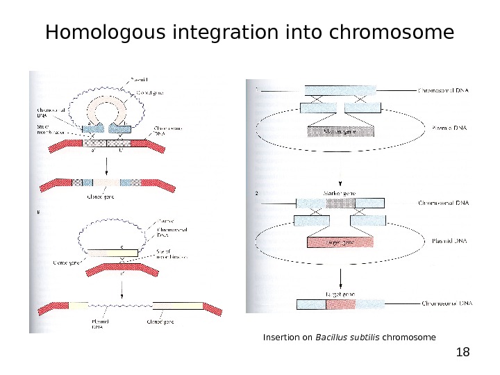
18 Homologous integration into chromosome Insertion on Bacillus subtilis chromosome
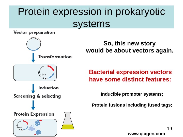
19 Protein expression in prokaryotic systems www. qiagen. com. So, this new story would be about vectors again. Bacterial expression vectors have some distinct features: Inducible promoter systems; Protein fusions including fused tags;
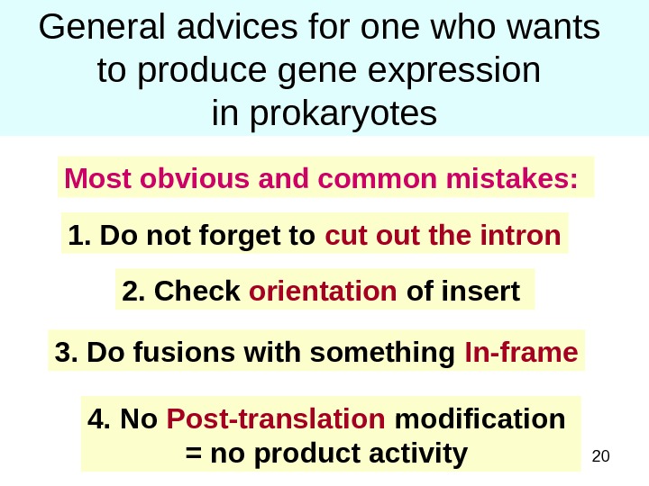
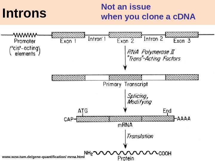
20 General advices for one who wants to produce gene expression in prokaryotes 1. Do not forget to cut out the intron 2. Check orientation of insert 3. Do fusions with something In-frame. Most obvious and common mistakes: 4. No Post-translation modification = no product activity
21 www. wzw. tum. de/gene-quantification/ mrna. html Introns Not an issue when you clone a c. DN
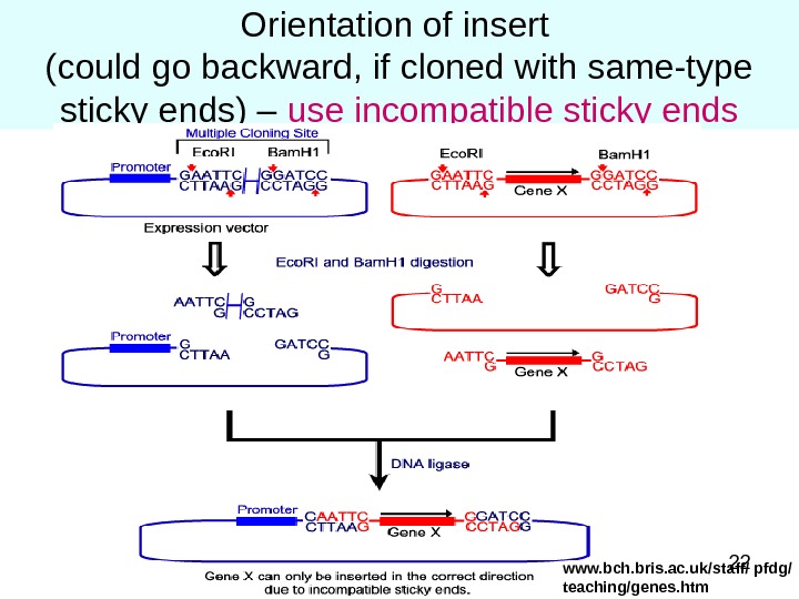
22 Orientation of insert (could go backward, if cloned with same-type sticky ends) – use incompatible sticky ends www. bch. bris. ac. uk/staff/ pfdg/ teaching/genes. htm
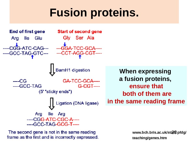
23 Fusion proteins. When expressing a fusion proteins, ensure that both of them are in the same reading frame www. bch. bris. ac. uk/staff/ pfdg/ teaching/genes. htm
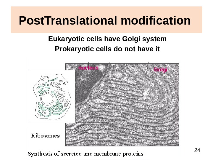
24 Post. Translational modification Eukaryotic cells have Golgi system Prokaryotic cells do not have it nucleus Golgi
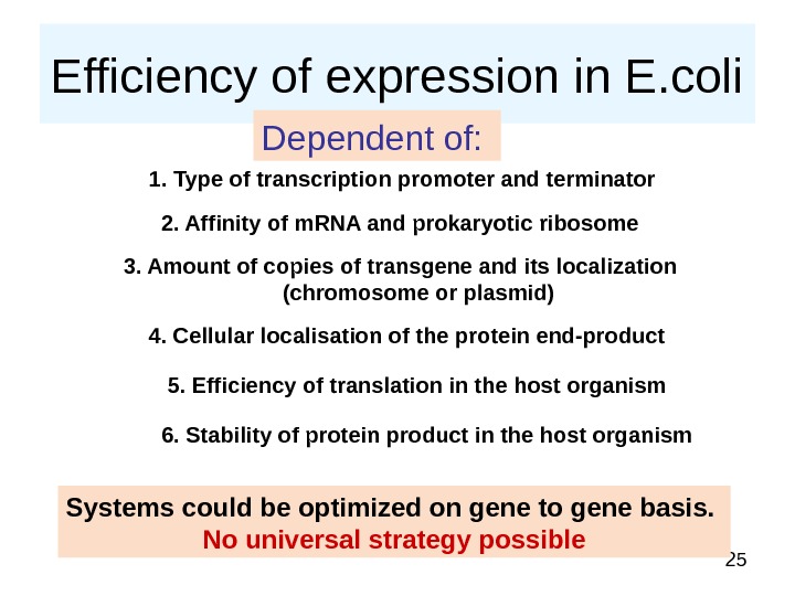
25 Efficiency of expression in E. coli Dependent of: 1. Type of transcription promoter and terminator 2. Affinity of m. RNA and prokaryotic ribosome 3. Amount of copies of transgene and its localization (chromosome or plasmid) 4. Cellular localisation of the protein end-product 5. Efficiency of translation in the host organism 6. Stability of protein product in the host organism Systems could be optimized on gene to gene basis. No universal strategy possible
26 Factors affecting transcription 1. Promoters (including regulated ones) PROKARYOTIC!!!! 2. Terminators PROKARYOTIC!!!!
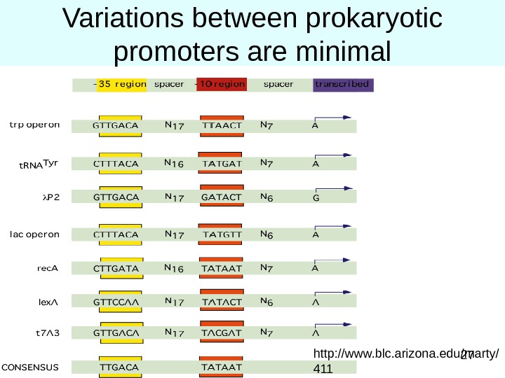
27 Variations between prokaryotic promoters are minimal http: //www. blc. arizona. edu/marty/
28 Factors affecting translation 1. Ribosome binding site (RBS) 2. Codon bias 3. Stability of the transcript
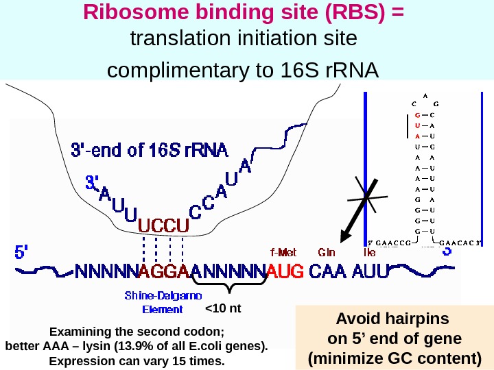
29 Ribosome binding site (RBS) = translation initiation site complimentary to 16 S r. RNA <10 nt Avoid hairpins on 5’ end of gene (minimize GC content)Examining the second codon; better AAA – lysin (13. 9% of all E. coli genes). Expression can vary 15 times.
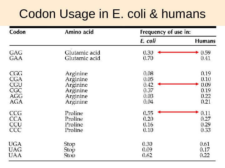
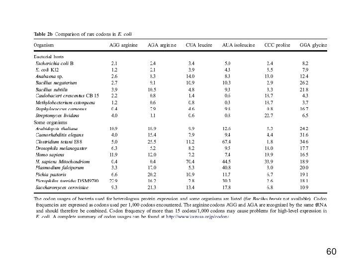
30 Codon Usage in E. coli & humans
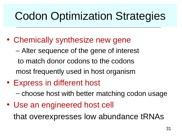
31 Codon Optimization Strategies • Chemically synthesize new gene – Alter sequence of the gene of interest to match donor codons to the codons most frequently used in host organism • Express in different host – choose host with better matching codon usage • Use an engineered host cell that overexpresses low abundance t. RNAs
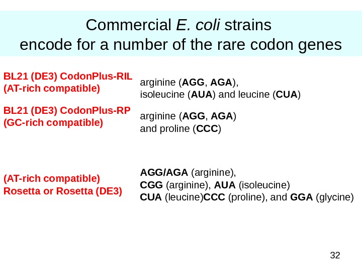
32 Commercial E. coli strains encode for a number of the rare codon genes AGG/AGA (arginine), CGG (arginine), AUA (isoleucine) CUA (leucine) CCC (proline), and GGA (glycine)(AT-rich compatible) Rosetta or Rosetta (DE 3) arginine ( AGG , AGA ) and proline ( CCC )BL 21 (DE 3) Codon. Plus-RP (GC-rich compatible) arginine ( AGG , AGA ), isoleucine ( AUA ) and leucine ( CUA )BL 21 (DE 3) Codon. Plus-RIL (AT-rich compatible)
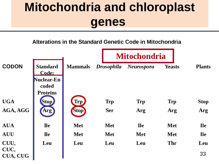
33 Mitochondria and chloroplast genes Alterations in the Standard Genetic Code in Mitochondria Leu. Thr. Leu. CUU, CUC, CUA, CUG Ile. Met. Ile. AUU Ile. Met. Ile. AUA Arg. Ser. Stop. Arg. AGA, AGG Stop. Trp. Stop. UGA Plants. Yeasts. Neurospora Drosophila Mammals. Standard Code: Nuclear-En coded Proteins. CODON Mitochondria
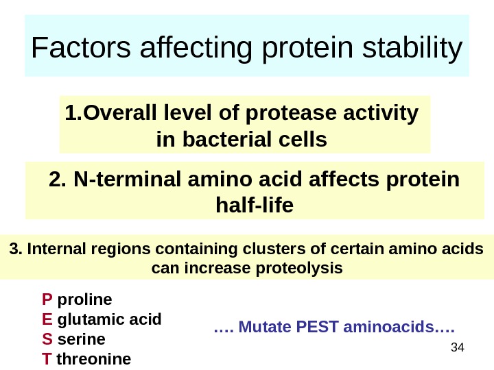
34 Factors affecting protein stability 1. Overall level of protease activity in bacterial cells 2. N-terminal amino acid affects protein half-life 3. Internal regions containing clusters of certain amino acids can increase proteolysis P proline E glutamic acid S serine T threonine …. Mutate PEST aminoacids….
35 Protease-deficient host strains BL 21, the work horse of E. coli expression, is deficient in two proteases encoded by the lon (cytoplasmic) and omp T (periplasmic) genes. It is dangerous to kill proteases, it makes E. coli grow much slowly as proteases needed for proper metabolism
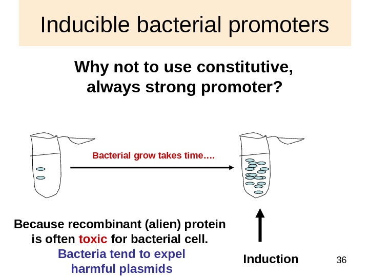
36 Inducible bacterial promoters Why not to use constitutive, always strong promoter? Induction Because recombinant (alien) protein is often toxic for bacterial cell. Bacteria tend to expel harmful plasmids Bacterial grow takes time….
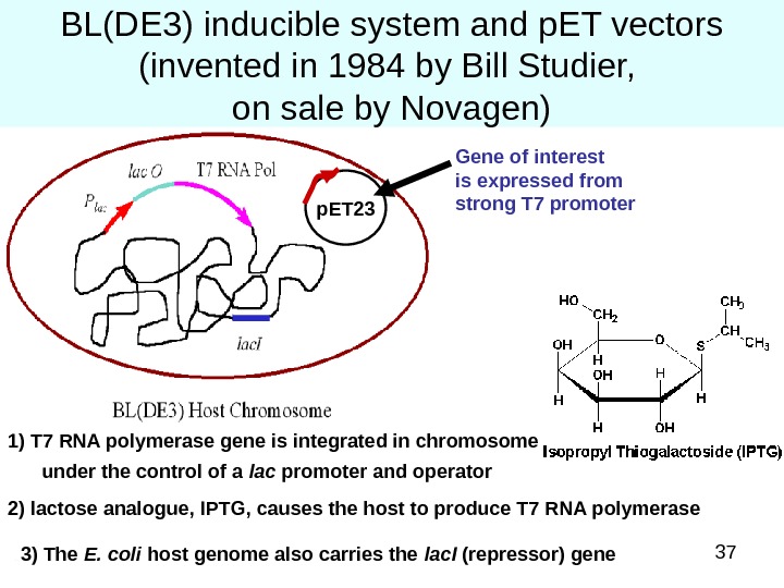
37 BL(DE 3) inducible system and p. ET vectors (invented in 1984 by Bill Studier, on sale by Novagen) 1) T 7 RNA polymerase gene is integrated in chromosome under the control of a lac promoter and operator 2) lactose analogue, IPTG, causes the host to produce T 7 RNA polymerase 3) The E. coli host genome also carries the lac. I (repressor) gene p. ET 23 Gene of interest is expressed from strong T 7 promoter
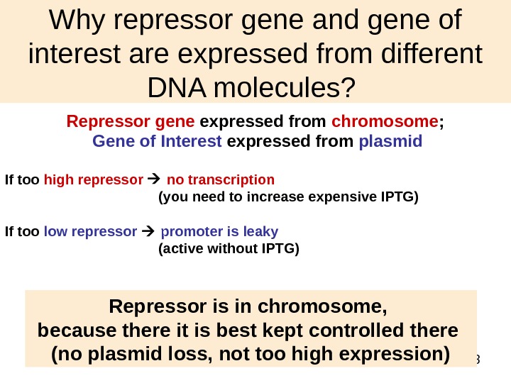
38 Why repressor gene and gene of interest are expressed from different DNA molecules? Repressor gene expressed from chromosome ; Gene of Interest expressed from plasmid If too high repressor no transcription (you need to increase expensive IPTG) If too low repressor promoter is leaky (active without IPTG) Repressor is in chromosome, because there it is best kept controlled there (no plasmid loss, not too high expression)
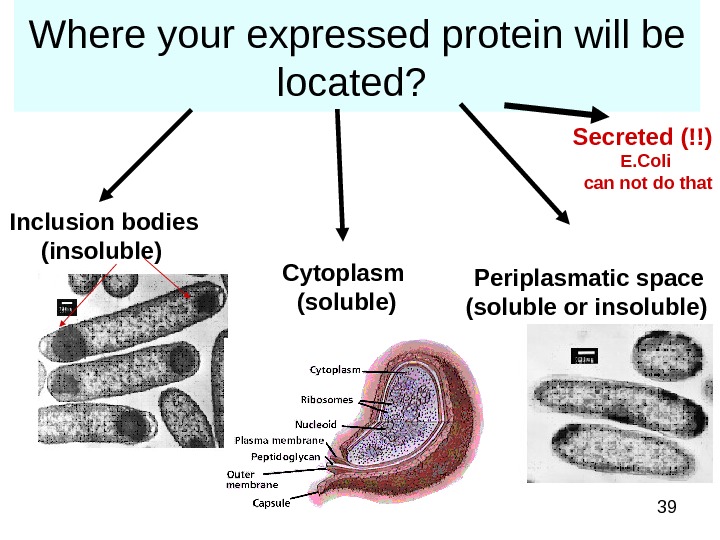
39 Where your expressed protein will be located? Inclusion bodies (insoluble) Cytoplasm (soluble) Periplasmatic space (soluble or insoluble) Secreted (!!) E. Coli can not do that
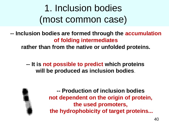
401. Inclusion bodies (most common case) — Inclusion bodies are formed through the accumulation of folding intermediates rather than from the native or unfolded proteins. — It is not possible to predict which proteins will be produced as inclusion bodies. — Production of inclusion bodies not dependent on the origin of protein, the used promoters, the hydrophobicity of target proteins. . .
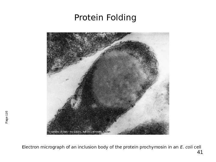
41 Electron micrograph of an inclusion body of the protein prochymosin in an E. coli cell. Page 116 Protein Folding
42 Good side of inclusion bodies 1) inclusion bodies can be accumulated in the cytoplasm to much higher level (greater than 25%) than production as soluble form; 2) inclusion bodies is initially isolated in a highly purified, solid, and concentrated state by simple physical operation (centrifugation). 3) inclusion bodies have no biological activity. For toxic proteins it may be the only one available; 4) inclusion bodies are resistant to proteolysis That results in the high yield of protein production.
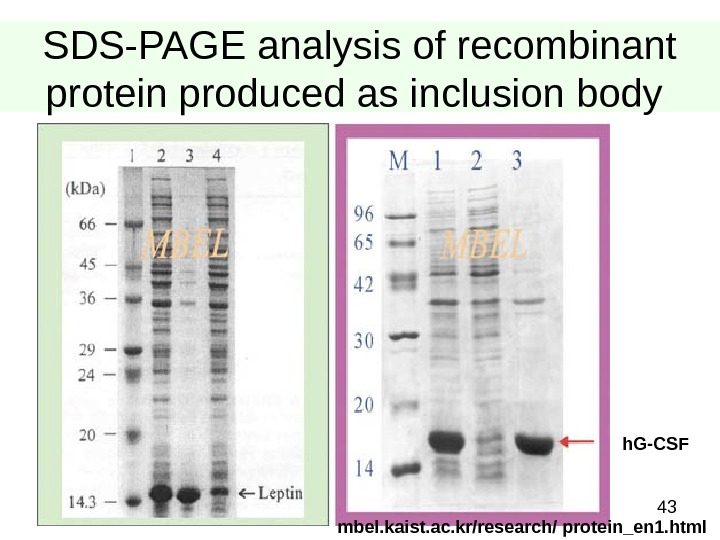
43 SDS-PAGE analysis of recombinant protein produced as inclusion body h. G-CSF mbel. kaist. ac. kr/research/ protein_en 1. html
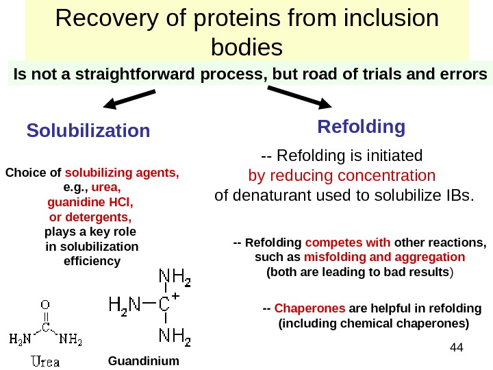
44 Recovery of proteins from inclusion bodies Is not a straightforward process, but road of trials and errors Solubilization Refolding Choice of solubilizing agents, e. g. , urea, guanidine HCl, or detergents, plays a key role in solubilization efficiency — Refolding is initiated by reducing concentration of denaturant used to solubilize IBs. Guandinium — Refolding competes with other reactions, such as misfolding and aggregation (both are leading to bad results ) — Chaperones are helpful in refolding (including chemical chaperones)
45 Question of questions – how to purify your protein?
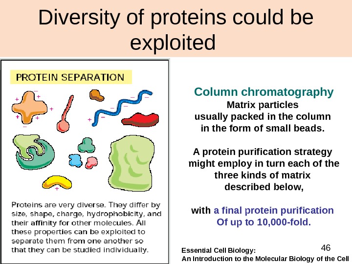
46 Diversity of proteins could be exploited Column chromatography Matrix particles usually packed in the column in the form of small beads. A protein purification strategy might employ in turn each of the three kinds of matrix described below, with a final protein purification Of up to 10, 000 -fold. Essential Cell Biology: An Introduction to the Molecular Biology of the Cell
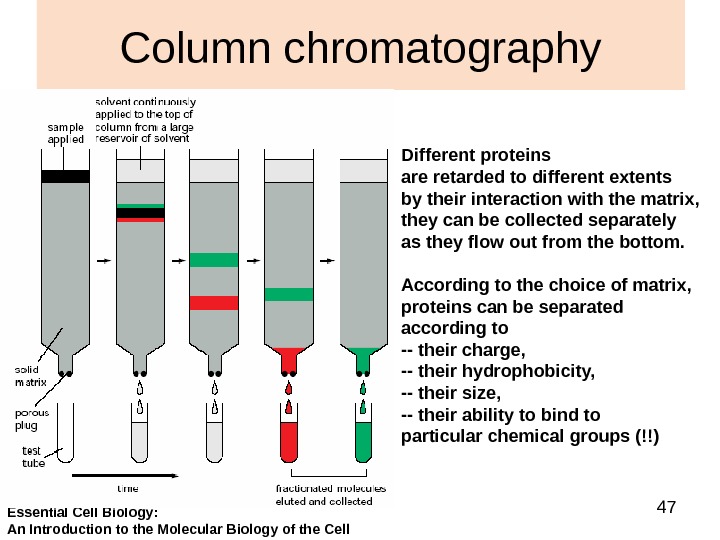
47 Column chromatography Different proteins are retarded to different extents by their interaction with the matrix, they can be collected separately as they flow out from the bottom. According to the choice of matrix, proteins can be separated according to — their charge, — their hydrophobicity, — their size, — their ability to bind to particular chemical groups (!!) Essential Cell Biology: An Introduction to the Molecular Biology of the Cell
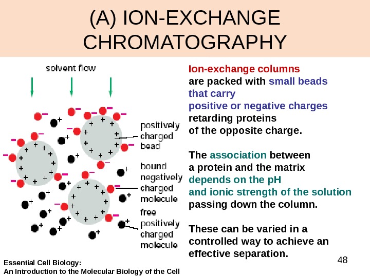
48(A) ION-EXCHANGE CHROMATOGRAPHY Ion-exchange columns are packed with small beads that carry positive or negative charges retarding proteins of the opposite charge. The association between a protein and the matrix depends on the p. H and ionic strength of the solution passing down the column. These can be varied in a controlled way to achieve an effective separation. Essential Cell Biology: An Introduction to the Molecular Biology of the Cell
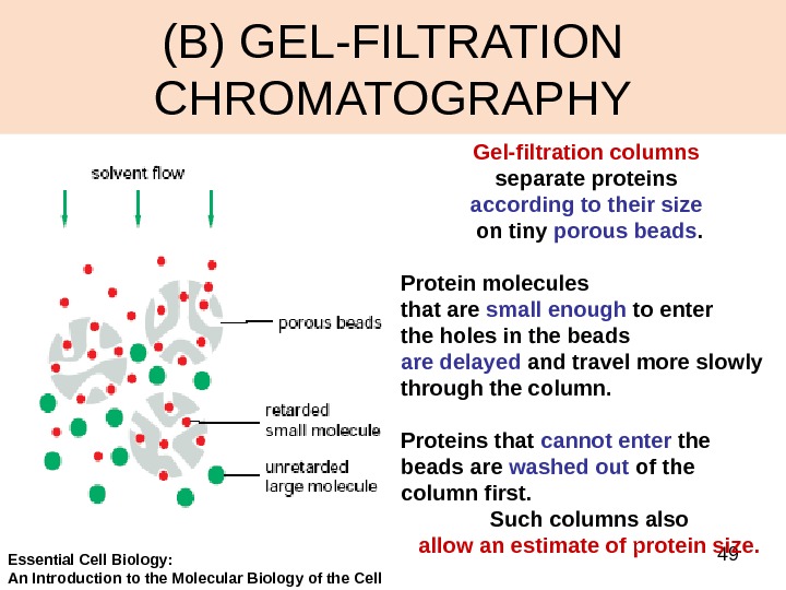
49(B) GEL-FILTRATION CHROMATOGRAPHY Gel-filtration columns separate proteins according to their size on tiny porous beads. Protein molecules that are small enough to enter the holes in the beads are delayed and travel more slowly through the column. Proteins that cannot enter the beads are washed out of the column first. Such columns also allow an estimate of protein size. Essential Cell Biology: An Introduction to the Molecular Biology of the Cell
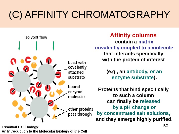
50(C) AFFINITY CHROMATOGRAPHY Affinity columns contain a matrix covalently coupled to a molecule that interacts specifically with the protein of interest (e. g. , an antibody, or an enzyme substrate ). Proteins that bind specifically to such a column can finally be released by a p. H change or by concentrated salt solutions, and they emerge highly purified. Essential Cell Biology: An Introduction to the Molecular Biology of the Cell
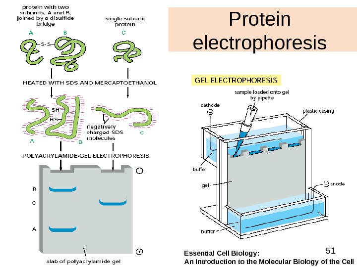
51 Protein electrophoresis Essential Cell Biology: An Introduction to the Molecular Biology of the Cell
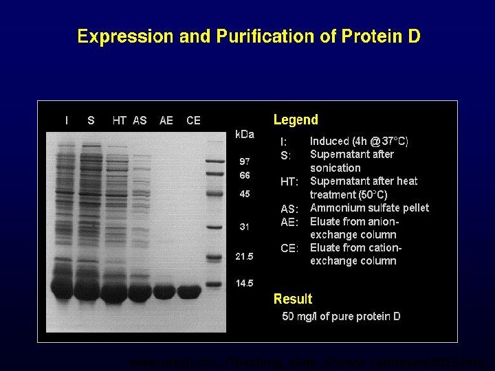
52 www. unizh. ch/. . . /Teaching_slide_shows/ Lambda/sld 015. htm
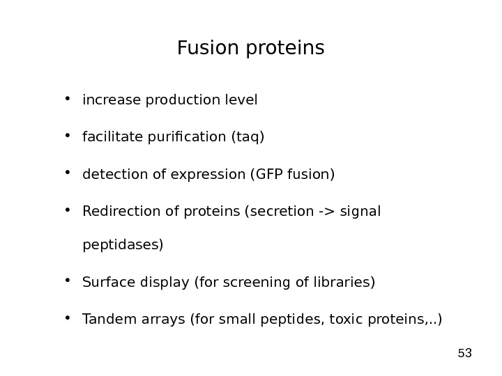
53 Fusion proteins • increase production level • facilitate purification (taq) • detection of expression (GFP fusion) • Redirection of proteins (secretion -> signal peptidases) • Surface display (for screening of libraries) • Tandem arrays (for small peptides, toxic proteins, . . )
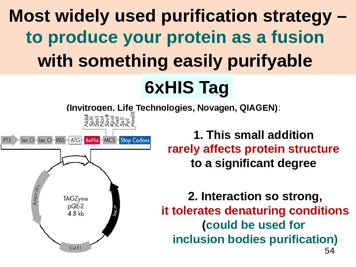
54 Most widely used purification strategy – to produce your protein as a fusion with something easily purifyable (Invitrogen, Life Technologies, Novagen, QIAGEN) : 6 x. HIS Tag 1. This small addition rarely affects protein structure to a significant degree 2. Interaction so strong, it tolerates denaturing conditions ( could be used for inclusion bodies purification)
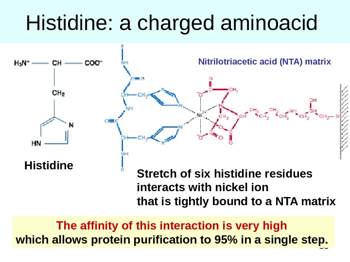
55 Histidine: a charged aminoacid The affinity of this interaction is very high which allows protein purification to 95% in a single step. Stretch of six histidine residues interacts with nickel ion that is tightly bound to a NTA matrix Nitrilotriacetic acid (NTA) matrix Histidine
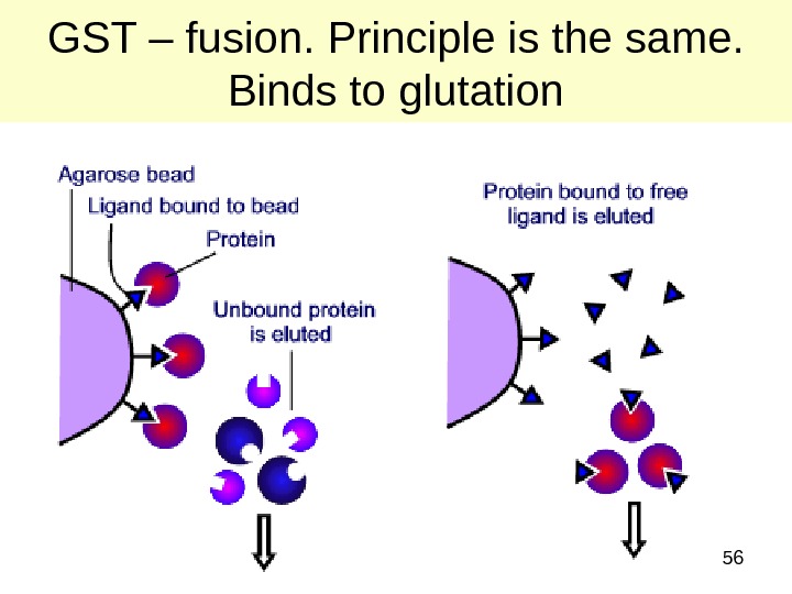
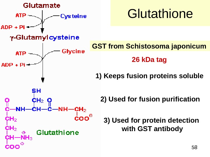
56 GST – fusion. Principle is the same. Binds to glutation
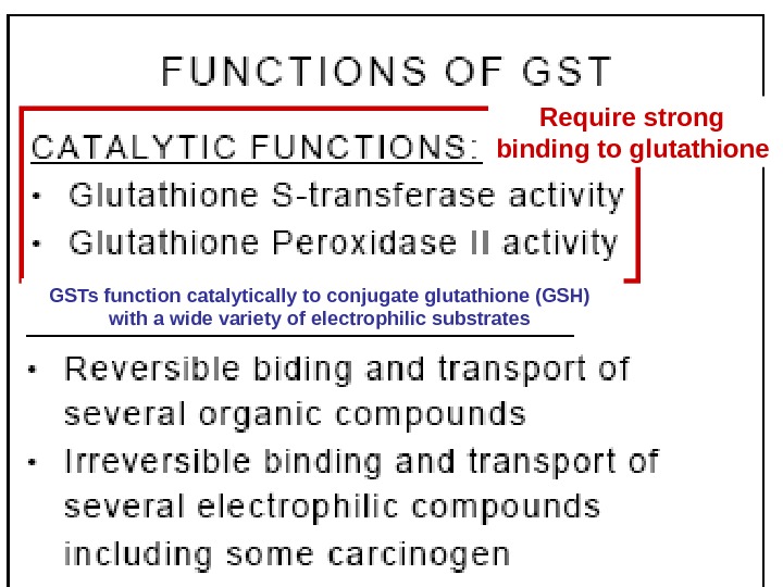
57 Require strong binding to glutathione GSTs function catalytically to conjugate glutathione (GSH) with a wide variety of electrophilic substrates
58 Glutathione GST from Schistosoma japonicum 1) Keeps fusion proteins soluble 2) Used for fusion purification 3) Used for protein detection with GST antibody 26 k. Da tag
59 FUSION PROTEIN BOUND TO GLUTATHIONE SEPHAROSE Glutathione GSTFOREIGN PEPTIDE SEPHAROSE Purification is simple : — WASH COLUMN EXTENSIVELY — ELUTE WITH REDUCED GLUTATHIONE — RESULTS IN PURE GST FUSION PROTEIN
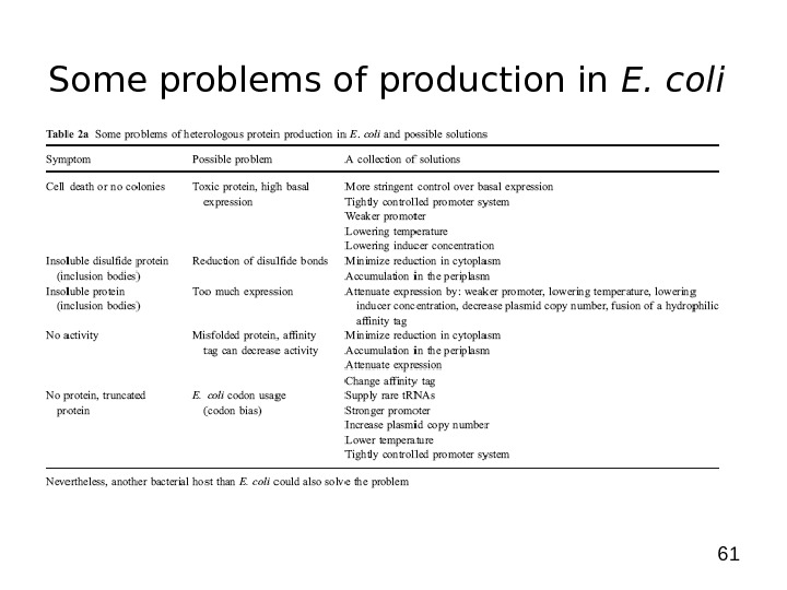
61 Some problems of production in E. coli
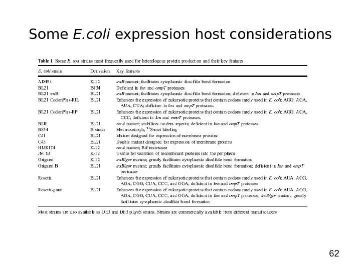
62 Some E. coli expression host considerations
63 Principal factors in bacterial expression
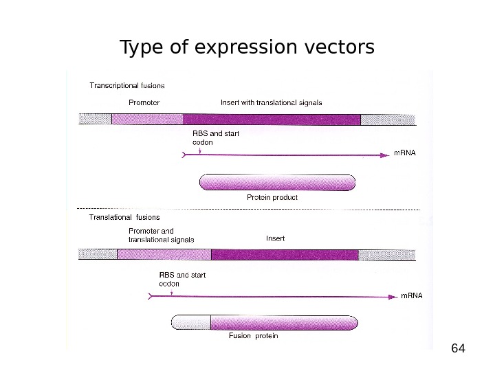
64 Type of expression vectors
65 Initiation of Transcription Promoters for Expression in Prokaryotes • In Escherichia coli — Lac system — p lac — Trp system — synthetic systems – p tac , p trc • In Bacillus
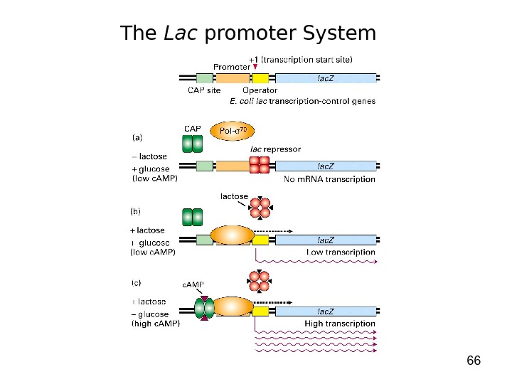
66 The Lac promoter System
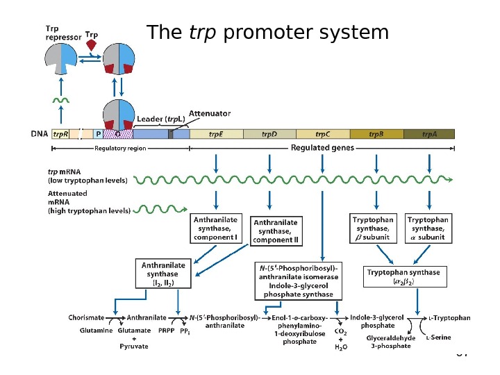
67 The trp promoter system
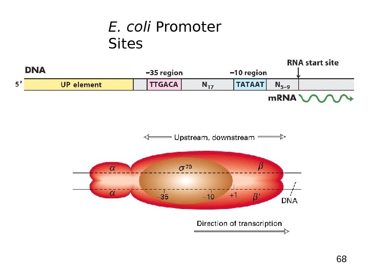
68 E. coli Promoter Sites
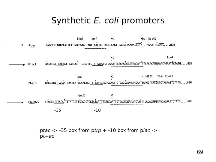
69 Synthetic E. coli promoters -35 -10 p tac -> -35 box from p trp + -10 box from p lac -> p t+ac
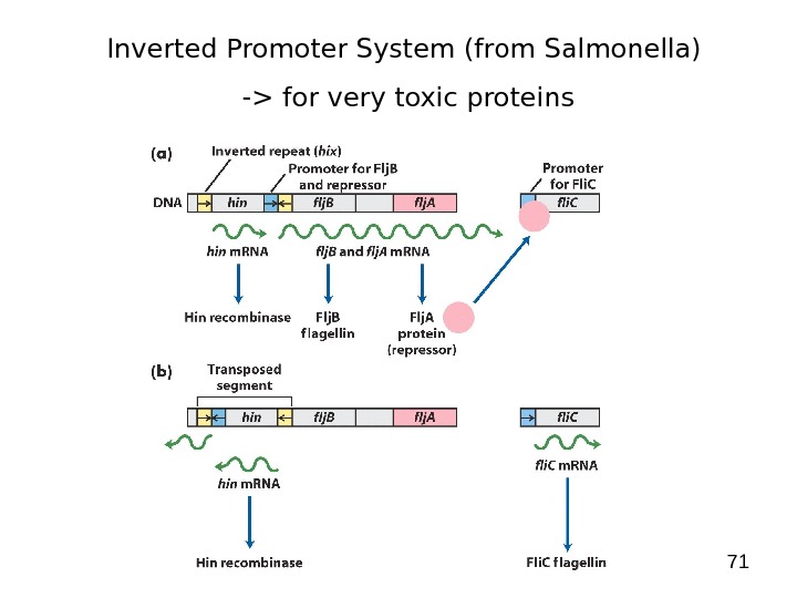
71 Inverted Promoter System (from Salmonella) -> for very toxic proteins
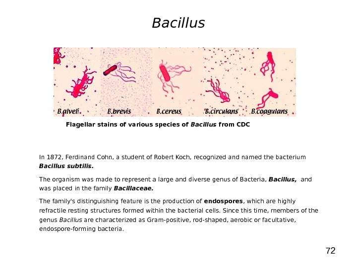
72 Bacillus In 1872, Ferdinand Cohn, a student of Robert Koch, recognized and named the bacterium Bacillus subtilis. The organism was made to represent a large and diverse genus of Bacteria, Bacillus, and was placed in the family Bacillaceae. The family’s distinguishing feature is the production of endospores , which are highly refractile resting structures formed within the bacterial cells. Since this time, members of the genus Bacillus are characterized as Gram-positive, rod-shaped, aerobic or facultative, endospore-forming bacteria. Flagellar stains of various species of Bacillus from
73 Bacillus • Antibiotic Producers : B. brevis (e. g. gramicidin, tyrothricin), B. cereus (e. g. cerexin, zwittermicin), B. circulans (e. g. circulin), B. laterosporus (e. g. laterosporin), B. licheniformis (e. g. bacitracin), B. polymyxa (e. g. polymyxin, colistin), B. pumilus (e. g. pumulin) B. subtilis (e. g. polymyxin, difficidin, subtilin, mycobacillin). • Pathogens of Insects : B. larvae, B. lentimorbis, and B. popilliae are invasive pathogens. B. thuringiensis forms a parasporal crystal that is toxic to beetles. • Pathogens of Animals : B. anthracis, and B. cereus. B. alvei, B. megaterium, B. coagulans, B. laterosporus, B. subtilis, B. sphaericus, B. circulans, B. brevis, B. licheniformis, B. macerans, B. pumilus, and B. thuringiensis have been isolated from human infections. • The Genus Bacillus includes two bacteria of significant medical importance, B. anthracis, the causative agent of anthrax, and B. cereus , which causes food poisoning. Nonanthrax Bacillus species can also cause a wide variety of other infections, and they are being recognized with increasing frequency as pathogens in humans.
74 Bacillus • Bacillus strains used as production organisms: — B. subtilis — B. brevis — B. licheniformis • Transformation systems: — via competent cells (during transition from vegetative cells -> sporulation, cell can take up DNA (ss) when population reaches a metabolic state called competence) — protoplast — bacteriophage-mediated transduction • Vectors: — replicating plasmids (p. UB 110, p. E 194, p. C 194, p. HP 13, shuttle vectors) -> replicating plasmids with temperature-sensitive origin of replication (replication stops above certain temp. -> p. E 194 stops above 45ºC) — integrative vectors (normally shuttle vectors) • Promoters: — apr. E promoter -> induction with onset of sporulation — amylase promoter -> growth-phase and nutrition regulated promoter (induction at end of exponential growth + repression by glucose) — sac. B promoter (levansurase) -> not regulated — spac promoter -> hybrid promoter (subtilis phage + lac operator) -> induction with IPTG
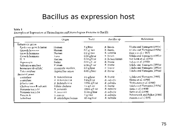
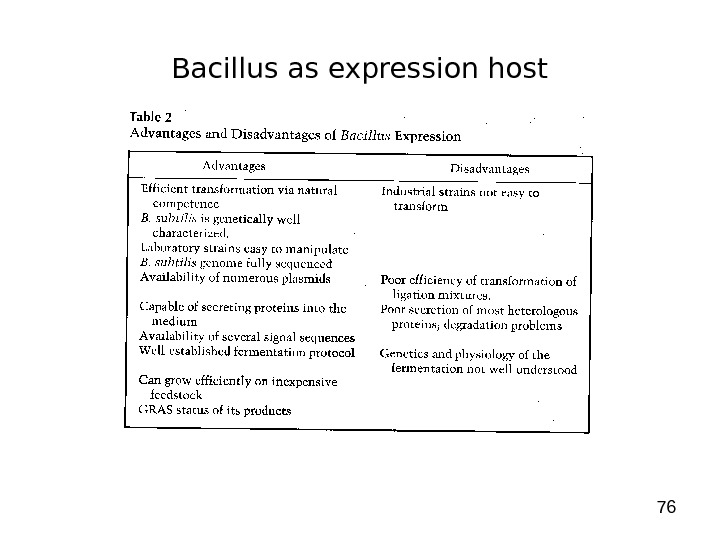
75 Bacillus as expression host
76 Bacillus as expression host
77 Products produced in Prokaryotic Systems • Restriction Endonucleases -> produced in E. coli • L- Ascorbic Acid (Vitamin C) -> recombinant Erwinia herbicola (gram-negative bacterium) • Synthesis of Indigo (blue pigment -> dye cotton /jeans) -> produced in E. coli • Amino Acids -> produced in Corynebacterium glutamicum (gram-positive bacterium) • Lipases (laundry industry) -> from Pseudomonas alcaligenes produced in Pseudomonas alcaligenes • Antibiotica (most of them from Streptomyces , other gram-positive bacteria, fungi) -> produced in recombinant Streptomyces and fungi ( Penicillium ) • Biopolymers (PHB -> biodegradable plastics) -> produced in E. coli (stabilized with par. B)
78 Expression in Eukaryotic Systems • Yeast — Saccharomyces cerevisiae (baker’s yeast) — Pichia pastoris • Insect Cells – Baculovirus • Mammalian Cells
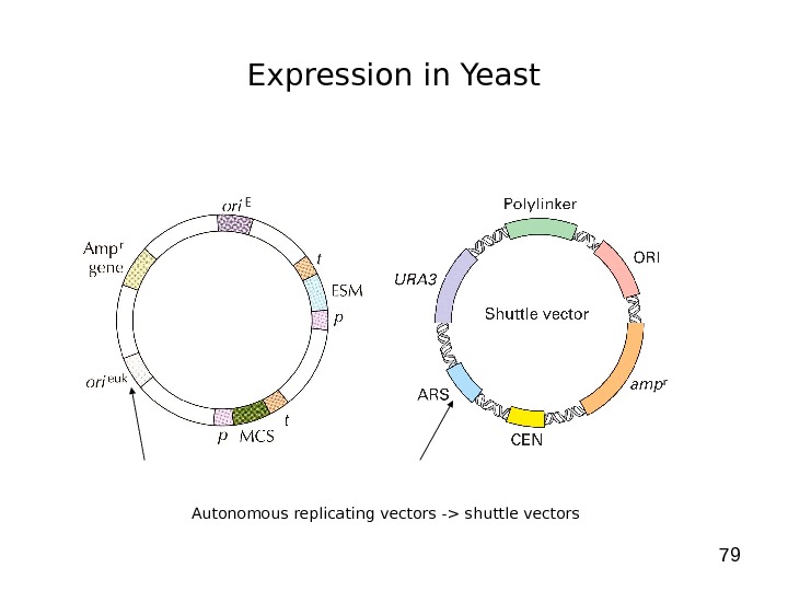
79 Expression in Yeast Autonomous replicating vectors -> shuttle vectors
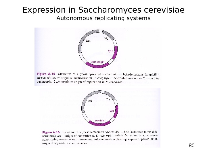
80 Expression in Saccharomyces cerevisiae Autonomous replicating systems
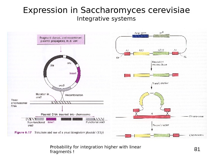
81 Expression in Saccharomyces cerevisiae Integrative systems Probability for integration higher with linear fragments !
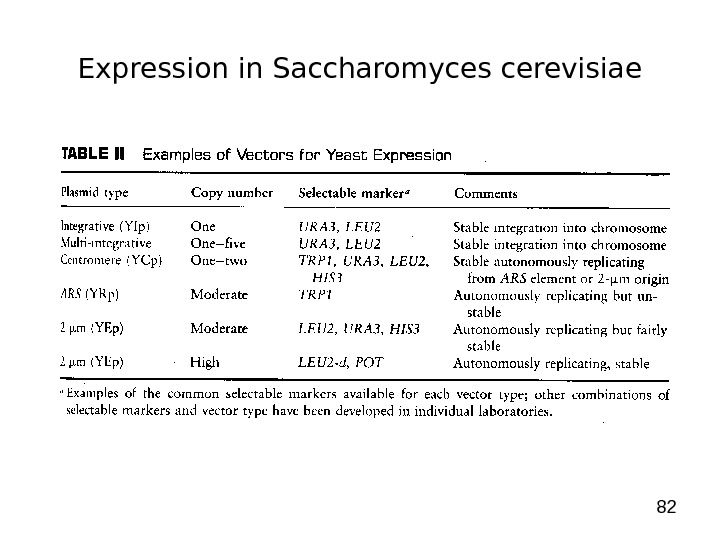
82 Expression in Saccharomyces cerevisiae
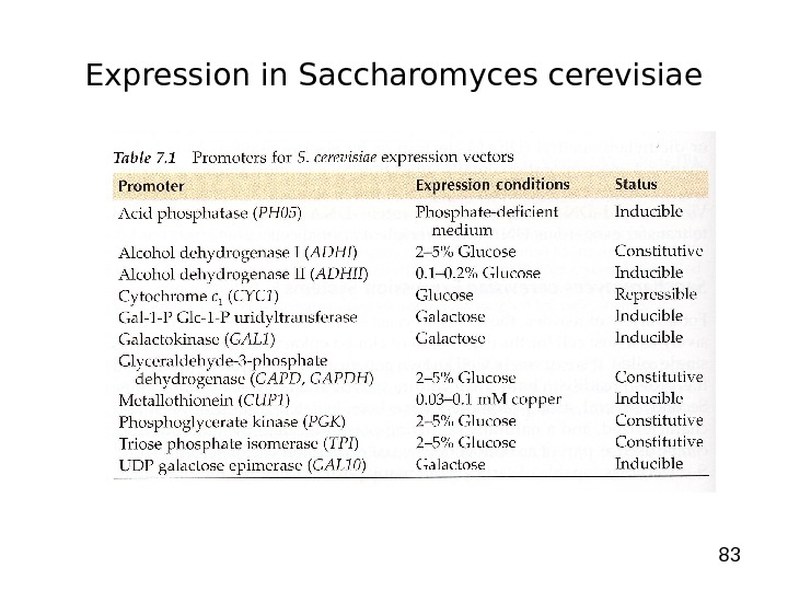
83 Expression in Saccharomyces cerevisiae
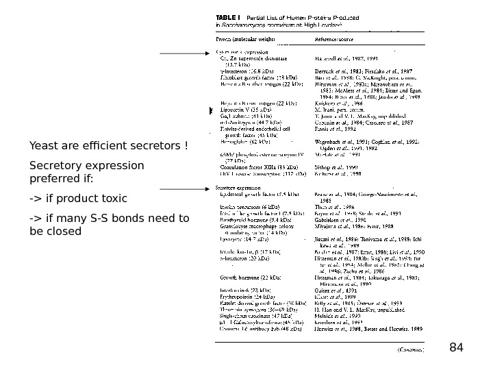
84 Yeast are efficient secretors ! Secretory expression preferred if: -> if product toxic -> if many S-S bonds need to be closed
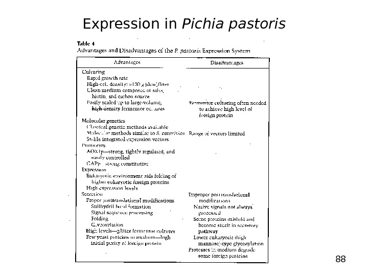
85 Expression in S. cerevisiae – Pichia pastoris Problems with production in S. cerevisiae: • For some proteins production level low • Hyperglycosylation (more than 100 mannose residues in N-glycosylation) • Sometimes secretion not good -> protein stack in cells (periplasma) • S. cerevisiae produces high amount of Et. OH -> toxic for the cells -> effects level of production Advantages of production in Pichia pastoris : • Highly efficient promoter, tightly regulated (alcohol oxidase -> AOX, induced by Me. OH) • Produces no Et. OH -> very high cell density -> secretion very efficient • Secretes very few proteins -> simplification of purification of secreted proteins
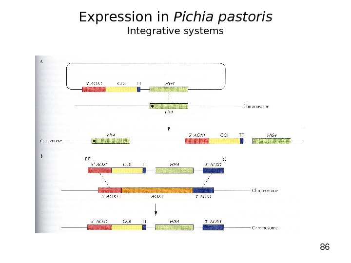
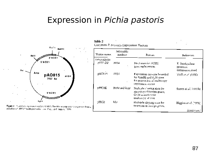
86 Expression in Pichia pastoris Integrative systems
87 Expression in Pichia pastoris
88 Expression in Pichia pastoris
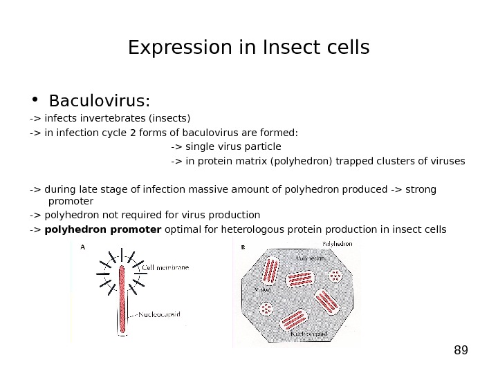
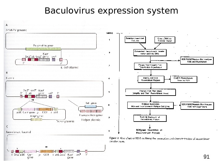
89 Expression in Insect cells • Baculovirus: -> infects invertebrates (insects) -> in infection cycle 2 forms of baculovirus are formed: -> single virus particle -> in protein matrix (polyhedron) trapped clusters of viruses -> during late stage of infection massive amount of polyhedron produced -> strong promoter -> polyhedron not required for virus production -> polyhedron promoter optimal for heterologous protein production in insect cells
90 Expression in Insect cells • Baculovirus: -> Autographa californica multiple nuclear polyhedrosis virus ( Ac. MNPV ) many used as expression vector -> Production of recombinant baculovirus: 1. create a transfer vector ( E. coli based plasmid with Ac. MNPV DNA – polyhedrin promoter/terminator + flanking sequences) -> gene of interest cloned downstream of promoter 2. Insect cells are cotransfected with virus (Ac. MNPV) + transfer vector -> in some double infected cells -> double crossover event (recombination) -> produce recombinant virus (bacmid -> E. coli — insect cell baculovirus shuttle vector) -> cells infected with recombinant virus -> produce plaques (lack of polyhedrin) 3. DNA hydridisation + PCR used to identify recombinant virus 4. Infection of insect cells with concentrated stock of verified recombinant virus -> 4 -5 days later protein harvested
91 Baculovirus expression system
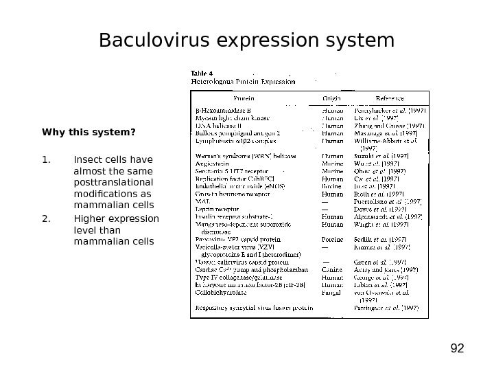
92 Why this system? 1. Insect cells have almost the same posttranslational modifications as mammalian cells 2. Higher expression level than mammalian cells Baculovirus expression system
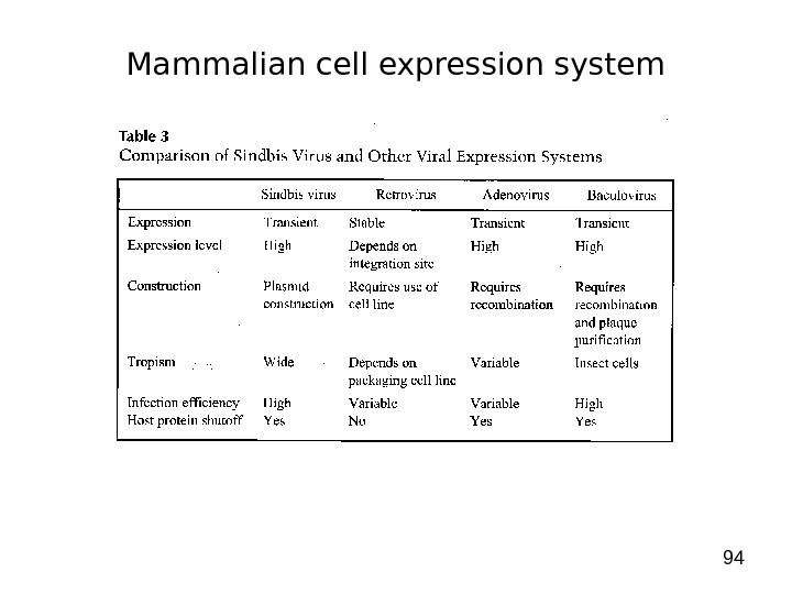
93 Mammalian cell expression system 1. Why do we use that system? -> to get full complement of posttranslational modifications on proteins 2. Developed cell lines: -> short term (transient) expression -> autonomous replicating systems -> viral origins (SV 40) — African green monkey kidney (COS) — baby hamster kidney (BHK) — human embryonic kidney (HEK-239) -> long term (stable) expression -> integration into chromosome -> viral origins — chinese hamster ovary (CHO)
94 Mammalian cell expression system
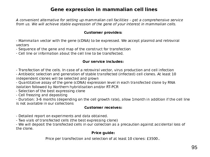
95 Gene expression in mammalian cell lines A convenient alternative for setting up mammalian cell facilities – get a comprehensive service from us. We will achieve stable expression of the gene of your interest in mammalian cells. Customer provides: — Mammalian vector with the gene (c. DNA) to be expressed. We accept plasmid and retroviral vectors — Sequence of the gene and map of the construct for transfection — Cell line or information about the cell line to be transfected. Our service includes: — Transfection of the cells. In case of a retroviral vector, virus production and cell infection — Antibiotic selection and generation of stable transfected (infected) cell clones. At least 10 independent clones will be selected and grown — Quantitative assay of the gene (c. DNA) expression level in each transfected clone by RNA isolation followed by Northern hybridisation and/or RT-PCR — Selection of the best expressing clone — Cell freezing and depositing — Duration: 3 -6 months (depending on the cell growth rate), allow 1 month in addition if the cell line is not available in our collections Customer receives: — Detailed report on experiments and data obtained. — Two vials of transfected cells (the best expressing clone) — We will deposit the transfected cells in our collection as a precaution against accidental loss of the clone. Price guide: Price per transfection and selection of at least 10 clones: £ 3500.
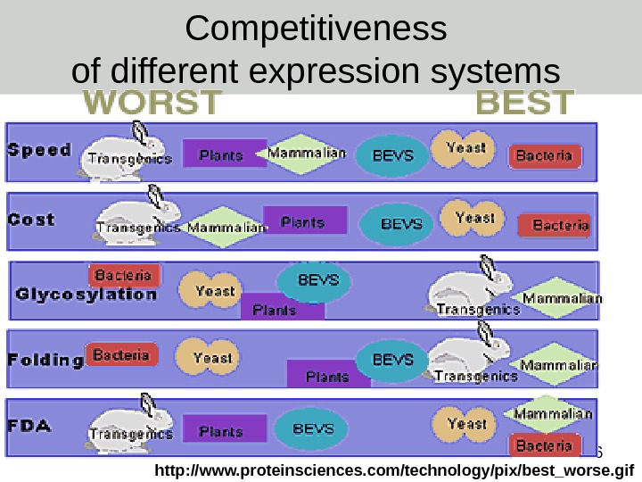
96 Competitiveness of different expression systems http: //www. proteinsciences. com/technology/pix/best_worse. gif