

Кисть english.ppt
- Количество слайдов: 30
 Topographical anatomy of the hand fingers. Incisions.
 The meaning of hand fingers is enormous for life. Person can’t fulfill a lot of jobs without hand or fingers. So, operative surgery of hand is a very important unit in general medicine.
 Thenar line 2) transverse") Look at your hand! You can see some lines. 1) Thenar line 2) transverse proximal line 3) transverse distal line In operative surgery and topographical anatomy this lines are used for determination of arteries and nerves projection.
Look at your hand! You can see some lines. 1) Thenar line 2) transverse proximal line 3) transverse distal line In operative surgery and topographical anatomy this lines are used for determination of arteries and nerves projection.
 If you divide thenar line into three parts and draw a square, where a proximal third is one side of square, you’ll get Kanavel’s prohibited area – the brunsh of n. medianus to m. opponens pollicis is located here. Incisions in this area are forbidden, because m. opponens pollicis may lost its function.
 Transverse proximal line is projection of arcus palmaris superficialis
 Transverse distal line is projection of metacarpophalangeal joint Proximal interdigital line is the middle of proximal phalanx Medium and distal interdigital line is interphalangeal [digital] joint
 Scin and its characteristics: 1. 2. 3. Epithelial tissue consist of more then 100 rows of cells Pigmentation, sebaceous glands, hair are absent There a lot of sudoriferous [sudoriparous] glands
 Characteristics of subcutaneous fat 1. 2. 3. 4. Slow subcutaneous fat regeneration. Lack of fascia superficialis. Subcutaneous fat in the fold area is considerably less. Subcutaneous fat has cellular structure.
 Characteristics of cellular structure • Purulent process tends to spreading inside • Infiltration anesthesia is very difficult – block anesthesia is used
 Palmar aponeurosis has triangular shape. One of the illnesses of palmar aponeurosis is Dupuytren's contracture. • Dupuytren's contracture
 • Dupuytren's contracture (also known as "Morbus Dupuytren, " "Dupuytren's disease, " or "Palmar fibromatosis", and sometimes misspelled as Dupuytren's constricture) is a fixed flexion contracture of the hand where the fingers bend towards the palm and cannot be fully extended (straightened). It is named after Baron Guillaume Dupuytren, the surgeon who described an operation to correct the affliction. • Dupuytren's contracture is caused by underlying contractures of the palmar fascia. The ring finger and little finger are the fingers most commonly affected. The middle finger may be affected in advanced cases, but the index finger and the thumb are nearly always spared. Dupuytren's contracture progresses slowly and is usually painless. In patients with this condition, the tissues under the skin on the palm of the hand thicken and shorten so that the tendons connected to the fingers cannot move freely. The palmar aponeurosis becomes hyperplastic and undergoes contracture.
![It’s operative [surgical] technique for Dupuytren's contracture](https://present5.com/presentation/3/149661395_156922900.pdf-img/149661395_156922900.pdf-12.jpg "It’s operative [surgical] technique for Dupuytren's contracture") It’s operative [surgical] technique for Dupuytren's contracture
It’s operative [surgical] technique for Dupuytren's contracture
 Skin innervation 1 – ulnaris, 2 – medianus 3 - radialis Lack of sensitivity in different parts of hand is a result of damage of nervs
 Innervations of muscles Symptoms of nerves damage: • N. medianus – m. m. «Gynecologist’s of thenar , 1, 2 hand» lumbrical muscles • N. ulnaris– • N. ulnaris m. m. of «clawhand» , and/or hypothenar, 3, 4 symptom «sheet of lumbrical muscles, all paper» m. m. interossea • N. radialis – flexors on • N. radialis– «chicken hand» fore arm
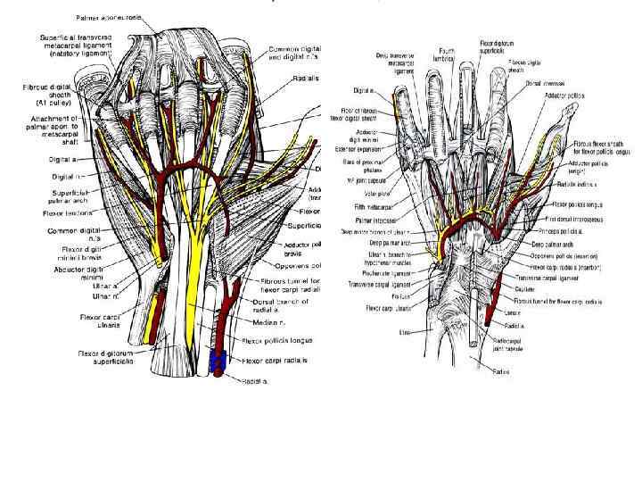
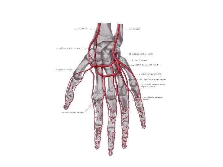
 Blood supply
 Tendons and ligaments of fingers Every finger has m. flexor digitorum superficialis and m. flexor digitorum profundus Tendons are surrounded with bursa synovialis begin from distal phalanx and ends: Bursa of thumb ends on the fore arm Bursa of 2, third, fourth fingers end on the metacarpophalangeal jonts Bursa of fifth fingers takes tendo of m. flexor 4, 3, 2 fingers and ends on the fore arm Also, There is a mesenterium near the basis of tendo m. flexors Mesenterium contains vessels and nervs If there is a pus inside bursa synovialis the mesenterium vessels is comressed and pressure necrosis develops
 The finger can’t be bended because of tendon necrosis. It needs endoplastic. Usually tendon of m. palmaris longus is used.
 There are the different structure variants of bursa synovialis
 Panaritium – purulent inflammation of finger. Agent –streptococcus and staphilococcus
 Localisation • Intradermal • Subdermal • Paronychia – paraungual panaritium • subungual • joint • osteal • tendinous • Pandactylitis is purulent inflammation of all finger tissues
 Paronychia
 Pandactylitis
 Anesthesia is local (Oberst’s Anesthesia) 1. Injection 2 -4 ml of Lidocain") Ttreatment (operation) Anesthesia is local (Oberst’s Anesthesia) 1. Injection 2 -4 ml of Lidocain (Novocain) to finger basis on each side. 2. Tourniquet to finger basis 3. Anesthesia begins in 4 -7 min.
Ttreatment (operation) Anesthesia is local (Oberst’s Anesthesia) 1. Injection 2 -4 ml of Lidocain (Novocain) to finger basis on each side. 2. Tourniquet to finger basis 3. Anesthesia begins in 4 -7 min.
 Incisions in case of Intradermal localisation It is necessary to cut spalled Epithelial tissue
 Incisions in case of subdermal localisation are fulfilled on each side between interdigital line and drainage.
 Incisions in case of tendinous localisation are fulfilled on each side between interdigital line and drainage. Drainage doesn’t have to be through to not damage the mesenterium.
![Operative [surgical] technique in case of osteal localisation may be different: necr(os)ectomy or amputation.](https://present5.com/presentation/3/149661395_156922900.pdf-img/149661395_156922900.pdf-29.jpg "Operative [surgical] technique in case of osteal localisation may be different: necr(os)ectomy or amputation.") Operative [surgical] technique in case of osteal localisation may be different: necr(os)ectomy or amputation.
Operative [surgical] technique in case of osteal localisation may be different: necr(os)ectomy or amputation.
![Also Operative [surgical] technique in case of jont localisation may be different: puncture, necr(os)ectomy](https://present5.com/presentation/3/149661395_156922900.pdf-img/149661395_156922900.pdf-30.jpg "Also Operative [surgical] technique in case of jont localisation may be different: puncture, necr(os)ectomy") Also Operative [surgical] technique in case of jont localisation may be different: puncture, necr(os)ectomy or osteoarthrotomy
Also Operative [surgical] technique in case of jont localisation may be different: puncture, necr(os)ectomy or osteoarthrotomy