
Theme: General anatomy of the опорно-impellent device. A


2_lekciya_general_anatomy.ppt
- Размер: 10.6 Mегабайта
- Количество слайдов: 11
Описание презентации Theme: General anatomy of the опорно-impellent device. A по слайдам
Theme: General anatomy of the опорно-impellent device. A bone as body. A structure and development of bones. Classification of bones. A role of social and biological factors in development and a structure of a skeleton.
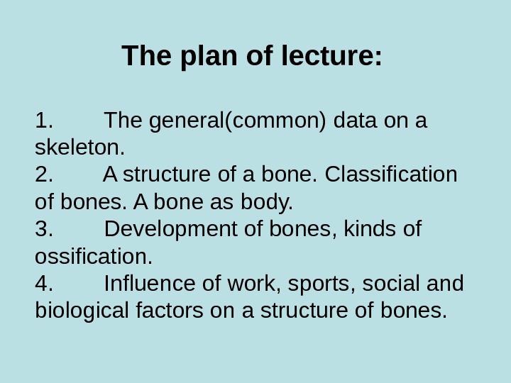
The plan of lecture: 1. The general(common) data on a skeleton. 2. A structure of a bone. Classification of bones. A bone as body. 3. Development of bones, kinds of ossification. 4. Influence of work, sports, social and biological factors on a structure of bones.
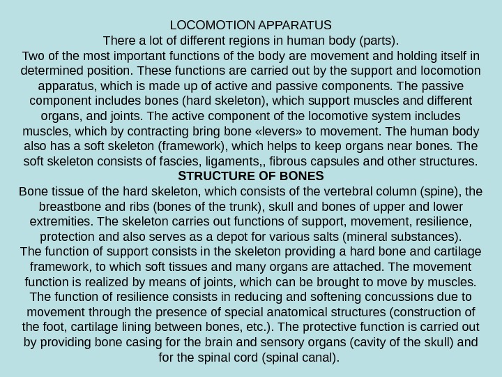
LOCOMOTION APPARATUS There a lot of different regions in human body (parts). Two of the most important functions of the body are movement and holding itself in determined position. These functions are carried out by the support and locomotion apparatus, which is made up of active and passive components. The passive component includes bones (hard skele ton), which support muscles and different organs, and joints. The active component of the locomotive system includes muscles, which by contracting bring bone «levers» to movement. The human body also has a soft skeleton (framework), which helps to keep organs near bones. The soft skeleton consists of fascies, ligaments, , fibrous capsules and other structures. STRUCTURE OF BONES Bone tissue of the hard skeleton, which consists of the vertebral col umn (spine), the breastbone and ribs (bones of the trunk), skull and bones of upper and lower extremities. The skeleton carries out functions of sup port, movement, resilience, protection and also serves as a depot for var ious salts (mineral substances). The function of support consists in the skeleton providing a hard bone and cartilage framework, to which soft tissues and many organs are at tached. The movement function is realized by means of joints, which can be brought to move by muscles. The function of resilience consists in reducing and softening concussions due to movement through the pres ence of special anatomical structures (construction of the foot, cartilage lining between bones, etc. ). The protective function is carried out by pro viding bone casing for the brain and sensory organs (cavity of the skull) and for the spinal cord (spinal canal).
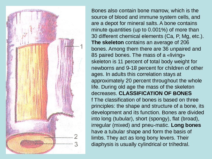
Bones also contain bone marrow, which is the source of blood and immune system cells, and are a depot for mineral salts. A bone contains minute quantities (up to 0. 001%) of more than 30 different chemical elements (Ca, P, Mg, etc. ). The skeleton contains an average of 206 bones. Among them there are 36 unpaired and 85 paired bones. The mass of a «living» skeleton is 11 percent of total body weight for newborns and 9 -18 percent for children of other ages. In adults this correlation stays at approximately 20 percent throughout the whole life. During old age the mass of the skeleton de creases. CLASSIFICATION OF BONES f The classification of bones is based on three principles: the shape and structure of a bone, its development and its function. Bones are divided into long (tubular), short (spongy), flat (broad), irregular (mixed) and pneu-matic. Long bones have a tubular shape and form the basis of limbs. They act as long bony levers. Their diaphysis is usually cylindrical or trihedral.
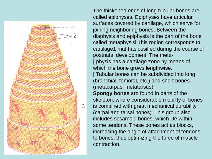
The thickened ends of long tubular bones are called epiphyses. Epiphyses have articular surfaces covered by cartilage, which serve for joining neighboring bones. Between the diaphysis and epiphysis is the part of the bone called metaphysisi This region corresponds to cartilage 1 mat has ossified during the course of postnatal development. The meta- [ physis has a cartilage zone by means of which the bone grows lengthwise. [ Tubular bones can be subdivided into long (branchial, femoral, etc. ) and short bones (metacarpus, metatarsus). Spongy bones are found in parts of the skeleton, where considerable mobility of bones is combined with great mechanical durability (carpal and tarsal bones). This group also includes sesamoid bones, which Ue within some tendons. These bones act as blocks, increasing the angle of attach ment of tendons to bones, thus opti mizing the force of muscle contrac tion.
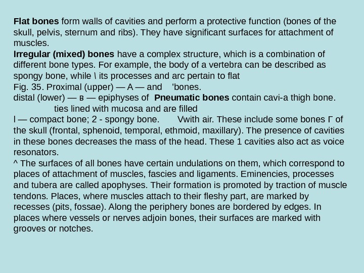
Flat bones form walls of cavi ties and perform a protective func tion (bones of the skull, pelvis, ster num and ribs). They have significant surfaces for attachment of muscles. Irregular (mixed) bones have a complex structure, which is a com bination of different bone types. For example, the body of a vertebra can be described as spongy bone, while \ its processes and arc pertain to flat Fig. 35. Proximal (upper) — A — and ‘bones. distal (lower) — в — epiphyses of Pneumatic bones contain cavi-a thigh bone. ties lined with mucosa and are filled l — compact bone; 2 — spongy bone. Vwith air. These include some bones Г of the skull (frontal, sphenoid, temporal, ethmoid, maxillary). The pres ence of cavities in these bones decreases the mass of the head. These 1 cavities also act as voice resonators. ^ The surfaces of all bones have certain undulations on them, which correspond to places of attachment of muscles, fascies and ligaments. Eminencies, processes and tubera are called apophyses. Their formation is promoted by traction of muscle tendons. Places, where muscles attach to their fleshy part, are marked by recesses (pits, fossae). Along the pe riphery bones are bordered by edges. In places where vessels or nerves adjoin bones, their surfaces are marked with grooves or notches.
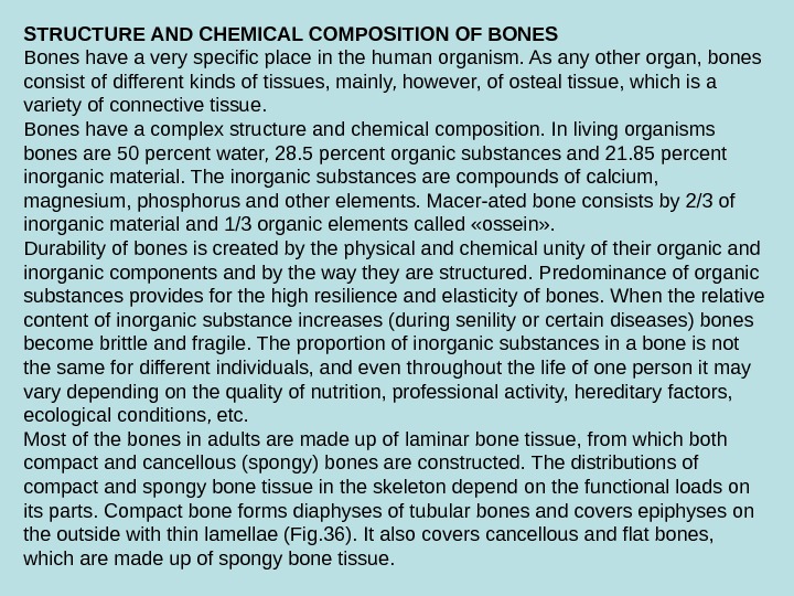
STRUCTURE AND CHEMICAL COMPOSITION OF BONES Bones have a very specific place in the human organism. As any oth er organ, bones consist of different kinds of tissues, mainly, however, of osteal tissue, which is a variety of connective tissue. Bones have a complex structure and chemical composition. In living organisms bones are 50 percent water, 28. 5 percent organic substances and 21. 85 percent inorganic material. The inorganic substances are com pounds of calcium, magnesium, phosphorus and other elements. Macer-ated bone consists by 2/3 of inorganic material and 1/3 organic elements called «ossein» . Durability of bones is created by the physical and chemical unity of their or ganic and inorganic components and by the way they are structured. Predomi nance of organic substances provides for the high resilience and elasticity of bones. When the relative content of in organic substance increases (during se nility or certain diseases) bones become brittle and fragile. The proportion of in organic substances in a bone is not the same for different individuals, and even throughout the life of one person it may vary depending on the quality of nutri tion, professional activity, hereditary fac tors, ecological conditions, etc. Most of the bones in adults are made up of laminar bone tissue, from which both compact and cancellous (spongy) bones are constructed. The distributions of compact and spongy bone tissue in the skeleton depend on the functional loads on its parts. Compact bone forms diaphyses of tubular bones and covers epiphyses on the outside with thin lamellae (Fig. 36). It also covers cancel lous and flat bones, which are made up of spongy bone tissue.
Compact bone tissue is perforated by thin canals, which contain blood vessels and nerves. Some canals run parallel with the sur face of the bone (central or Haversian canals). Others open onto the bone sur face in the form of nutrient foramens, through which arteries and nerves en ter and veins leave the bone. Walls of the central (Haversian) ca nals are formed by concentric lamellae 4 -15 mm thick, which are as if inserted one into another. One canal can be encircled by 4 -20 such lamellae. The central canals with the surrounding lamellae are called osteons, or Haversian systems (Fig. 37). An osteon is a structural unit of compact substance of a bone (Fig. 38). The space be tween osteons is filled by intercalary lamellae. The external layer of com pact bone is formed by outer lamellae. The internal layer, which limits the medullary cavity, is formed by inner lamellae. Spongy (cancellous, trabecular) bone has the appearance of a sponge and is formed by bone trabeculae with spaces between them. The size and positioning of the trabeculae depends on the force exerted on the bone when it is stretched or compressed. Compression and strain curves are hypothetical lines that correspond to the orientation of the trabeculae (Fig. 39). Positioning of the trabeculae at an angle to each other results in more even distribution of pressure (from muscle traction) on the bone. This type of structure determines the durability of bones with a minimum of bone matter used. Plasticity of bone tissue and its active reconstruction is realized through constant formation of new bone cells and extracellular matrix and parallel destruction (resorption) of old bone. Resorption is a result of osteoclast activity. In place of destroyed bone formation of new lamellae and os- teons takes place.
DEVELOPMENT AND GROWTH OF BONES In its development the skeleton of a fetus passes through several stages, namely mesenchymal (connective tissue, membranous), cartilaginous and osseous. There are two ways of development of bone tissue, depending on the bone’s origin. Some bones form directly from embryonic connective tissue, skipping the cartilage stage. Bones of the vault of the skull, for example, are formed in this way (intramembranous ossification). Other bones pass through both membranous and cartilaginous stages. Bones of the trunk, limbs and base of the skull all develop from a cartilage model. In this case bone formation can be endochondral, perichondral and peri-osteal. Endochondral ossification takes place deep within cartilage; peri chondral ossification takes place at the periphery of cartilage (with partic ipation of the perichondrium). Ossification begins in one or several points inside the cartilage model. Around connective fibers and blood vessels that penetrate the cartilage young bone cells (osteoblasts) form trabecu-lae, which begin to increase in size and grow in different directions. Grad ually, osteoblasts develop into mature osteocytes and bone tissue is formed. Depending on the time period when bone tissue appears in the carti lage model it can be called a primary (main) or a secondary (accessory) center of ossification. Primary centers of ossification appear in diaphyses of tubular bones and most spongy and irregular bones during the first half of the prenatal period. Secondary ossification centers form in epiphyses of tubular bones at the end of prenatal development and after birth (until age of 17 -18).
These accessory centers of ossification provide forma tion of processes, protuberances and crests. A layer of cartilage (epiphyseal plate) remains between ossification centers of the diaphysis and epiphysis after their formation and is replaced by bone tissue only by age of 18 -20. Growth of bone in thickness is pro moted by the deep layer of the periosteum. The medullary canal of tubular bones forms inside the diaphysis by means of resorption of endochondrally formed bone. First signs of aging of bones manifest themselves as bone protrusions that appear at the periphery of articular surfaces. These are called margin al osteophytes. In hands they most typically appear on caputs of middle phalanges. As aging progresses they also appear in the base of the middle and distal phalanges. Diaphyses tend to widen due to an increase of peri-osteal osteogenesis. Growth and aging of bones depend on many factors, including the general state of the organism (lifestyle), as well as environ mental influences.