
Theme : Concept about connections of bones.


4_lekciya_concept_about_connections_of_bones..ppt
- Размер: 9.2 Mегабайта
- Количество слайдов: 25
Описание презентации Theme : Concept about connections of bones. по слайдам
Theme : Concept about connections of bones. Development of connections. Kinds of connections of bones.
The plan of lecture: 1. Introduction. 2. Development connection of bones. 3. Continuous connections of bones. 4. Прерывные connections of bones — joints. 5. Симфиз.
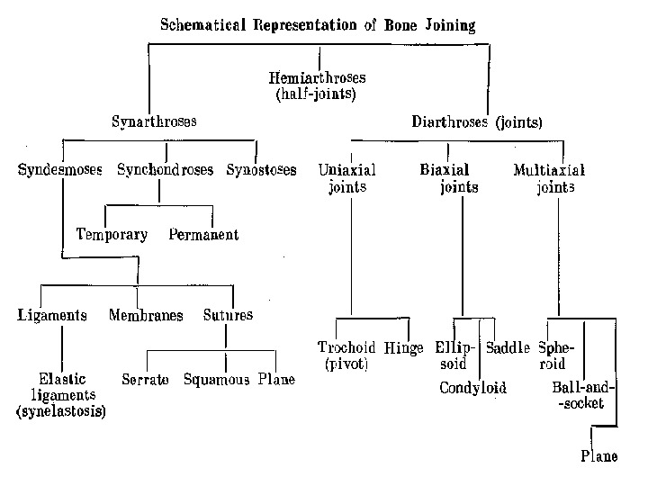
JOINTS Joints are part of the support and locomotion apparatus. They retain bones close to each other and provide their mobility during various move ments. CLASSIFICATION OF JOINTS All joints are subdivided into three large groups: continuous articula tions, symphyses and discontinuous (synovial) joints. Continuous articulations can be formed by different kinds of con nective tissue.
Synostosis and semi-joint. A — syndesmosis: 1 — interosseus membrane of forearm; В — synchondrosis: 2 — intervertebral disk; С — symphysis (secondary cartilaginous joint): 3 — pubic symphisis.
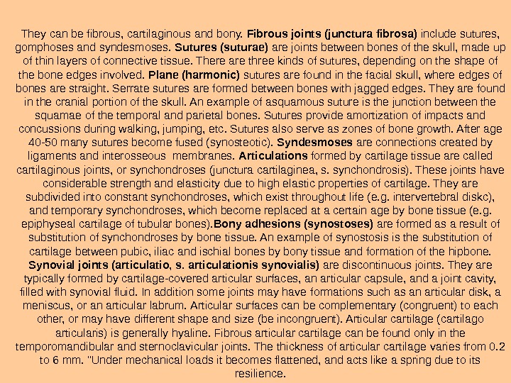
They can be fibrous, cartilaginous and bony. Fibrous joints (junctura fibrosa) include sutures, gomphoses and syndesmoses. Sutures (suturae) are joints between bones of the skull, made up of thin layers of con nective tissue. There are three kinds of sutures, depending on the shape of the bone edges involved. Plane (harmonic) sutures are found in the facial skull, where edges of bones are straight. Serrate sutures are formed between bones with jagged edges. They are found in the crani al portion of the skull. An example of asquamous suture is the junc tion between the squamae of the temporal and parietal bones. Sutures pro vide amortization of impacts and concussions during walking, jumping, etc. Sutures also serve as zones of bone growth. After age 40 -50 many sutures become fused (synosteotic). Syndesmoses are connections created by ligaments and interosseous membranes. Articulations formed by cartilage tissue are called cartilaginous joints, or synchondroses (junctura cartilaginea, s. synchondrosis). These joints have considerable strength and elasticity due to high elastic properties of cartilage. They are subdivided into constant synchondroses, which exist throughout life (e. g. intervertebral diskc), and temporary synchondroses, which become replaced at a certain age by bone tissue (e. g. epiphyseal cartilage of tubular bones). Bony adhesions (synostoses) are formed as a result of substitution of synchondroses by bone tissue. An example of synostosis is the substi tution of cartilage between pubic, iliac and ischial bones by bony tissue and formation of the hipbone. Synovial joints (articulatio, s. articulationis synovialis) are discon tinuous joints. They are typically formed by cartilage-covered articular surfaces, an articular capsule, and a joint cavity, filled with syn ovial fluid. In addition some joints may have formations such as an articular disk, a meniscus, or an articular labrum. Articular surfaces can be complementary (congruent) to each other, or may have different shape and size (be incongruent). Articular cartilage (cartilago articularis) is generally hyaline. Fibrous articular cartilage can be found only in the temporomandibular and sternoclavicular joints. The thickness of articular cartilage varies from 0. 2 to 6 mm. «Under mechanical loads it becomes flattened, and acts like a spring due to its resilience.
Transverse section of articular cartilage. 1 — superficial layer; 2 — cartilaginous basic substance; 3 — profound layer (groups of chondrocytes); 4 — cartilage, brandied with Ca; 5 — bone. Structure of joint. 1 — periosteum; 2 — bone; 3 —joint capsule; 4 — epiphysial cartilage; 5 — articular cavity.
The articular capsule (capsula articularis) is attached to the edges of the articular cartilage or at some distance from it. It ac cretes firmly with the periosteum, forming a closed articular cavity, the pressure inside which is maintained below atmospheric. The capsule ismade up of two layers: a fibrous membrane on the outside and a synovial membrane inside. The fibrous membrane (membrana fibrosa) is thick and very durable. It is formed by fibrous connective tissue. In some places the fibrous membrane is thickened by ligaments, which strengthen the capsule. These ligaments are called capsularif they are situated within the capsule wall, and extracapsular when they are situated outside the capsule. Some joint have intracapsular ligaments inside the joint cavity, which are covered by the synovial membrane (e. g. the cruciate ligaments of the knee joint). The thin synovial membrane (membrana synovialis) lines the fibrous membrane from the inside, forming microprotrusions called synovial villi, which greatly increase its sur face area. Often the synovial membrane forms folds (e. g. in the knee joint), in the base of which there accumulations of fatty tissue.
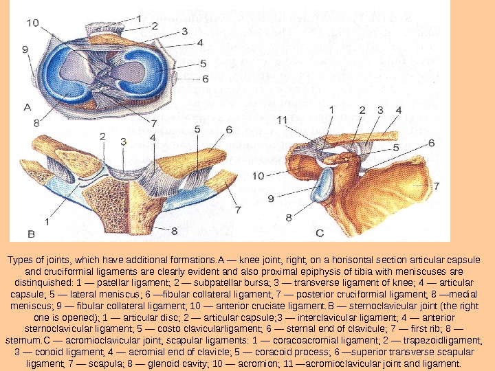
Types of joints, which have additional formations. A — knee joint, right; on a horisontal section articular capsule and cruciformial ligaments are clearly evident and also proximal epiphysis of tibia with meniscuses are distinquished: 1 — patellar ligament; 2 — subpatellar bursa; 3 — transverse ligament of knee; 4 — articular capsule; 5 — lateral meniscus; 6 —fibular collateral ligament; 7 — posterior cruciformial ligament; 8 —medial meniscus; 9 — fibular collateral ligament; 10 — anterior cruciate ligament. В — sternoclavicular joint (the right one is opened); 1 — articular disc; 2 — articular capsule; 3 — interclavicular ligament; 4 — anterior sternoclavicular ligament; 5 — costo clavicularligament; 6 — sternal end of clavicule; 7 — first rib; 8 — sternum. С — acromioclavicular joint; scapular ligaments: 1 — coracoacromial ligament; 2 — trapezoidligament; 3 — conoid ligament; 4 — acromial end of clavicle; 5 — coracoid process; 6 —superior transverse scapular ligament; 7 — scapula; 8 — glenoid cavity; 10 — acromion; 11 —acromioclavicular joint and ligament.
The articular cavity (cavum articulare) is a closed fis sure-like space confined by the articular surfaces and capsule. It is filled with synovial fluid, a mucoid lubricant, which moistens the articular surfaces, allowing them to glide freely against each other. Synovial fluid also provides nutrients to the articular cartilage. Articular discs and menisci (disci et menisci articulares) are intraarticular cartilaginous plates of different shapes, which reduce or elim- s inate incongruity between articular surfaces. CLASSIFICATION OF JOINTS (CONTINUATION) Joints can be classified based on their anatomy or on their biome-chanic properties. According to the anatomic classification, joints are sub divided into simple and compound (composite), depending on the num ber of bones involved, and into complex and combination joints. Sim ple joints are formed by two articular surfaces (e. g. the shoulder joint, the hip joint and many other joints). Compound joints (e. g. the radiocarpal joint, etc. ) are formed by three or more articular surfaces. ; Complex joints have an articular disc or meniscus (the sternoclavicular, temporomandibular and knee joints). Combination joints are groups of articulations, which are isolated anatomically, but function simultaneously. An example of this is the temporomandibular joints. According to the biomechanic classification, joints are subdivided | into uniaxial, biaxial and multiaxial, depending on the number of rotation axes they have. Uniaxial joints have one rotation axis, around which they can perform flexion and extension or abduction and adduction movements, or rotation to the outside (supination) and inside (pronation). Uniaxial articulations include hinge (elbow joint) and pivot joints (proximal and distal radioulnar articulations) (Fig. 69). Biaxial joints have two axes of rotations, for example, flexion-extension and abduction-ad duction. Such joints are the radiocarpal articulation (el l i p s о i d), the car-pometacarpal joint of the thumb and the atlanto-occipital joint ( с о n d у l a r). Multiaxial (triaxial) joints (shoulder and hip joints) are formed by spherical articular surfaces. They can perform all types of movement: flexion-extension, abduction-adduction and supination-pronation (rotation).
AGE CHARACTERISTICS OF JOINTS In embryogenesis all articulations are at first formed as continuous joints. Later, as bones are gradually drawn closer together, the mesen-chyme layer between them becomes thinner and is replaced by fibrous or cartilage tissue. Synovial joints start forming during weeks 6 -11 of em bryogenesis. A slit appears in the mesenchyme layer between bones and the surrounding mesenchyme forms the articular capsule and ligaments. The deep layer of the capsule transforms into the synovial membrane. In complex articulations such as the knee and temporomandibular joints two articular slits are formed, and the mesenchyme between them transforms into an articular disk or meniscus. The cartilaginous glenoid labrum is formed from articular cartilage, when its central part resolves and becomes thin, while the peripheral part accretes to the borders of the articular sur face. Symphyses are formed by transformation of mesenchyme between bones into cartilage, and generation of a narrow slit in its thickness.
General myology. A muscle as body. Development of muscles in ontogenesis. Classification of muscles. The auxiliary apparatus of muscles. Biomechanics of muscles: anatomic and physiological diameters of muscles. Elevating force. The external and internal forces working on muscles and joints.
THE MUSCULAR SYSTEM Skeletal muscles are attached to bones and, by contracting, cause them to move (Fig. 84). Muscles participate in formation of body cavities and have influence upon organs of sight, hearing and equilibrium. They help to retain the body in balance, provide support and movement, ac complish respiration and swallowing and perform facial gestures. The human body contains about 400 muscles, which contract voluntarily, when a person wills them to. STRUCTURE OF THE SKELETAL MUSCLES The principle elements of the skeletal muscles are striated muscle fibers. These fibers are surrounded by loose connective tissue called en -domysium. Bundles, or fascicles, of fibers are separated from one an other by connective tissue layers called perimysium, and the whole mus cle is surrounded by the epimy sium (outer perimysium). Loose connective tissue found in muscles performs functions of support and de marcation between functional elements. It also holds the blood vessels, which nourish muscle fibers, and nerves. Muscle fibers form the fleshy part of the muscle called the v e n t e r, which continues into the muscle tendon. The muscle fascicles or the tendon attach to bones.
CLASSIFICATION OF MUSCLES There are several ways of classification of skeletal muscles. Muscles can be classified according to topography, shape, orientation of fibers, function and position relative to joints. They are divided into superficial and deep, lateral and medial, and internal and external. Muscles can be of many possible shapes (Fig. 85). Walls of the abdo men are formed by broad strap muscles. Fusiform muscles are typical for i the extremities. They attach to bones and move them like levers. Fascicles \ in a fusiform muscle are oriented parallel to its longitudinal axis. Muscles jin which fiber fascicles are situated only on one side of the tendon are /called unipennate. If they are situated on two sides, the muscle is called j bipennate. And if fascicles extend from several sides of the tendon, the 1 ni. Uj. Scle is called multipennate. Some muscles have two or more heads of origin whiclvcbnnect with each other into a common venter that continues into one insertion tendon. These are named according to the number of their heads, for example, biceps, triceps, etc.
Forms of muscles. A — fusiform muscle; В — unipennate muscle; С — bipennate muscle; D — two-headed muscle; E — strap (band-shaped) muscle; F — two-bellied muscle; G — sphincter.
Many muscles of the body are named according to their shape. Thus, / there is a rhomboid muscle, the orbicular muscles, etc. Some muscles / were named based on the orientation of their fibers (transverse muscle of \ the abdomen) or according to their function (levator scapulae muscle, pr-\ onator teres, pronator quadratus, etc). Finally, muscles are divided according to function into synergists and I antagonists. Synergists are muscles, which cause movement in the same Idirection, while antagonists cause movement in opposite directions. THE AUXILIARY APPARATUS OF MUSCLES The work of muscles is directed by specialized anatomic formations that compose the auxiliary apparatus of muscles. These include fasciae, fibrous canals, tendon sheaths, synovial bursae and trochleae of muscles. Fasciae are connective tissue encasements of muscles. They separate muscles from each other, support them during contraction, and for some muscles they serve as a point of origin. In pathological processes fasciae can restrict the spreading of pus or blood (during hemorrhage). Fasciae are divided into superficial and deep. Superficial fasciae are situated beneath. i the skin separating muscles from subcutaneous fat. Deep fasciae are situated between adjacent muscle layers. Between retinacula of muscles and underlying bones there are osteofibrous or fibrous canals, divided into sectors with connective tissue I partitions. These canals contain synovial tendon sheaths Tendons of some muscles contain sesamoid bones in the region of a joint. These serve to increase the angle between the tendon and the bone. The largest sesamoid bone found in the body is the patella. Sometimes there is a synovial bursa situated between the bone and the muscle, tendon or skin, which decreases friction and eases the sliding of the muscle.
Synovial sheath of tendon. A — transverse seeflon; В — longitudinal section. 1 — fibrous sheath; 2 — synovial sheath; 3 — tendon; 4 — synovial cavity; 5 — mesotendon.
WORK OF MUSCLES The work of a muscle depends on its size, shape and structure. A single muscle fiber can develop a force of approximately 0. 1 -0. 2 g. Anabsolute force produced by a muscle is on the average 10 kg per 1 mm 2, and varies for different muscles between 6. 24 and 16. 8 kg/mm 2. The strength of a muscle is directly proportional to the number of muscle fi bers. The total area of all muscle fibers on a transverse section, cut perpen dicularly to the long axis of the muscle, is called the anatomical cross section. The size of the physiological cross-section depends on the structure of the muscle. The more fibers there are per unit on the transverse cut, the greater is the physiological cross section. Unipennate and bipennate muscles, which have a large number of short fibers obliquely attached to the tendon, have a greater physiological cross section than strap or fusiform muscles of the same size. Anatomic (continuous line) and physiologic (dot line) diameters of muscles of various shapes. A — flat muscle; В — fusiform muscle; С — unipennate muscle.
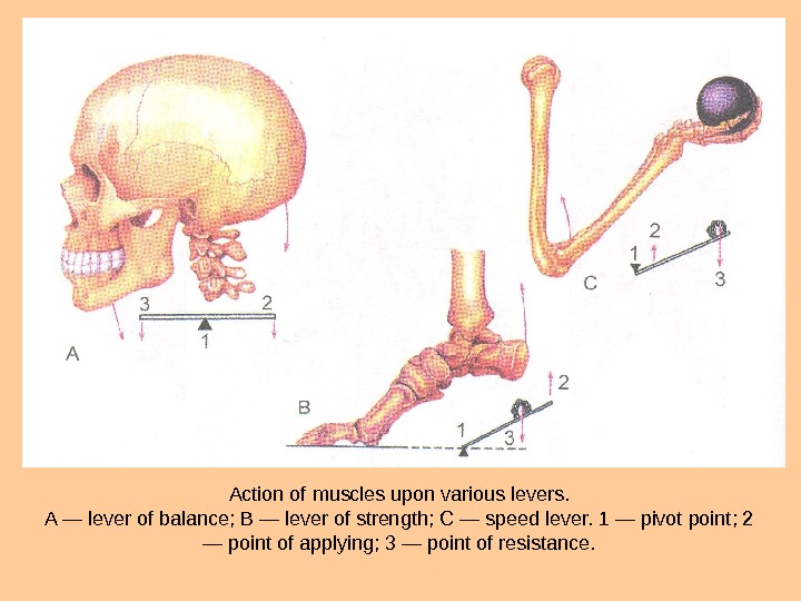
Action of muscles upon various levers. A — lever of balance; В — lever of strength; С — speed lever. 1 — pivot point; 2 — point of applying; 3 — point of resistance.
Overcoming work is carried out when muscle contraction changes the positions of the body or its part, with or without an extra load, moving i it against gravity. Work is called yielding when muscle force succumbs to the force of weight of the body part (extremity) or a load. The muscle is carrying out work, but its length increases instead of decreasing. An example of this is when the load on a muscle is heavier than it is capable of lifting or holding in place. In this case the load is lowered, even though the muscle is exert ing force. Fixating work is carried out when the force of contraction is used to hold a load in a certain position, without displacement. For example, when a person is standing or sitting still or holding an object. The force of mus cle contraction is equal to the gravity force of the body or the object. In this case the length of the muscle is not changing (isometric contraction). Overcoming and yielding types of work are also called dynamic work, since the force of muscle contraction is causing the body or its parts to move. Fixating work is also called static, because there is no movement-taking place. A first-class lever is a two-armed system, and is also called a «bal ance lever» . A second-class lever is a one-armed lever. There are two types of sec ond-class levers, depending on where the force application and resisting weight are situated relative to each other. The first type is considered to be a power lever. In such a system the lever arm of force application is longer than that of the weight resistance. An example of this is the foot, in which the heads of metatarsal bones serve as the pivot point, the force is applied at the calcaneus (by the triceps of the leg), and the weight of the body acts upon the talocrural joint.
This lever system has an advantage in power, but a disadvantage in speed. The other type of one-armed system (otherwise called a third-class lever) is considered a speed lever. The lever arm force in this system is shorter than the lever arm of the counteracting weight. In case of the elbow joint, much greater force is required from the flexor muscles for acting upon the weight, which is situated at a considerable distance fromthe fulcrum. This lever has an advantage in speed and amplitude of move ment of a longer lever, but there is a disadvantage in power. DEVELOPMENT OF MUSCLES The origin of skeletal muscle in embryogenesis is the middle embryon ic layer called mesoderm, which contains somites. The dorsomedial section of a somite, called the myotome. Initially contains a cavity — the myocele. As the myotome grows and turns into a syncytium mass, its cavity gradual ly disappears. Then, the cell mass differentiates into striated muscle fibers with a metameric arrangement. The myotome divides into cylindrical sec tions of muscle fibers. The dorsal sections of myotomes form the deep (prop er) muscles of the back. The ventral sections form the deep muscles of the chest and anterior and lateral abdominal walls.
Muscles of the head and some muscles of the neck are formed from the ventral non-metameric part of the mesoderm of the cranial end of the em bryo. This group of muscles (visceral muscles) includes the masticatory muscles, some muscles of the neck, which form through transformation of the first visceral arch; the muscles of facial expression (including the platys-ma) and some other muscles that form from the second visceral arch. The sternocleidomastoid and trapezius muscles develop from the brachial arch musculature. This group also includes some muscles of the perineum, for example the levator ani muscle. Some muscles develop from myotomes of cranial somites. These include the muscles that move the eyeball. Some muscles develop from mesenchymal rudiments of the limbs, and their proximal ends later move onto the body. This group of muscles includes the pectoralis major, pectoralis minor, latissimus dorsi and psoas major muscles. There is also a group of muscles, which move from the body onto a limb. These include the trapezius, rhomboid, sternocleido mastoid, serratus anterior, omohyoid and levator scapuli muscles. The muscles of this group develop from the ventral sections of myotomes and from the brachial musculature. Their distal ends move from the body onto the bones of the skull or extremities. There also muscles, which devel op from mesenchyme of the limbs and remain on the extremities.