

Жанов Дамир.pptx
- Количество слайдов: 10
 process equipment for medical textile industry ZHANOV DAMIR KAZAN NATIONAL RESEARCH TECHNOLOGICAL UNIVERSITY DEPARTMENT OF PROCESS EQUIPMENT FOR MEDICAL TEXTILE INDUSTRY
 Brief History of X-Ray Discovered in 1895 by Wilhelm Konrad Roentgen Electromagnetic wave Travels 186, 000 miles/sec Short wavelength Penetrates solid objects Reacts with photographic film
 Fluoroscopy It is used for viewing organs or passage of substances through organs
 is a medical imaging modality where tomographic images or") Computed tomography (CT scanning) is a medical imaging modality where tomographic images or slices of specific areas of the body are obtained from a large series of two-dimensional X-ray images taken in different directions. These cross-sectional images can be combined into a three-dimensional image of the inside of the body and used for diagnostic and therapeutic purposes in various medical disciplines.
Computed tomography (CT scanning) is a medical imaging modality where tomographic images or slices of specific areas of the body are obtained from a large series of two-dimensional X-ray images taken in different directions. These cross-sectional images can be combined into a three-dimensional image of the inside of the body and used for diagnostic and therapeutic purposes in various medical disciplines.
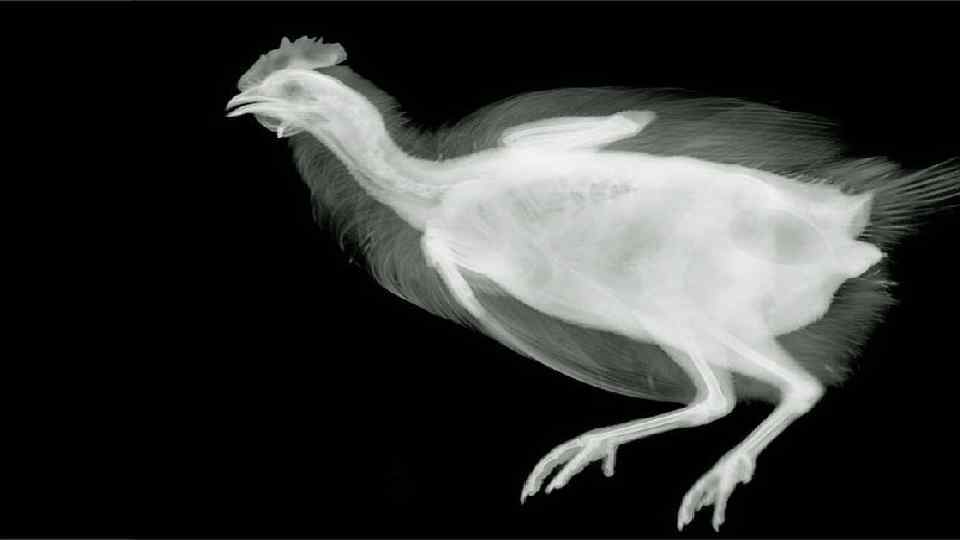
 Radiograph Bones contain much calcium , which due to its relatively high atomic number absorbs x-rays efficiently. This reduces the amount of X-rays reaching the detector in the shadow of the bones, making them clearly visible on the radiograph.
") Adverse effects Diagnostic X-rays (primarily from CT scans due to the large dose used) increase the risk of developmental problems and cancer in those exposed. X rays are classified as carcinogenic ones.
Adverse effects Diagnostic X-rays (primarily from CT scans due to the large dose used) increase the risk of developmental problems and cancer in those exposed. X rays are classified as carcinogenic ones.
 Radiation Safety and Dose Reducing patient exposure -Advances in technology -Assessment of benefit-to-risk ratio -Prevent serious damage from radiation by limiting radiation dose levels -Individual dose limits set
 Words can be like X-rays if you use them properly--they'll go through anything. You read and you're pierced. ” ~ Aldous Huxley
 Thank you for attention