
Презентация суставы позвоночник


- Размер: 1.6 Mегабайта
- Количество слайдов: 79
Описание презентации Презентация суставы позвоночник по слайдам
Болезни суставов и позвоночника
Классификация болезней суставов и позвоночника ► Воспалительные Ревматоидный артрит Анкилозирующий спондилит Реактивный артрит (болезнь Рейтера) Псориатический артрит ► Дегенеративные Остеоартроз остеохондроз ► Метаболические Подагра Пирофосфатная артропатия
Rheumatoid Arthritis: Background ► Ревматоидный артрит (( РАРА ) ) – хроническое системное аутоиммунное заболевание синовиальных суставов неизвестной этиологии, характеризующееся прогрессирующим симметричным эрозивным полиартритом. . ► Constitutional symptoms, including fatigue, malaise, and morning stiffness, are common. ► Extra-articular involvement of organs such as the skin, heart, lungs, and eyes can be significant. ► RA causes joint destruction and thus often leads to considerable morbidity and mortality. ► The treatment of RA is rapidly advancing with the recent addition of new and innovative therapies.
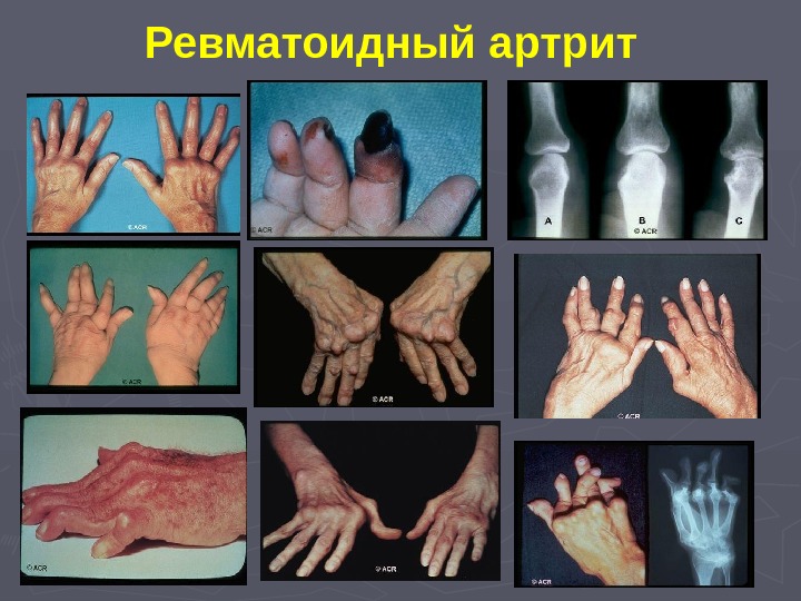
Ревматоидный артрит
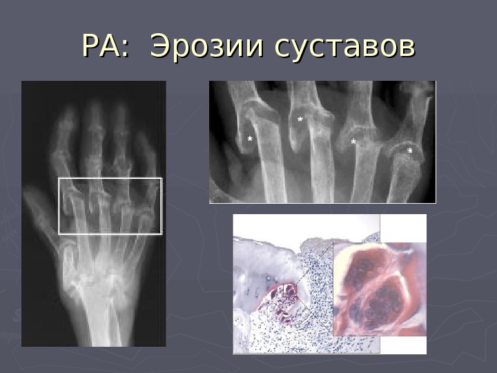
РА: Эрозии суставов
► Частота : : примерно 3 3 случая на 10, 000 населения (распространеность примерно 1%1% )) ► Ассоциируется со значительной смертностью/заболеваемостью и нетрудоспособностью. Через 5 5 лет ~~ 33% больных нетрудоспособны ; ; через 10 10 лет – приблизительно 50 % ► Спонтанные ремисии редки (( приблизительно 5-10%). ► Продолжительность жизни сокращается на 5-10 летлет ► Пол : : женщины болеют в 2-3 раза чаще мужчин ► Вораст : : чаще в возрасте 35-50 летлет. Ревматоидный артрит
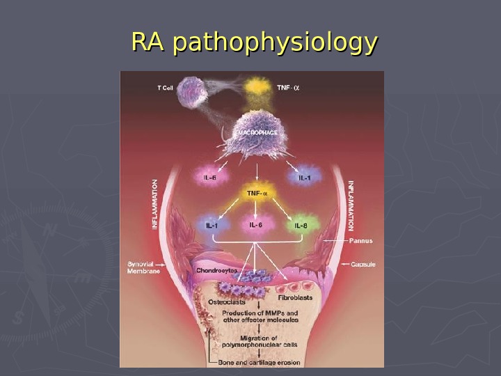
► RA has a significant genetic component, and the so-called shared epitope of the HLA-DR 4/DR 1 cluster is present in up to 90% of patients with RA, although it is also present in more than 40% of controls. ► Synovial cell hyperplasia and endothelial cell activation are early events in the pathologic process that progresses to uncontrolled inflammation and consequent cartilage and bone destruction. Genetic factors and immune system abnormalities contribute to disease propagation. ► Major cellular roles are played by CD 4 T cells, mononuclear phagocytes, fibroblasts, osteoclasts, and neutrophils, while B lymphocytes produce autoantibodies (ie, rheumatoid factors [RFs]). Abnormal production of numerous cytokines, chemokines, and other inflammatory mediators (eg, tumor necrosis factor alpha [TNF-alpha, interleukin (IL)– 1, IL-6, transforming growth factor beta, IL-8, fibroblast growth factor, platelet-derived growth factor) have been demonstrated in patients with RA. ► Ultimately, inflammation and exuberant proliferation of synovium (ie, pannus) leads to destruction of various tissues such as cartilage, bone, tendons, ligaments, and blood vessels. Although the articular structures are the primary sites, other tissues are also affected. Rheumatoid Arthritis: Pathophysiology:
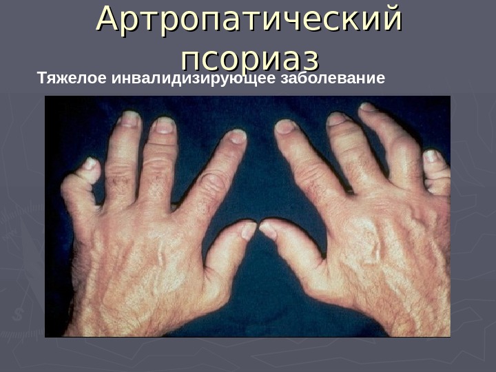
Артропатический псориаз Тяжелое инвалидизирующее заболевание
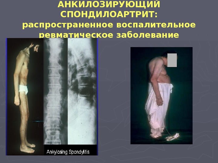
АНКИЛОЗИРУЮЩИЙ СПОНДИЛОАРТРИТ: распространенное воспалительное ревматическое заболевание
RA pathophysiology
Infliximab (( Remicade )) E. HSIA, K. Ruley, M. Rahman. APLAR Journal of Rheumatology, 2006; 8: 107-118 Mouse Ig. G 1 (25%) Human Ig. G 1 ( 7 5%) TNF Infliximab TNF receptor► Chimeric Ig. G 1 monoclonal antibody ► Connect to TNF with high specificity, affinity and avidity ► Neutralize soluble and transmembranic TN
TNF α Effects
► The American College of Rheumatology (1987) developed the following criteria for the classification of RA. . 1. 1. Morning stiffness : This occurs in and around the joints and lasts at least 1 hour before maximal improvement. 2. 2. Arthritis of 3 or more joint areas: At least 3 joint areas simultaneously have soft tissue swelling or fluid (not bony overgrowth) observed by a physician. The 14 possible areas are right or left proximal interphalangeal (PIP), metacarpophalangeal (MCP), wrist, elbow, knee, ankle, and metatarsophalangeal (MTP) joints. 3. 3. Arthritis of hand joints of at least one area swollen in a wrist, MCP, or PIP joint 4. 4. Symmetric arthritis with simultaneous involvement of the same joint areas on both sides of the body: Bilateral involvement of PIPs, MCPs, and MTPs is acceptable without absolute symmetry. 5. 5. Rheumatoid nodules: Subcutaneous nodules are present over bony prominences or extensor surfaces or in juxta-articular regions. 6. 6. Serum RF: Abnormal amounts of serum RF are demonstrated by any method for which the result has been positive in fewer than 5% of healthy control subjects. 7. 7. Radiographic changes typical of RA on posteroanterior hand and wrist radiographs, which must include erosions or unequivocal bony decalcification localized in or most marked adjacent to the involved joints: Osteoarthritic changes alone do not qualify. ► A patient can be classified as having RA if 4 of 7 criteria are present. Rheumatoid Arthritis: diagnostic criteria (ACR, 1987)
► Patients often present with constitutional complaints including malaise, fever, fatigue, weight loss, and myalgias. They may report difficulty performing activities of daily living (eg, dressing, standing, walking, personal hygiene, using their hands). ► Effect of RA on organs and organ systems Skin: Subcutaneous nodules ( rheumatoid nodules ). Vasculitic lesions of the skin may manifest as palpable purpura or skin ulceration. . Cardiac: Myocardial infarction, myocardial dysfunction, and asymptomatic pericardial effusions are common; symptomatic pericarditis and constrictive pericarditis are rare. Myocarditis, coronary vasculitis, valvular disease, and conduction defects are occasionally observed. Pulmonary: pleural effusions, interstitial fibrosis, nodules (Caplan syndrome), and bronchiolitis obliterans-organizing pneumonia. GI: The liver is often affected in patients with Felty syndrome (ie, RA, splenomegaly, and neutropenia). Vascular: Vasculitic lesions can occur in any organ but are most commonly found in the skin. Lesions may present as palpable purpura, skin ulcers, or digital infarcts. . Hematologic: Most active patients have an anemia of chronic disease. Several hematologic parameters parallel disease activity, including normochromic-normocytic anemia, thrombocytosis. . Neurologic: Entrapment of nerves is common, such as with the median nerve in carpal tunnel syndrome. Vasculitic lesions, mononeuritis multiplex, and cervical myelopathy may cause serious neurological consequences. Ocular: Keratoconjunctivitis sicca is common in RA and is often the initial manifestation of secondary Sjögren syndrome. The eye may also have episcleritis, uveitis, and nodular scleritis that may lead to scleromalacia. Rheumatoid Arthritis: Clinical features
The cause(s) of RA is unknown. Genetic, environmental, hormonal, immunologic, and infectious factors may play significant roles. Socioeconomic, psychological, and lifestyle factors may influence disease outcome. ► Genetic Approximately 60% of US patients with RA carry the so-called shared epitope of the HLA-DR 4 cluster, which constitutes one of the peptide binding sites of certain HLA-DR molecules associated with RA (eg, HLA-DR beta *0401, 0404, or 0405); in addition, HLA-DR 1 (HLA-DR beta *0101) also carries this shared epitope and confers risk, particularly in certain southern European areas. ► Immunologic All of the major immunologic elements play fundamental roles in the initiation, propagation, and maintenance of the autoimmune process of RA. The exact orchestration of the cellular and cytokine events that lead to pathologic consequences, such as synovial proliferation and subsequent joint destruction , is complex. It involves T and B lymphocytes, antigen-presenting cells (eg, B cells, macrophages, dendritic cells), and numerous cytokines. Aberrant production and regulation of both pro- and anti-inflammatory cytokines and cytokine pathways are found in RA. T cells are assumed to play a pivotal role in the initiation of RA, and the key player in this respect is assumed to be the Th 1 CD 4 cells. (T helper 1 cells produce IL-2 and interferon gamma. ) These cells may subsequently activate macrophages and other cell populations, including synovial fibroblasts. The latter 2 populations are the main producers of the proinflammatory cytokines TNF-alpha and IL-1 that appear to be the major driving forces of inflammation. B cells are important in the pathologic process because they may serve as antigen-presenting cells and activated T cells, produce numerous autoantibodies (eg, RF, to citrullinated proteins ), and secrete cytokines. Elimination of populations of B cells with monoclonal antibodies (eg, rituximab) offers another effective therapeutic option. Experimental models suggest that synovial macrophages and fibroblasts may become autonomous and thus lose responsiveness to T-cell activities in the course of the disease. In patients with RA, autoantibodies are not only directed against immunoglobulin G (Ig. G), ie, RFs, but also against a variety of other antigens such as nuclear antigens (RA 33, EBNA), citrullinated proteins (anti-CCP antibodies), collagen, and glucose-6-phosphate isomerase. Rheumatoid Arthritis: Causes
Medical Care: Optimal care of patients with RA requires an integrated approach of pharmacologic and nonpharmacologic therapies. ► Nonpharmacologic Education is important in helping patients to understand their disease and to learn how to cope with its consequences. Physiotherapy and physical therapy are initiated to help improve and sustain range of motion, increase muscle strength, and reduce pain. Occupational therapy is initiated (1) to help patients to use joints and tendons efficiently without stressing these structures, (2) to help patients decrease tension on the joints through the use of specially designed splints, and (3) to help patients to cope with daily life through the use of adaptations to the patients’ environment and the use of different aids. Orthopedic measures include reconstructive and replacement-type surgical measures. Rheumatoid Arthritis: Treatment
2003 2006 Akylosing Spondylitis (2003) Psoriatic Arthritis (2004) Ulcerative Colitis ( 2006) Ankylosing Spondylitis ( 2003) 2004 Crohn’s (2005) Enbrel (Wyeth) Humira (Abbott)Remicade (Schering – Plough) Psoriasis (2006)ASPIRE (2004)EUCrohn’s Maintenance (2003) Juvenile Rheumatoid Arthritis (2005) Pediatric Crohn’s ( 2006) Raptiva (Serono)Rheumatoid Arthritis ( 2003) Psoriasis (2004) 2005 Psoriatic Arthritis (2004) Ankylosing Spondylitis (2005) Psoriatic Arthritis (2006) Psoriasis (2005) Psoriasis (2006)Results of Biologic Treatment
► Pharmacologic The most important measure to successfully treat RA is the use of DMARDs can retard or prevent disease progression and, thus, joint destruction and subsequent loss of function. Until the full action of DMARDs takes effect, anti-inflammatory or analgesic medications may be required as bridging therapy to reduce pain and swelling. DMARDs can be classified into xenobiotic and biologic agents. . Xenobiotic agents ► The xenobiotic DMARDs, ie, methotrexate (MTX), sulfasalazine (SSZ), cyclosporin A, gold salts, D-penicillamine, chloroquine and hydroxychloroquine, etc. ► MTX and SSZ are the most active compounds in terms of frequency of remissions and time to onset of action and provide the best risk-benefit ratios. MTX alone or in combination with other agents has become the standard of care for moderate-to-severe RA. Rheumatoid Arthritis: Treatment
Biologic agents : : ► The recognition of TNF-alpha and IL-1 as central proinflammatory cytokines has led to the development of agents that block these cytokines or their effects. The TNF blockers include etanercept, infliximab, and adalimumab. ► In clinical trials, up to 70% of patients achieve significant responses, but remissions are not usually observed. ► These agents bind TNF and thus prevent its interaction with its receptors; infliximab binds to cells that express membrane TNF, while etanercept binds lymphotoxin (formerly termed TNF-beta) in addition to soluble TNF-alpha. Failure to respond to one TNF blocker does not preclude response to another. As with xenobiotics, the decision to continue or stop biologic agents can often be made within 3 months after initiation of therapy. Rheumatoid Arthritis: Treatment
Glucocorticoids : : ► Glucocorticoids are potent anti-inflammatory drugs and are commonly used in patients with RA to bridge the time until DMARDs are effective. ► Doses of up to 10 mg of prednisone per day are typically used, but some patients may require higher doses. ► Timely dose reductions and cessation are important because of the adverse effects associated with long-term steroid use. Nonsteroidal anti-inflammatory drugs : : ► NSAIDs interfere with prostaglandin synthesis through inhibition of the enzyme cyclooxygenase (COX), thus reducing swelling and pain. However, they do not retard joint destruction and, therefore, when used alone, are not sufficient to treat RA. Similar to glucocorticoids, they can be reduced in dose or discontinued with successful DMARD therapy. Rheumatoid Arthritis: Treatment
Surgical Care: ► Cervical spine involvement usually affects C 1-C 2 and may potentially cause serious neurological consequences. Patients who are to undergo intubation or procedures that may involve manipulation of the neck should have careful evaluation of the cervical spine. ► Patients with RA often need multiple operations over time (eg, synovectomy, tendon corrections, joint replacements). Consultations: ► Orthopedists ► Physical and rehabilitative medicine specialists Rheumatoid Arthritis: Treatment
Osteoarthritis ► Background: Osteoarthritis (OA) is the most common articular disease worldwide, affecting over 20 million individuals in the United States alone. ► Its high prevalence entails significant costs to society. ► Costs can be particularly significant for the elderly, who face potential loss of independence and who may need help with daily living activities. ► As the populations of developed nations age over the next few decades, the need for better understanding of OA and for improved therapeutic alternatives will continue to grow.
Pathophysiology ► Traditionally, OA has been considered a disease of articular cartilage. The current concept holds that OA involves the entire joint organ, including the subchondral bone and synovium. ► OA has always been classified as a noninflammatory arthritis; yet, there is increasing evidence for inflammation occurring with cytokine and metalloproteinase release into the joint. Therefore, the term degenerative joint disease is no longer appropriate when referring to OA. ► The joints predominantly involved are weight bearing and include the knees, hips, cervical and lumbosacral spine, and feet. Other commonly affected joints include the distal interphalangeal (DIP) and proximal interphalangeal (PIP) joints of the hands. ► Cartilage is grossly affected. Focal ulcerations eventually lead to cartilage loss and eburnation. Subchondral bone formation occurs as well, with development of bony osteophytes.
Frequency ► In the US: OA affects over 20 million individuals. Radiologic definition indicates that more than half of the population older than 65 years is affected. ► Internationally: OA is the most common articular disease. Estimates vary among different populations.
Age ► OA can be defined epidemiologically (ie, using radiographic criteria) or clinically (eg, radiographs plus clinical symptoms). Radiographic criteria indicate that OA occurs in 30% of affected individuals aged 45-65 years and in more than 80% by their eighth decade of life, although most are asymptomatic.
The etiopathogenesis of OA ► Stage 1: Proteolytic breakdown of the cartilage matrix occurs. Chondrocyte metabolism is affected, leading to an increased production of enzymes, which includes metalloproteinases (eg, collagenase, stromelysin) that destroy the cartilage matrix. Chondrocytes also produce protease inhibitors, including tissue inhibitors of metalloproteinases (TIMP) 1 and 2 but in amounts insufficient to counteract the proteolytic effect. ► Stage 2: This stage involves the fibrillation and erosion of the cartilage surface, with a subsequent release of proteoglycan and collagen fragments into the synovial fluid. ► Stage 3: The breakdown products of cartilage induce a chronic inflammatory response in the synovium. Synovial macrophage production of cytokines, such as interleukin 1 (IL-1), tumor necrosis factor-alpha, and metalloproteinases, occurs. These can diffuse back into the cartilage and directly destroy tissue or stimulate chondrocytes to produce more metalloproteinases. Other pro-inflammatory molecules (eg, nitric oxide [NO], an inorganic free radical) also may be a factor. Eventually, these events alter the joint architecture, and compensatory bone overgrowth occurs in an attempt to stabilize the joint. As the joint architecture is changed and further mechanical and inflammatory stress occurs on the articular surfaces, the disease progresses unchecked.
Diagnosis ► Diagnosis usually can be made on clinical grounds. The history and physical examination findings are sufficient. Radiographic findings confirm the initial impression, and laboratory values typically are within the normal range. The initial goal is to differentiate OA from other arthritides (eg, rheumatoid arthritis [RA]). ► Secondary OA must be considered in individuals with chondrocalcinosis, joint trauma, metabolic bone disorders, hypermobility syndromes, and neuropathic diseases.
Lab Studies/ Imaging Studies ► Lab Studies: ► No specific laboratory abnormalities are associated with OA. ► The acute-phase reactants and erythrocyte sedimentation rate are not elevated. ► Synovial fluid analysis usually indicates a white cell count less than 2000 per mm 3 with a mononuclear predominance. ► Imaging Studies: ► Radiography ► Conduct imaging studies of the affected joint. ► The presence of osteophytes (ie, spurs at the joint margins) is the most characteristic findings. ► Other findings include asymmetric joint space narrowing, subchondral sclerosis, and subchondral cyst formation. ► Roentgenographic findings are often poor predictors of the degree of symptomatology in a particular patient.
Medical Care ► Medical Care (patient education, temperature modalities, weight loss, exercise, physical therapy, occupational therapy) ► Reduction of joint stress ► Physical therapy
Pharmacologic therapy ► Begin treatment with acetaminophen for mild or moderate pain without apparent inflammation. ► If clinical response to acetaminophen is not satisfactory or if clinical presentation is inflammatory, consider nonsteroidal anti-inflammatory drug (NSAIDs). ► In patients with highly resistant pain, consider the analgesic tramadol. ► Contemplate intraarticular injections of glucocorticoids to improve symptoms. It is recommended that no more than 4 glucocorticoid injections be administered to a single joint per year because of the concerns with long-term damage to cartilage.
► The goals of pharmacotherapy are to reduce morbidity and to prevent complications. Pay careful attention to a particular pharmacologic regimen’s adverse event profile. ► Pain control is essential in the care of the patient with OA. The goals of treatment are pain alleviation and improvement of functional status. No proven disease/structure-modifying intervention presently exists. ► COX-2 inhibitors (Celecoxib) — Although increased cost can be a negative factor, the incidence of costly and potentially fatal GI bleeds is less in nonaspirin users receiving COX-2 inhibitors than with traditional NSAIDs.
Surgical Care ► Closed-needle joint lavage ► Arthroscopy ► Osteotomy ► Arthroplasty
Reactive Arthritis and Reiter Syndrome
Reiter Syndrome ► • Complicates 1-2% of cases of NGU • Most common peripheral inflammatory arthritis in young men • 60-96% of cases associated with HLA-B 27 • 2 types of inciting infections: ► – NGU (Chlamydiatrachomatis) ► – Bacterial gastroenteritis (Salmonella, Shigella, Yersinia, Campylobacter)
Reiter Syndrome • Urethritis: initial manifestation in 80%; other features develop 1-5 weeks later • Arthritis: ► – Begins within 4 weeks of urethritis onset ► – Knees (followed by ankles) are most commonly involved • Conjunctivitis, iritis, uveitis
Reiter Syndrome ► • Skin lesions: ► – Waxy papules on soles, ► palms; epithelialize& ► thicken to produce ► keratoderma ► blenorrhagicum ► – Circinate balanitis: ► occurs in 25-40%
Reiter Syndrome • Initial episode usually lasts 2-6 months • >50% will relapse • Nearly half develop some degree of permanent disability • Treatment: ► – Doxycycline x 7 days ► – NSAIDs ► – Cytotoxicagents in recalcitrant cases
Management of Sex Partners • Chlamydia is isolated from 30-60% of female partners of men with GC or NGU • Partners should be evaluated, tested & treated if they had sexual contact with the patient during the 60 days preceding symptom onset or diagnosis • Treat the most recent sex partner even if >60 days before onset or diagnosis • Instruct to avoid sexual contact until treatment is completed and both partners are asymptomatic
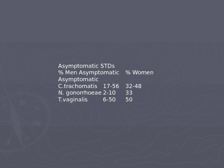
Asymptomatic STDs % Men Asymptomatic % Women Asymptomatic C. trachomatis 17-56 32-48 N. gonorrhoeae 2-10 33 T. vaginalis 6-
NGU Clinical Manifestations • Incubation period 2-35 days (50% develop symptoms within 4 days) • Discharge mucopurulentor completely clear; may be scant • In some men discharge may only be detected in the morning • Onset of symptoms may be less acute than in GCU
Tests for Chlamydia trachomatis ► Test Site Sensitivity (%) Specificity (%) ► Cervix 56-93 100 ► Culture Urethra (male) 40-81 NA ► DFA Cervix 50-81 88-99 ► Cervix 52-100 95-100 ► Urethra 82-100 ► EIA Urine 19-97 99-100 ► Cervix 65-97 94-98 ► NAH Urine 71-95 98-100 ► Cervix 64-97 97-100 ► PCR Urine 81-100 97-100 ► Cervix 94-95 100 ► Urethra 95 100 ► LCR Urine 87-
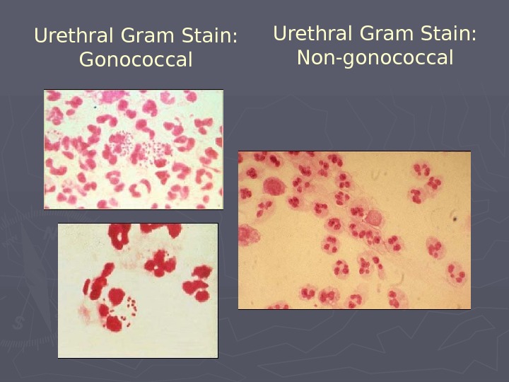
Urethral Gram Stain: Non-gonococcal. Urethral Gram Stain: Gonococcal
► The syndrome has been associated with gastrointestinal infections with Shigella, Salmonella, and Campylobacter species and other microorganisms, along with genitourinary infections (especially with Chlamydia trachomatis). ► In 1967, the term reactive arthritis was first used in cases associated with Yersinia gastroenteritis. ► A strong association with human leukocyte antigen (HLA)–B 27 was found.
► Mortality/Morbidity: Reactive arthritis typically follows a self-limited course, with resolution of symptoms by 3-12 months, even in patients who are acutely incapacitated. However, the condition has a high tendency to recur, particularly with ocular and urogenital inflammation. Individuals who are HLA-B 27 positive have a higher frequency of recurrence. A new infection or other stress factor could cause a reactivation of the disease. ► Sex: Reactive arthritis following food-borne enteric infections affects males and females with the same frequency. Disease associated with venereally acquired infections occurs in a male-to-female ratio of 9: 1. ► Age: Most patients are aged 20-40 years.
► History: Reactive arthritis usually develops 2-4 weeks after a genitourinary or gastrointestinal infection. ► The onset is most often acute, with malaise, fatigue, and fever. An asymmetrical predominately lower extremity oligoarthritis is the major presenting symptom. Low back pain occurs in 50% of patients. Heel pain is common because of enthesopathies at the Achilles or plantar aponeurosis insertion on the calcaneus. The complete Reiter triad of urethritis, conjunctivitis, and arthritis may occur.
► Physical: ► Joints, axial skeleton, entheses. Peripheral joint involvement is typically asymmetric and most frequently affects the weight-bearing joints (ie, knees, ankles, hips), but the shoulders, wrists, and elbows are also affected. ► Ocular findings. Conjunctivitis is part of the classic triad of Reiter syndrome, and it can occur before or at the onset of arthritis.
Medical Care ► Nonsteroidal anti-inflammatory drugs ► Corticosteroids ► Antibiotics (Doxycycline) ► Disease-modifying antirheumatic drugs (Sulfasalazine, methotrexate)
Ankylosing Spondylitis ► Frequency: In the US: The prevalence of ankylosing spondylitis is 0. 1-0. 2% overall but is higher in certain Native American populations and lower in African Americans. ► The male-to-female ratio of ankylosing spondylitis is 3: 1. ► he age of onset of ankylosing spondylitis usually is from the late teens to age 40 years.
► General symptoms ► Symptoms include those related to inflammatory back pain, peripheral enthesitis, arthropathy, and constitutional and organ-specific extra-articular manifestations. ► Because ankylosing spondylitis is a systemic inflammatory disease, systemic features are common. ► Morning stiffness is characteristic, and fatigue is common. ► Fever and weight loss may occur during periods of active disease.
► Extraarticular manifestations : : ► Uveitis (also called iritis or iridocyclitis) ► Cardiovascular involvement (distortion of the aortic ring, resulting in aortic valve insufficiency) ► Pulmonary involvement (Restrictive lung disease, Bilateral apical pulmonary fibrosis)
► HLA-B 27 positivity is present in 92% of white patients with ankylosing spondylitis and is present less commonly in patients of other ethnicities. ► Determining HLA-B 27 status is not a necessary part of the clinical evaluation.
Criteria for Diagnosis of Ankylosing Spondylitis New York Criteria (1984) Rome Criteria (1961) Low back pain with inflammatory characteristics Limitation of lumbar spine motion in sagittal and frontal planes Decreased chest expansion Bilateral sacroiliitis grade 2 or higher Unilateral sacroiliitis grade 3 or higher Low back pain and stiffness for >3 months that is not relieved by rest Pain and stiffness in the thoracic region Limited motion in the lumbar spine Limited chest expansion History of uveitis Definite ankylosing spondylitis when the fourth or fifth criterion mentioned presents with any clinical criteria Diagnosis of ankylosing spondylitis when any clinical criteria present with bilateral sacroiliitis grade 2 or higher
Imaging Studies ► Standard radiographs (sacroiliitis is usually bilateral, symmetric, and gradually progressive over years) ► Magnetic resonance imaging and computed tomography scan (evidence of early sacroiliitis, erosions, and enthesitis that are not apparent on standard radiographs)
Medical Care ► Nonsteroidal anti-inflammatory drugs ► Sulfasalazine ► The TNF-a antagonists have been shown to be effective in the treatment of ankylosing spondylitis, and etanercept, infliximab, and adalimumab are approved therapies. ► Corticosteroids
MRI during Infliximab Treatment 0 неделя 12 неделя 54 неделя
This radiograph of the pelvis of a patient with ankylosing spondylitis shows bilateral sacroiliitis with sclerosis and narrowing of the sacroiliac joints
This radiograph of the lumbar spine of a patient with end-stage ankylosing spondylitis shows bridging syndesmophytes, resulting in bamboo spine
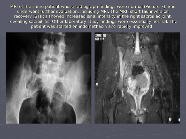
MRI of the same patient whose radiograph findings were normal (Picture 7). She underwent further evaluation, including MRI. The MRI (short tau inversion recovery [STIR]) showed increased sinal intensity in the right sacroiliac joint, revealing sacroiliitis. Other laboratory study findings were essentially normal. The patient was started on indomethacin and rapidly improved.
Gout ► Gout is a common disorder of uric acid metabolism that can lead to recurrent episodes of joint inflammation, tissue deposition of uric acid crystals, and joint destruction if left untreated. Unlike many other rheumatic diseases, gout is very treatable. A definitive diagnosis can be made using joint aspiration and synovial fluid analysis. Early diagnosis and treatment have made a significant impact in this disorder, as evidenced by the declining incidence of chronic tophaceous gout. However, tophaceous gout may occur due to misdiagnosis, poor management, and poor patient compliance.
Gout: Pathophysiology ► Gout is caused by excess stores of uric acid that accumulate in tissues, including the synovium. Lowering serum uric acid levels can prevent attacks of gouty arthritis. However, urate crystals also can be found in synovial fluid in the absence of joint inflammation. Thus, the mere presence of intrasynovial urate crystals is not sufficient to cause flares of gouty arthritis.
Gout: Pathophysiology ► Gout flares can be triggered by fluxes in uric acid levels or by microtrauma, each of which can lead to shedding of uncoated crystals. When bare areas of the urate crystals are exposed, they can bind immunoglobulin G (Ig. G). Such binding stimulates neutrophils to engulf and phagocytose the crystals. Once this occurs, there is a brisk influx of neutrophils associated with elevated levels of interleukin-1, interleukin-6, interleukin-8, tumor necrosis factor-alpha, prostaglandin E 2, leukotriene B 4, kinins, C 5a, and other inflammatory mediators. It is also conceivable is that recoating of uric acid crystals by apo E or apo B could lead to the spontaneous resolution of attacks.
Gout: Frequency ► Gout is present in approximately 1% of the general population. ► Gout has a worldwide distribution; regional differences may reflect environmental, dietary, and genetic influences. ► The prevalence for men is 13. 6 cases per 1000 men, and the prevalence for women is 6. 4 cases per 1000 women.
Gout: History ► Acute monoarticular arthritis is the initial presentation of gout in 90% of patients. In early gout, usually only 1 or 2 joints are involved. Typically, they are the smaller, lower-extremity joints. Podagra (inflammation of the first metatarsophalangeal joint) is the initial joint manifestation involved in 50% of cases. Eventually, it is involved in 90% of cases. Podagra is not synonymous with gout. Podagra can be observed in patients with pseudogout, sarcoidosis, gonococcal arthritis, psoriatic arthritis, and reactive arthritis. The attacks begin abruptly and reach maximum intensity in 8-12 hours. The joints are red, hot, and exquisitely tender; even a bed sheet on the swollen joint is uncomfortable. Untreated, the first attacks resolve spontaneously in less than 2 weeks. Intermittent inflammatory arthritis, in which the joints return to normal between attacks, typically is caused by crystalline disorders and is true of gouty arthritis early in its course. Gout can initially present as a polyarticular arthritis in 10% of patients. Elderly women, particularly women with renal insufficiency and taking a thiazide diuretic, often develop polyarticular arthritis as their first manifestation of gout. These attacks may occur in coexisting Heberden and Bouchard nodes. Such patients also may develop tophi more quickly, occasionally without prior episodes of acute gouty arthritis.
► Tophi are collections of uric acid crystals in the soft tissues. They occur in more than half of untreated patients. While the classic location is along the helix of the ear, they can be found in multiple locations, including the fingers, toes, in the olecranon bursae, and along the olecranon, where they can resemble rheumatoid nodules. The finding of a rheumatoid nodule in a patient with a negative rheumatoid factor should prompt the clinician to consider gout in the differential diagnosis. Finding tophi during the first episode of gout is unusual; they tend to develop after 10 years in untreated patients who develop chronic gouty arthritis.
► Patients with gout have a 1000-fold increased incidence of renal stones and therefore may have a history of renal colic. ► Patients with gout also are prone to develop urate nephropathy, in which uric acid crystals are deposited in the medullary interstitium and pyramids. While patients with gout have a higher incidence of renal impairment, whether hyperuricemia is an independent risk factor for this remains unclear because such patients have a higher incidence of hypertension, diabetes, and other risk factors for renal insufficiency.
► Causes: Gout can develop when excessive stores of uric acid are present. Uric acid is a byproduct of purine metabolism. Lacking uricase, humans remove uric acid primarily by renal excretion. Of patients with primary gout, 90% develop excess stores of uric acid because they are unable to excrete sufficient amounts of uric acid in their urine (underexcretion). The remaining patients produce excessive amounts of uric acid (overproduction). When uric acid levels exceed 6. 8 mg/d. L, with some variability depending on temperature and p. H, uric acid can crystallize.
► Individual attacks of gout often are triggered by acute fluxes in uric acid levels that may lead to the exposure or shedding of crystals that are not coated with apo B or apo E. This can result from alcohol ingestion, overindulgence in certain foods, starvation, trauma, hemorrhage, or medications such as diuretics. It also can result from situations that lower levels of uric acid, including the use of radiocontrast dyes and medications such as allopurinol.
Diagnosis ► Synovial fluid: When a patient presents with acute inflammatory monoarticular arthritis, aspiration of the involved joint is critical to rule out an infectious arthritis and to attempt to confirm a diagnosis of gout or pseudogout by crystal identification. ► Serum uric acid ► Uric acid in 24-hour urine sample
TREATMENT ► There are 3 stages in the management of gout: (1) treating the acute attack, (2) providing prophylaxis to prevent acute flares, and (3) lowering excess stores of uric acid to prevent flares of gouty arthritis and to prevent tissue deposition of uric crystals. ► Management of acute gout: Options for treatment of acute gout include nonsteroidal anti-inflammatory drugs (NSAIDs), corticosteroids, or colchicine (a classic treatment but now rarely indicated). The choice is based primarily on the patient’s other health problems, such as renal insufficiency and peptic ulcer disease. ► Lowering uric acid levels: The goal of therapy is to lower serum uric acid levels to approximately 5-6 mg/d. L.
Gout: Diet ► Diet modifications can only improve the serum uric acid levels by 1 mg/d. L and rarely are able to lower uric acid levels sufficiently to prevent further attacks and accumulation of uric acid. ► Patients should avoid alcohol because it elevates levels of uric acid and therefore can precipitate attacks of gout. Indeed, heavy drinkers are much more likely to have recurrent gout attacks, even with allopurinol therapy.
Gout. Acute podagra due to gout in an elderly man.
Gout. Tophaceous deposits in ear.
Gout. Tophaceous deposits on elbow.
Picture 4. Gout. Chronic tophaceous gout in an untreated patient with end-stage renal disease.
Gout. Fluid obtained from a tophaceous deposit in a patient with gout.
Gout. Strongly negative birefringent, needle-shaped crystals diagnostic of gout obtained from an acutely inflamed joint.
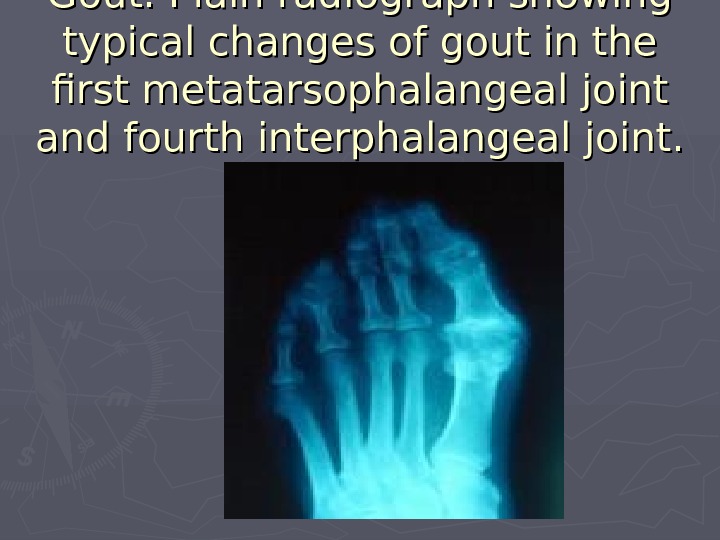
Gout. Plain radiograph showing typical changes of gout in the first metatarsophalangeal joint and fourth interphalangeal joint.
Gout. Plain radiograph showing chronic tophaceous gouty arthritis in the hands.
► Untreated chronic tophaceous gout can develop and lead to severe joint destruction. ► Hyperuricemia is associated with increased all-cause mortality. This is not due to gout, per se, but to diseases associated with gout, such as insulin resistance, type 2 diabetes mellitus, abdominal obesity, hypercholesterolemia, and hypertension.