
Презентация heart anatomy 1 Imangali


- Размер: 3.5 Mегабайта
- Количество слайдов: 18
Описание презентации Презентация heart anatomy 1 Imangali по слайдам
Anatomy of the heart work was made by I mangali Maira
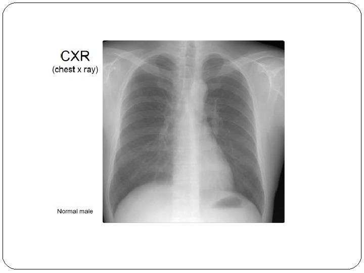
Plan : Size, Location, and Orientation Coverings Heart Wall Chambers Chest x ray
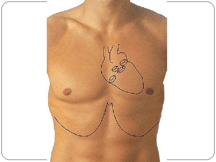
Heart Anatomy Size, Location, and Orientation Enclosed in the mediastinum Base (posteriorsuperior portion) and Apex (inferioranterior portion)
Heart Anatomy Coverings Pericardium protects the heart anchors the heart to surrounding structures such as the diaphragm and the great vessels prevents overfilling of the heart with blood
Heart Anatomy Coverings pericardial cavity contains a film of serous fluid pericarditis: inflammation of the pericardium which may lead to adhesions between the layers or the buildup of fluid in the pericardial cavity (cardiac tamponade)
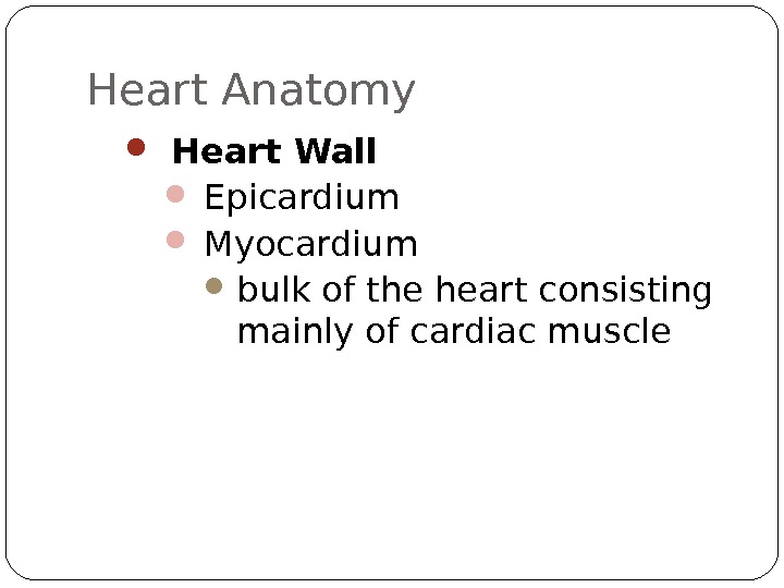
Heart Anatomy Heart Wall Epicardium Myocardium bulk of the heart consisting mainly of cardiac muscle
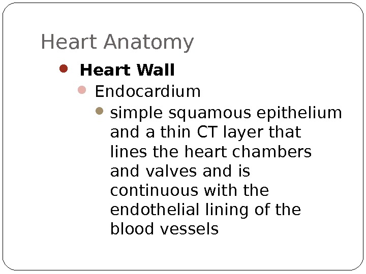
Heart Anatomy Heart Wall Endocardium simple squamous epithelium and a thin CT layer that lines the heart chambers and valves and is continuous with the endothelial lining of the blood vessels
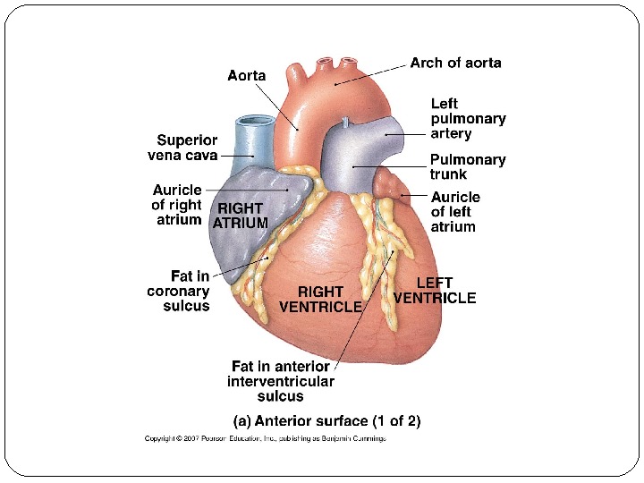
Heart Anatomy Chambers Atria Features small, thin-walled chambers Functions receiving chambers for blood returning to the heart from the circulation push the blood into the adjacent ventricles.
Heart Anatomy Chambers Atria Receive blood from right side Superior and Inferior Vena Cava Coronary Sinus (draining the myocardium) left side Pulmonary Veins
Heart Anatomy Chambers Ventricles Features make up most of the mass of the heart the walls of the left ventricle are 3 X thicker than those of the right
Heart Anatomy Chambers Ventricles Functions discharging chambers of the heart propel blood to Pulmonary Trunk (right ventricle), Aorta (left ventricle)
Normal female
List of bibliographies : http: //www. innerbody. com/image/card 01. html http: //www. texasheartinstitute. org/HIC/Anatomy/anatomy 2. cfm http: //www. cardioconsult. com/Anatomy/