
Презентация Еsophageal disease stricture diverticula


esophageal_disease_stricture_diverticula.ppt
- Размер: 3.2 Mегабайта
- Количество слайдов: 39
Описание презентации Презентация Еsophageal disease stricture diverticula по слайдам
ЕЕ sophageal disease (stricture, diverticula, achalasia) Surgery department № 2№ 2 , , DSM
Clinical picture of esophageal diverticula Food regurgitation might occur when standing, bending or lying down Patients might develop dysphagia (difficulty swallowing) CC hronic coughing CC hest pain HH eartburn WW eight loss
Esophageal diverticula classification By acuirence: congenital acquired By By quantity : : single mm ultiple By localisation crcr yy copharyngeal (or Zencer) midoesophageal epiphrenic
Clinical picture of esophageal diverticula As food collects in the pockets, it promotes bacteria in the esophagus, which also leads to halitosis (bad breath). A patient’s voice also might change.
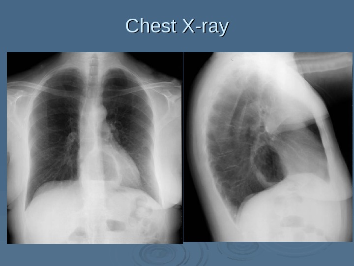
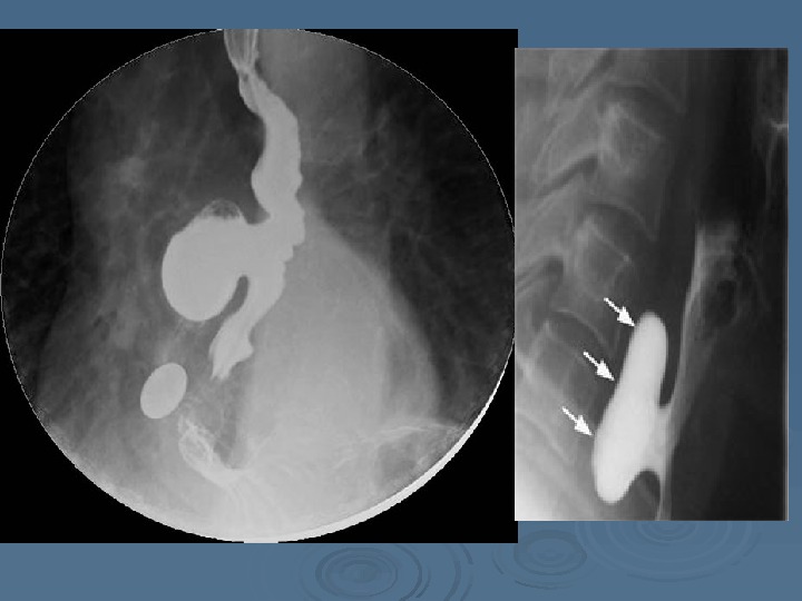
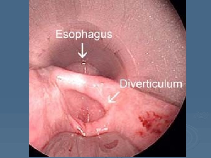
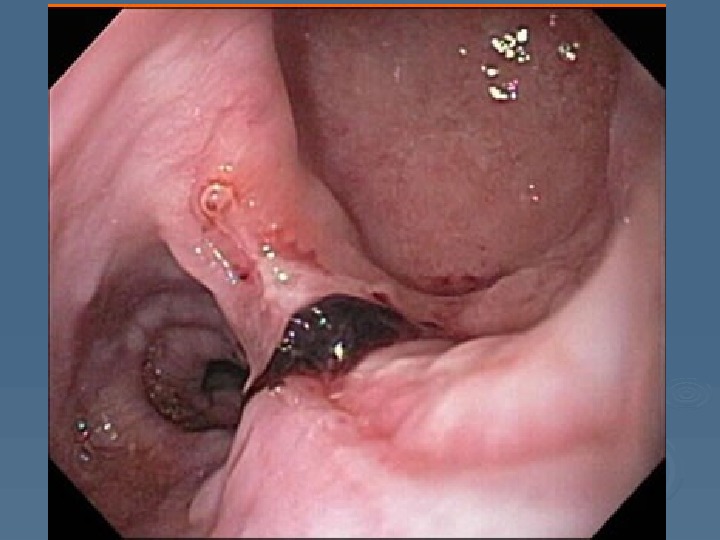
Diagnostic of esophageal diverticula Esophagoscopy Chest X-ray Contrast esophagography
Chest X-ray
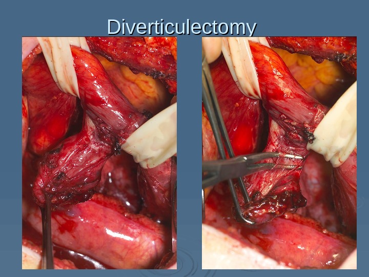
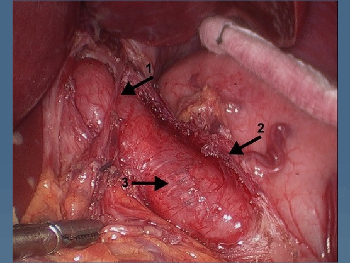
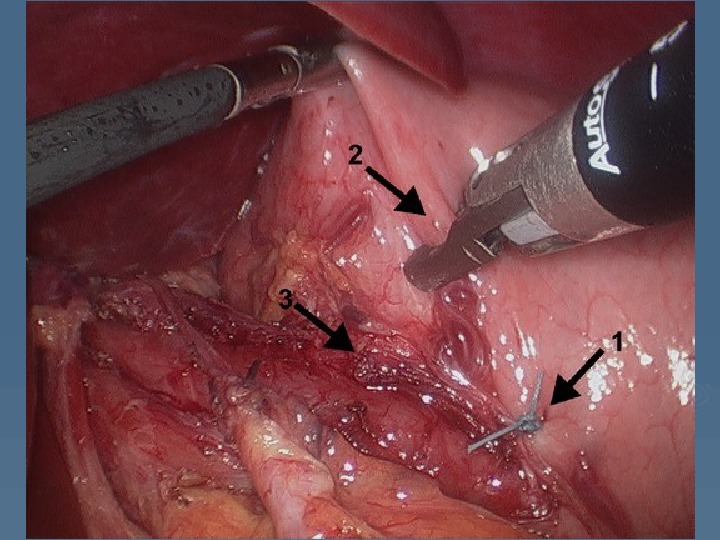
Diverticulectomy
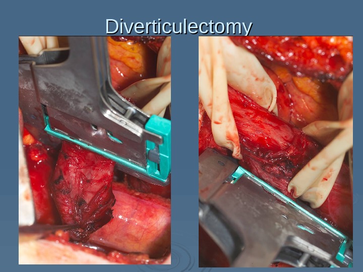
Diverticulectomy
Diverticulectomy
Esophageal achalasia A rare motor disorder of the esophagus characterized by inability of the lower esophageal sphincter and esophageal muscle to relax as well as dilation of the esophagus
Esophageal achalasia clinical picture Dysphagia (most common) Regurgitation Chest pain Heartburn Weight loss
Esophageal achalasia classification There are four stages: 1. 1. Functional spasm with out gullet extension 2. 2. Stable spasm with moderate gullet extension but peristalsis is saved 3. 3. Scar changes of esophageal wall with pronounced its dilatation, peristalsis is absent 4. 4. Pronounced gullet dilatation with its S-type bending and erosive esophagitis
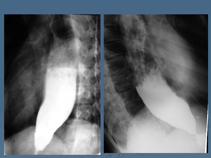
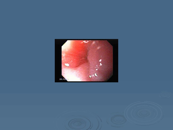
Esophageal achalasia diagnostic Contrast esophagography (barium swallowing) Fibroesophagoscopy Manometry Biopsy
Achalasia treatment In 1 and 2 stage – conservative treatment with spasmolitics or its combination with submucose botex injection In 1, 2 and 3 stage baloon dilatation is appropriable In 3 and 4 stage – just only myotomy by Heller or Petrovskiy could be provided
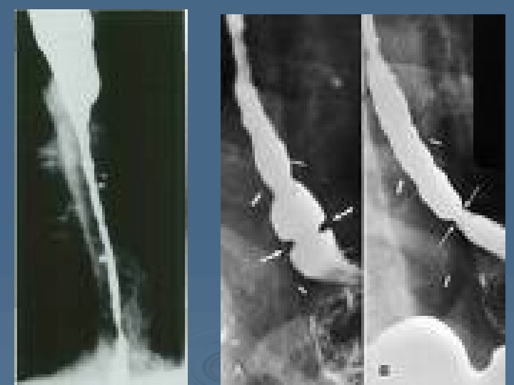
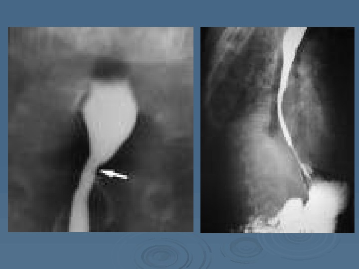
Esophageal stricture An esophageal stricture is a gradual narrowing of the esophagus, which can lead to swallowing difficulties. The strictures are caused by scar tissue that builds up in the esophagus.
When the lining of the esophagus is damaged, scarring develops. When scarring occurs, the lining of the esophagus becomes stiff. In time, as this scar tissue continues to build up, the esophagus begins to narrow in that area. Esophageal stricture
Esophageal stricture clinical picture Heartburn DD ysphagia OO dynophagia FF ood impaction WW eight loss CC hest pain.
Esophageal stricture diagnostic Contrast esophagography (barium swallowing) Fibroesophagoscopy Biopsy
Esophageal stricture treatment Dilation. The esophagus is stretched by passing a dilator or air-filled balloon is passed through a endoscope. Repeated dilation may be necessary to prevent the stricture from returning.
Esophageal stricture treatment If is performed if a stricture can’t be dilated enough to allow solid food to pass through. Surgery is also performed if repeated dilations do not keep these strictures from returning.
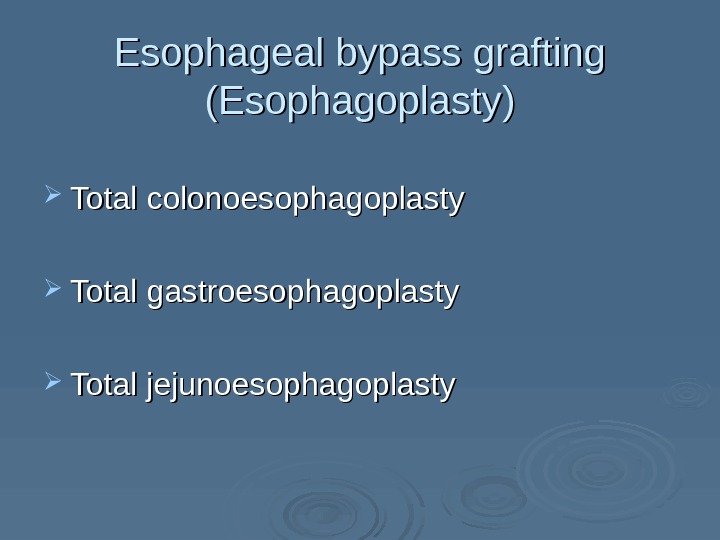
Esophageal bypass grafting (Esophagoplasty) Total colonoesophagoplasty Total gastroesophagoplasty Total jejunoesophagoplasty
Thanks for your attention