Percussion of the lungs Palpation of the chest








percussion_of_the_lungs_lecture_4_gorach.ppt
- Размер: 3.1 Mегабайта
- Количество слайдов: 34
Описание презентации Percussion of the lungs Palpation of the chest по слайдам
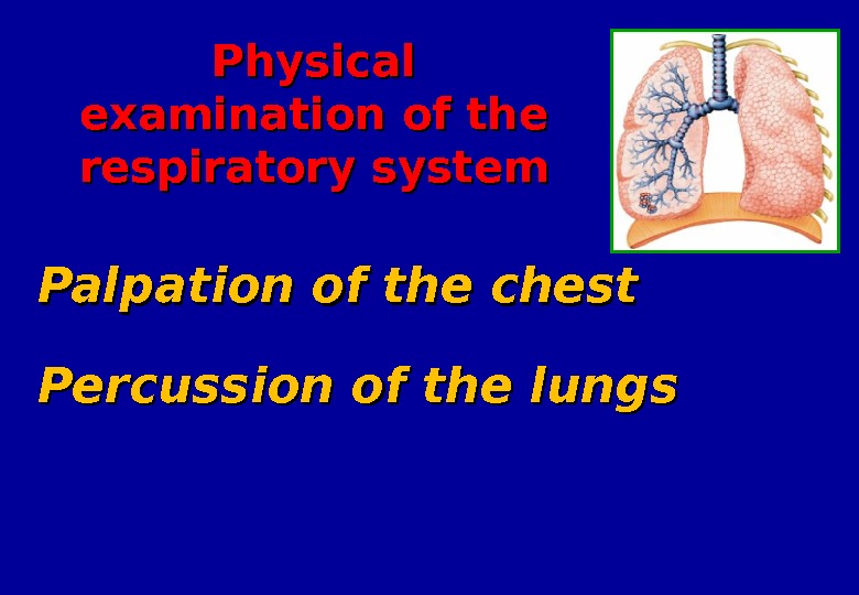
 Percussion of the lungs Palpation of the chest Physical examination of the respiratory system
 Inspection of the chest (( inspectio thoracis )) This is the objective method of examination based on visual evaluation of condition and pathological changes in thorax Static inspection – based on revelation of thorax features without taking into the act of breathing Dynamic inspection — based on revelation of thorax features with taking into the act of breathing
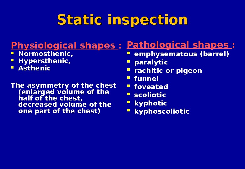
 Static inspection Physiological shapes : Normosthenic , Hypersthenic , Asthenic The asymmetry of the chest (enlarged volume of the half of the chest, decreased volume of the one part of the chest) Pathological shapes : emphysematous ( barrel ) paralytic rachitic or pigeon funnel foveated scoliotic kyphoscoliotic
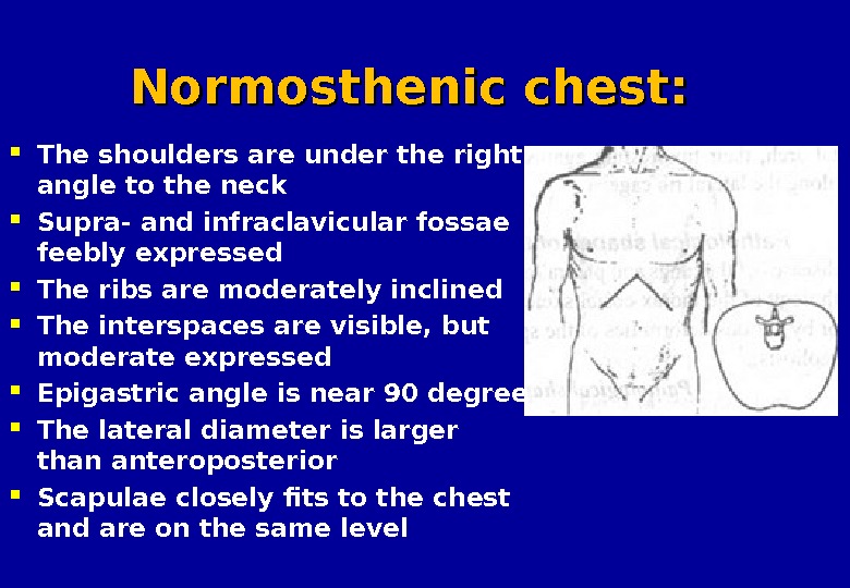
 Normosthenic chest: The shoulders are under the right angle to the neck Supra- and infraclavicular fossae feebly expressed The ribs are moderately inclined The interspaces are visible, but moderate expressed Epigastric angle is near 90 degree The lateral diameter is larger than anteroposterior Scapulae closely fits to the chest and are on the same level
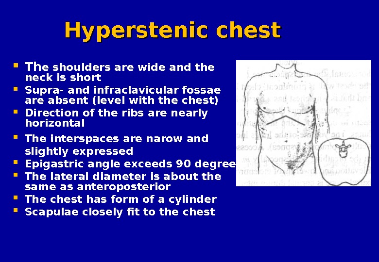
 Hyperstenic chest Th e shoulders are wide and the neck is short Supra- and infraclavicular fossae are absent (level with the chest) Direction of the ribs are nearly horizontal The interspaces are narow and slightly expressed Epigastric angle exceeds 90 degree The lateral diameter is about the same as anteroposterior The chest has form of a cylinder Scapulae closely fit to the chest
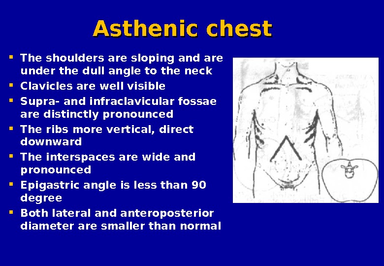
 Asthenic chest The shoulders are sloping and are under the dull angle to the neck Clavicles are well visible Supra- and infraclavicular fossae are distinctly pronounced The ribs more vertical, direct downward The interspaces are wide and pronounced Epigastric angle is less than 90 degree Both lateral and anteroposterior diameter are smaller than normal
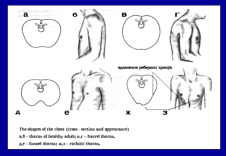
 The shapes of the chest ( cross — section and appearance ) а, б – thorax of healthy adult ; в, г – barrel thorax. д, е – funnel thorax ; ж, з – rachitic thorax.
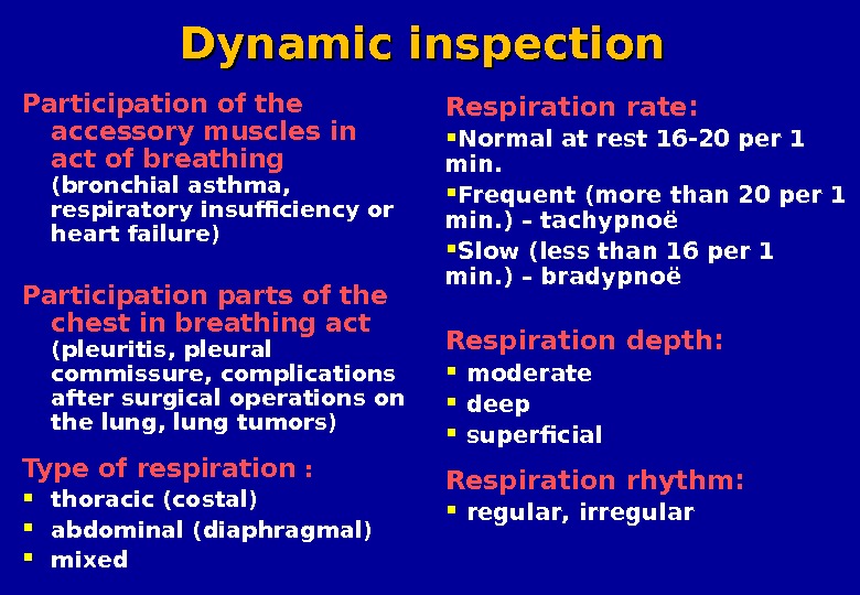
 Dynamic inspection Participation of the accessory muscles in act of breathing ( bronchial asthma , respiratory insufficiency or heart failure ) Participation parts of the chest in breathing act ( pleuritis , pleural commissure , complications after surgical operations on the lung , lung tumors ) Type of respiration : thoracic ( costal ) abdominal ( diaphragmal ) mixed Respiration rate : Normal at rest 16 -20 per 1 min. Frequent ( more than 20 per 1 min. ) – tachypno ë Slow ( less than 16 per 1 min. ) – bradypno ë Respiration depth : moderate deep superficial Respiration rhythm: regular , irregular
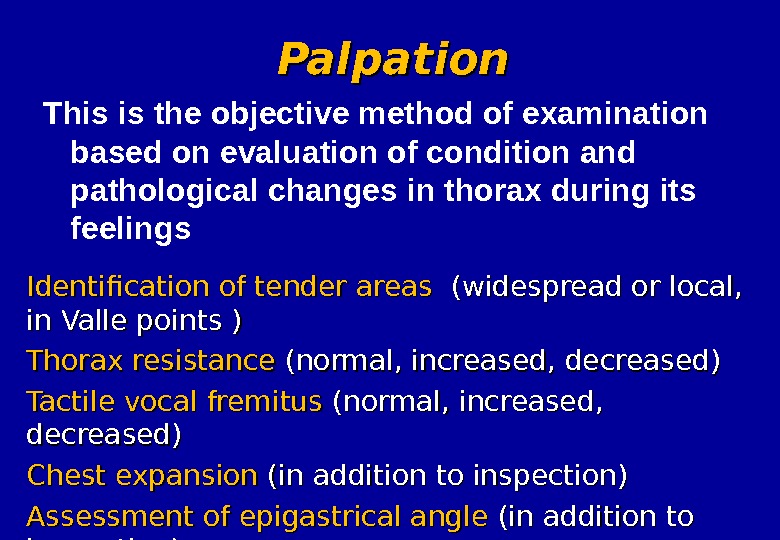
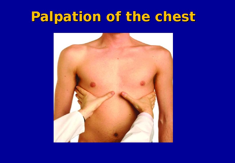
 Palpation Identification of tender areas (( widespread or local, in Valle points ) ) Thorax resistance (normal, increased, decreased) Tactile vocal fremitus (( normal , , increased , , decreased) Chest expansion (in addition to inspection) Assessment of epigastrical angle (in addition to inspection) This is the objective method of examination based on evaluation of condition and pathological changes in thorax during its feelings
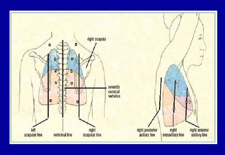
 Topographic regions of the chest Supraclavicular region – above clavicles Infraclavicular region – below clavicles Suprascapular regoin – above scapulae Interscapular region – between the scapulae Infrascapular region – below scapular
 Assessment of thorax elasticity ; а – antero-posterior , б – lateral.
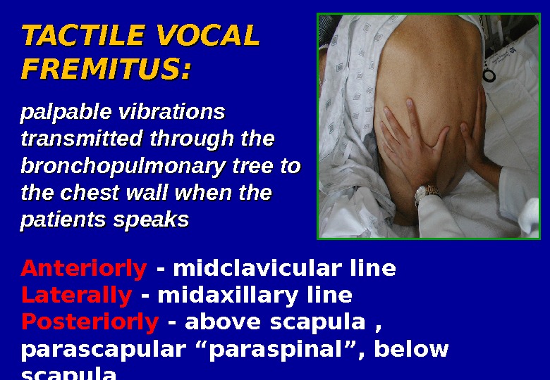
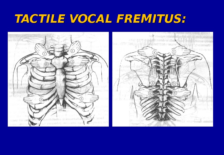
 TACTILE VOCAL FREMITUS: palpable vibrations transmitted through the bronchopulmonary tree to the chest wall when the patients speaks Anteriorly — midclavicular line Laterally — midaxillary line Posteriorly — above scapula , parascapular “paraspinal”, below scapula
 TACTILE VOCAL FREMITUS:
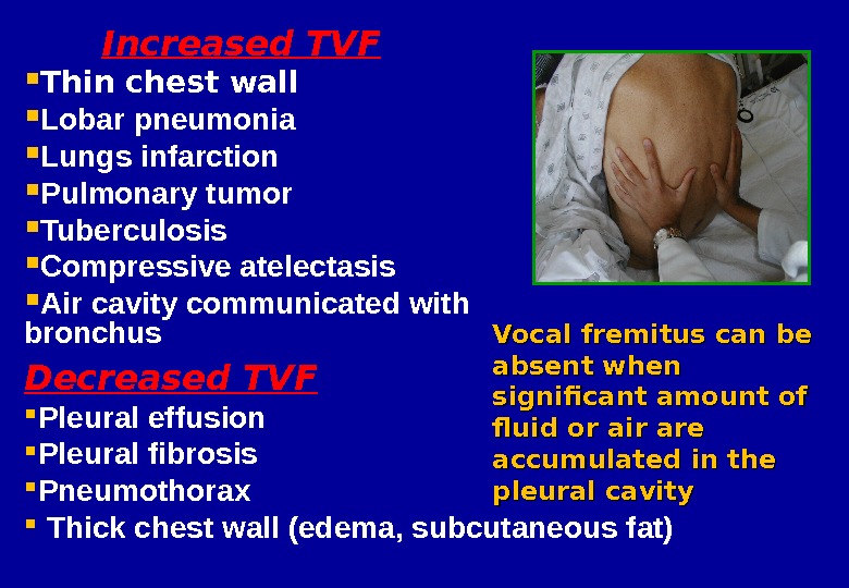
 Increased TVF Thin chest wall Lobar pneumonia Lungs infarction Pulmonary tumor Tuberculosis Compressive atelectasis Air cavity communicated with bronchus Decreased TVF Pleural effusion Pleural fibrosis Pneumothorax Thick chest wall (edema, subcutaneous fat) Vocal fremitus can be absent when significant amount of fluid or air are accumulated in the pleural cavity
 Palpation of the chest
 Palpation of the chest
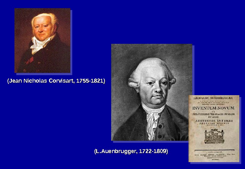
 (L. Auenbrugger, 1722 -1809) (Jean Nicholas Corvisart, 1755 -1821)
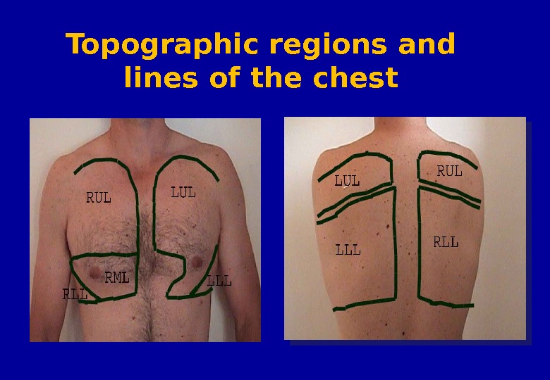
 Topographic regions and lines of the chest
 The left and right midaxillary lines – linea axillaris media dextra and sinistra The left and right posterior axillary lines – linea axillaris posterior dextra and sinistra The scapular left and right lines – linea scapularis dextra and sinistra The paraspinal lines dextra and sinistra – linea paravertebralis dextra and sinistra The vertebral line – linea vertebralis – linea mediana posterior. Topographic regions and lines of the chest

 Topographic regions and lines of the chest
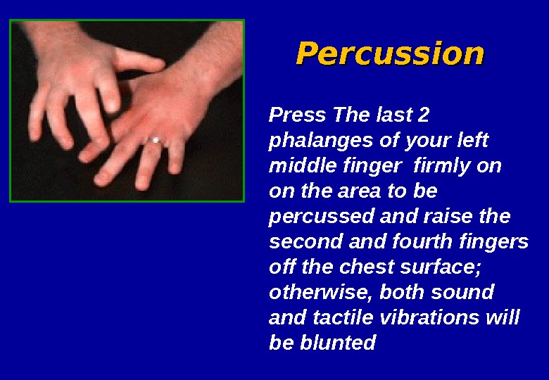
 Press The last 2 phalanges of your left middle finger firmly on on the area to be percussed and raise the second and fourth fingers off the chest surface; otherwise, both sound and tactile vibrations will be blunted Percussion
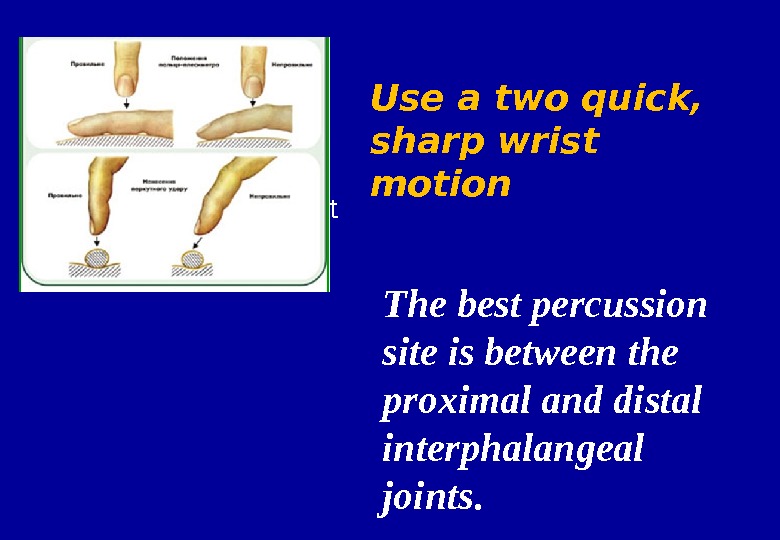
 • Movement from wrist The best percussion site is between the proximal and distal interphalangeal joints. Use a two quick, sharp wrist motion
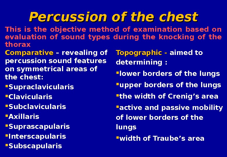
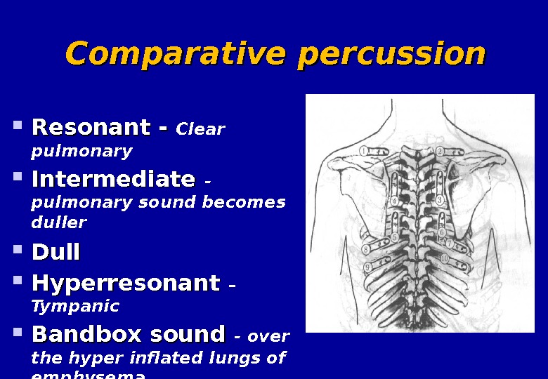
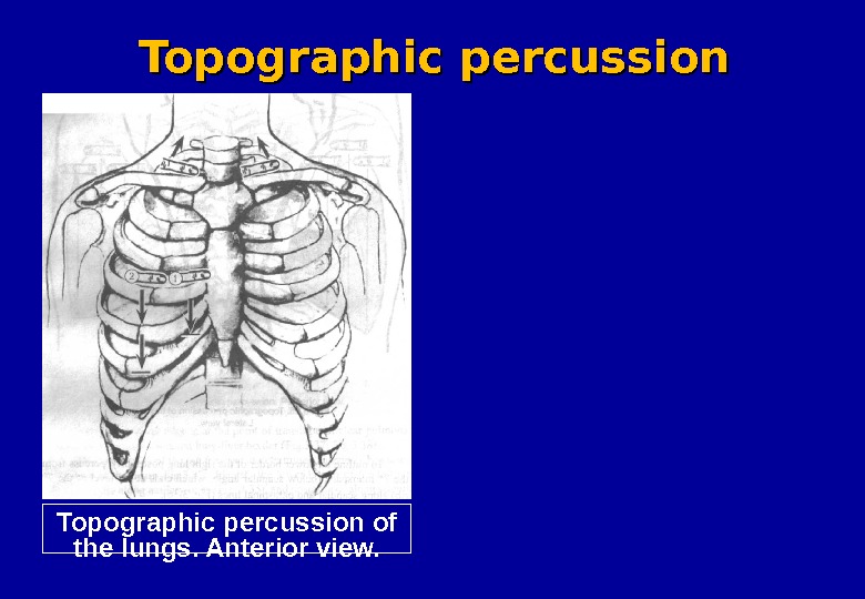
 Comparative – revealing of percussion sound features on symmetrical areas of the chest : Supraclavicularis Clavicularis Subclavicularis Axillaris Suprascapularis Interscapularis Subscapularis Topographic — — aimed to determining : lower borders of the lungs upper borders of the lungs the width of Crenig’s area active and passive mobility of lower borders of the lungs width of Traube’s area. This is the objective method of examination based on evaluation of sound types during the knocking of the thorax Percussion of the chest
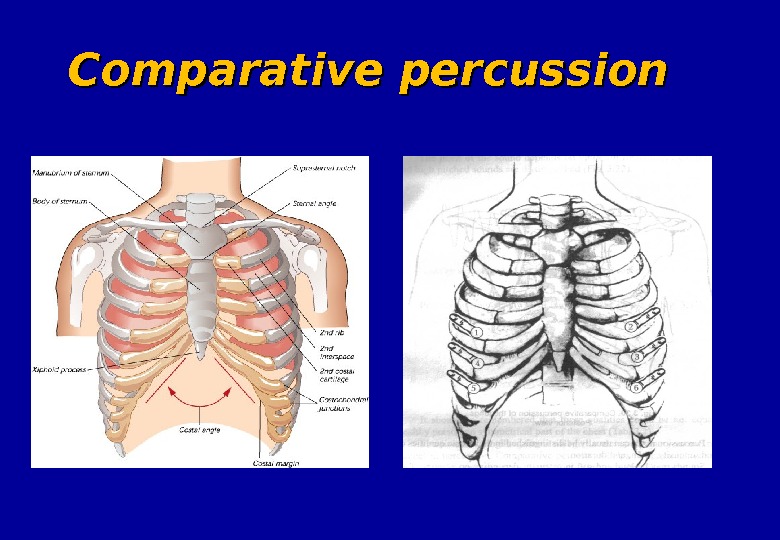
 Comparative percussion
 Comparative percussion Resonant — Clear pulmonary Intermediate — pulmonary sound becomes duller Dull Hyperresonant – Tympanic Bandbox sound — over the hyper inflated lungs of emphysema
 The main symptoms based on comparative percussion Percussion sound on the symmetric areas : Clear pulmonary ( in healthy persons ) Dullness ( dulling ) Infiltration of lung tissue ( tuberculosis , pneumonia , pneumosclerosis , lung cancer , abscess , lung gangrene ) Accumulation of liquid in pleural cavity Stony dull – large pleural effusion pleural thickening Tympanic Increasing the air capacity of lung tissue ( bronchial asthma, lung emphysema ) Formation the cavity with air in lung parenchyma ( released form contents caverns , abscess , bronchoectasis ) Accumulation of air in pleural cavity (pneumothorax )
 The main symptoms based on topographic percussion 1. Lower borders : Removal down ( lung emphysema , bronchial asthma , lower standing of diaphragm ) Removal upper ( athelectasis , surgical ablation the part of lung , higher standing of diaphragm , subdiaphragmal abscess ) 2. Upper borders : Removal down ( tuberculosis of lung apexes , pneumosclerosis , athelectasis of lung apexes ) Removal upper (lung emphysema , bronchial asthma ) 3. Width of Traube’s area: Increasing more than 6 sm — lung emphysema , bronchial asthma Decreasing less than 4 sm — tuberculosis of lung apexes , pneumosclerosis , athelectasis of lung apexes
 Topographic percussion of the lungs. Anterior view. Topographic percussion
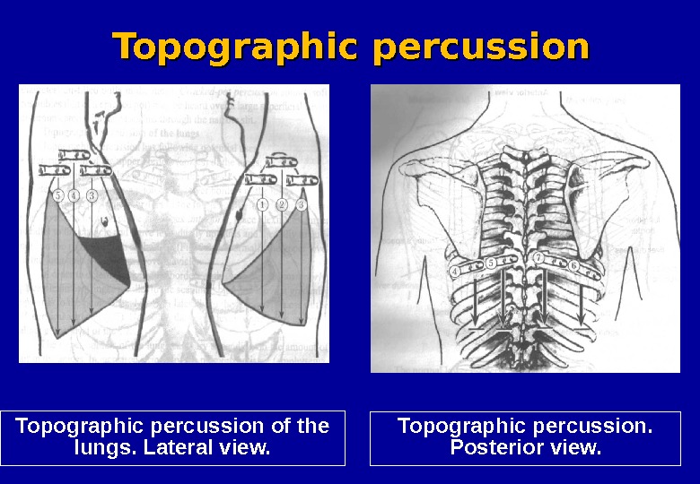
 Topographic percussion of the lungs. Lateral view. Topographic percussion. Posterior view. Topographic percussion
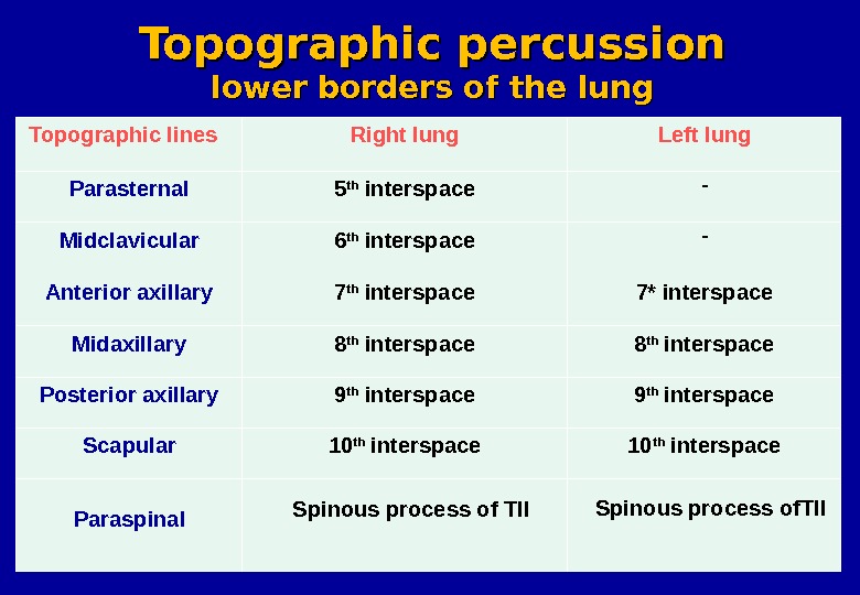
 Topographic lines Right lung Left lung Parasternal 5 th interspace — Midclavicular 6 th interspace — Anterior axillary 7 th interspace 7* interspace Midaxillary 8 th interspace Posterior axillary 9 th interspace Scapular 10 th interspace Paraspinal Spinous process of Tll Spinous process of. Tll. Topographic percussion lower borders of the lung
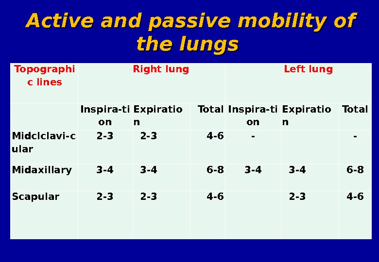
 4. Active and passive mobility of the lungs – – the significance of lung tissue elasticity state and the possible mobility of lower lung border : Enough (6 -8 sm ) by linea axillaris media, scapularis – normal Decreased ( less than 6 sm ) by linea scapularis — lung emphysema , bronchial asthma , pneumosclerosis , pleural commissural , sweating pleuritis 5. 5. The Traube’s area – – the area of tympanic sound under the left ribs arch. Diagnostically impotence –decreasing of area width : Cancer of cardial part of stomach Increasing of the liver Increasing of the spleen Left side sweating pleuritis
 Determining of the mobility of lower borders of the lungs
 Topographi c lin es Right lung Left lung Inspira-ti on Expiratio n Total Midc lclavi-c ular 2 -3 4 -6 — — Midaxillary 3 -4 3 -4 6 -8 Scapular 2 -3 4 -6 Active and passive mobility of the lungs

