
Occlusive diseases of arteries of lower extremities Prepared


lower_extre_diseases_zhagniyev_447.ppt
- Размер: 1.4 Mегабайта
- Количество слайдов: 41
Описание презентации Occlusive diseases of arteries of lower extremities Prepared по слайдам
Occlusive diseases of arteries of lower extremities Prepared by : Zhagniyev Zh. , 44 7 GM
It is a common disorder, characterized by a specific lesion of the arteries elastic and muscular-elastic types in the form of focal proliferation of connective tissue lipid infiltration of intima. Obliterative atherosclerosis of vessels of lower extremeties
Atherosclerosis of the aorta and great vessels of the lower extremities in the first place among the other peripheral arterial disease. Mostly common in men over 40 years, which often leads to severe limb ischemia, due to which patients lose their ability to work. Obliterative atherosclerosis of vessels of lower extremities
Etiology and pathogenesis Among the basic concepts of the development of atherosclerosis is theory of cholesterol-lipid infiltration. It is based on changes in the composition of blood plasma — hypercholesterolemia, dyslipoproteinemia — and a violation of the permeability of the arterial wall
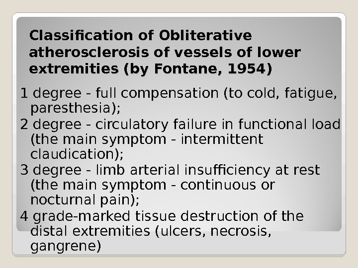
Classification of Obliterative atherosclerosis of vessels of lower extremities (by Fontane, 1954) 1 degree — full compensation (to cold, fatigue, paresthesia); 2 degree — circulatory failure in functional load (the main symptom — intermittent claudication); 3 degree — limb arterial insufficiency at rest (the main symptom — continuous or nocturnal pain); 4 grade-marked tissue destruction of the distal extremities (ulcers, necrosis, gangrene)
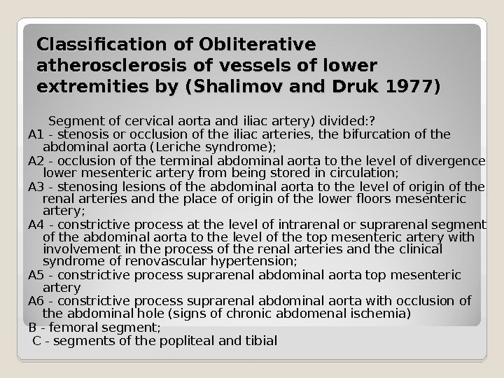
Classification of Obliterative atherosclerosis of vessels of lower extremities by (Shalimov and Druk 1977) Segment of cervical aorta and iliac artery) divided: ? A 1 — stenosis or occlusion of the iliac arteries, the bifurcation of the abdominal aorta (Leriche syndrome); A 2 — occlusion of the terminal abdominal aorta to the level of divergence lower mesenteric artery from being stored in circulation; A 3 — stenosing lesions of the abdominal aorta to the level of origin of the renal arteries and the place of origin of the lower floors mesenteric artery; A 4 — constrictive process at the level of intrarenal or suprarenal segment of the abdominal aorta to the level of the top mesenteric artery with involvement in the process of the renal arteries and the clinical syndrome of renovascular hypertension; A 5 — constrictive process suprarenal abdominal aorta top mesenteric artery A 6 — constrictive process suprarenal abdominal aorta with occlusion of the abdominal hole (signs of chronic abdomenal ischemia) B — femoral segment; C — segments of the popliteal and tibial
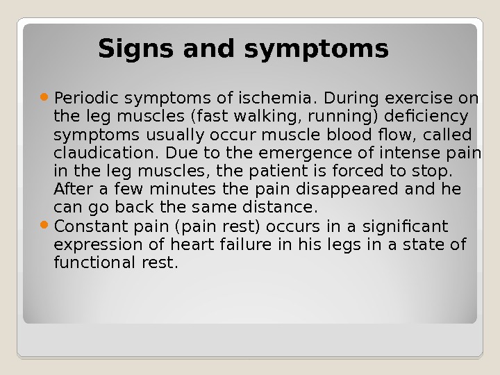
Signs and symptoms Periodic symptoms of ischemia. During exercise on the leg muscles (fast walking, running) deficiency symptoms usually occur muscle blood flow, called claudication. Due to the emergence of intense pain in the leg muscles, the patient is forced to stop. After a few minutes the pain disappeared and he can go back the same distance. Constant pain (pain rest) occurs in a significant expression of heart failure in his legs in a state of functional rest.
Destructive changes of the distal extremities should be considered the final manifestation of severe tissue ischemia. It can manifest as focal necrosis, trophic ulcers and gangrene of fingers or feet. Development of such changes usually precedes a long period of illness, with intermittent claudication, discoloration and skin temperature and trophic disorders in the form of its muscle atrophy feet and legs, hair loss, malnutrition and impaired growth of nails. Necrotic changes first, as a rule, occur on toes. Before that, the majority of patients can be seen spotted cyanosis of the skin, which does not change the color of the position of the limb.
Angiogramm
Treatment of Lerish’s syndrome
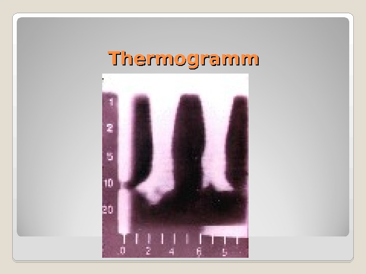
Laboratory and instrumental methods of diagnosis of arteriosclerosis obliterans of lower extremities 1. CBC. 2. Urine analysis. 3. Biochemical analysis of blood to the definition of lipid metabolism, total cholesterol, triglycerides. 4. Coagulogramma 5. Capillaroscopy. 6. Rheovasography. 7. Oscilloscope. 8. Aortoarteriography 9. Doppler. 10. Thermography. 11. ECG.
Angiogramm
Therapeutic tactics and choice of treatment. Conservative treatment It is advisable for the I-II degree of chronic ischemia, as well as patients at high risk for complications of atherosclerosis and arterial system of the lower extremities, which are not subject to surgery.
Surgical treatment Vascular reconstruction is carried out, usually at IІ-III degrees, sometimes-IV degree of ischemia. The evaluation criteria for surgical treatment is carried out on the basis of the aorto-arteriography, ultrasound great vessels and transoperative audit of vessels.
Surgical treatment With a combination of aorto-iliac (type A 1) and femoral-popliteal-Goleneva (type B i C) occlusion of aorto-femoral reconstruction wire segment provided cross one of the two femoral arteries or complete reconstruction of the main arteries of the thigh. To this type of operations include bypass surgery and prosthetics.
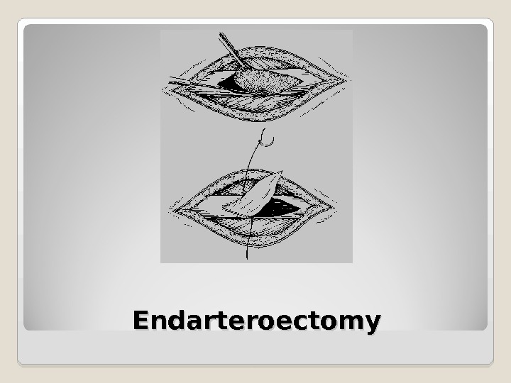
Surgical treatment When isolated, segmental, limited occlusion of the aorta and the bifurcation of the common iliac arteries, and other perform endarterectomy. Endarterectomy can be performed semi-open, and eversion techniques. It can also be done using ultrasound and laser technology. Regardless of the method of implementation, the side plastic endarterectomy end of the arterial wall through autovenous patches.
Surgical treatment Endarterectomy of deep femoral artery with further profundoplasty. Last performed with arteriotomy in a discharge of the deep femoral artery and after removing atherosclerotic plaque complete obliteration autovenous side plastic.
Surgical treatment In patients with necrotic changes of course have to give preference autoplastic reconstruction methods (endarterectomy autovenous bypass). In senior patients age in the presence of severe comorbidities reconstruction of aorto-femoral segment associated with high risk. Limb salvage with the threat of amputation in patients with severe ischemia (III, IV degree) will be achieved by suprapubic arterio-femoral or arterio-inguinal-femoral bypass.
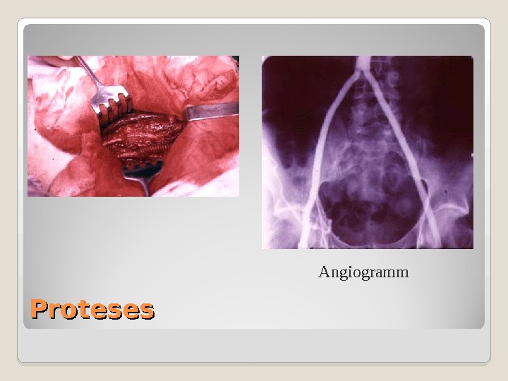
Surgical treatment. Isolation of the abdominal aorta implantation of the prosthesis
Proteses Angiogramm
Obliterative endarteriitis — a disease of vascular neurohumoral genesis which begins with peripheral channel, mainly arteries and leads to the obliteration of the lumen. Occlusive disease is the second most other peripheral arterial disease. The disease is more common in men aged 20 -30 years. Male to female ratio is 99: 1.
Etiology and pathogenesis Factors: low temperatures, nicotine intoxication, mechanical injury, etc. Changes in the walls of blood vessels lead to autoimmune processes that significantly enhance the proliferative processes of the vascular intima. The first affects the blood vessels that nourish the arteries, which leads to disruption of blood flow to the intracapillary leaky walls. There comes a spasm of regional vessels, slowing blood flow, hypercoagulability and the end result-tissue necrosis.
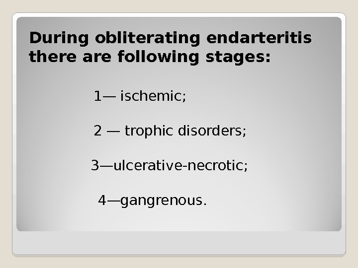
During obliterating endarteritis there are following stages: 1— ischemic ; 2 — trophic disorders ; 3— ulcerative-necrotic ; 4— gangrenous.
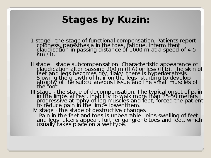
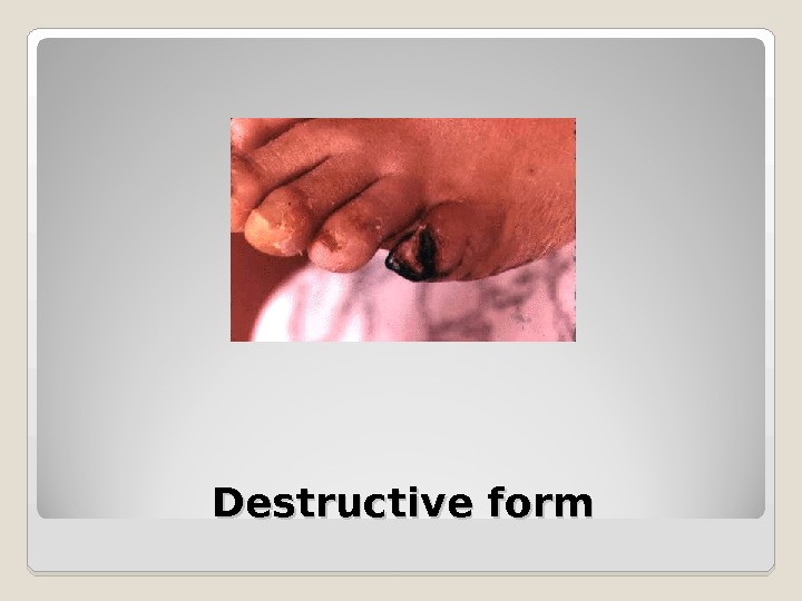
Stages by Kuzin: 1 stage — the stage of functional compensation. Patients report coldness, paresthesia in the toes, fatigue, intermittent claudication in passing distance of 1000 m at a speed of 4 -5 km / h. II stage — stage subcompensation. Characteristic appearance of claudication after passing 200 m (II A) or less (II b). The skin of feet and legs becomes dry, flaky, there is hyperkeratosis. Slowing the growth of hair on the legs, starting to develop atrophy of the subcutaneous tissue and the small muscles of the foot. III stage — the stage of decompensation. The typical onset of pain in the limbs at rest, inability to walk more than 25 -50 meters progressive atrophy of leg muscles and feet, forced the patient to reduce pain in the limbs lower them. IV stage — the stage of destructive changes Pain in the feet and toes is unbearable. Joins swelling of feet and legs, ulcers appear, further gangrene toes and feet, which usually takes place on a wet type.
Destructive form
Thermogramm
Functional probes Symptom of plantar ischemia Opel is blanching the sole of the foot of the affected limb, lift up. Depending on the rate of appearance of blanching can judge the degree of impaired circulation in the limbs, with severe ischemia, it occurs over the next 4. 6 seconds. Shamova sample. The patient was offered to lift up to 2 -3 minutes. straightened his leg at the knee joint and in the middle third of the thigh cuff is applied to the device Riva-Rocci, which creates a pressure that exceeds the systolic. After this step is lowered to a horizontal position, and after 4 -5 minutes. cuff removed. In healthy people, an average of 30 seconds. appears reactive hyperemia dorsum of fingers. If it comes in 1 -1. 5 min. , That is a relatively small limbs, circulatory failure, the delay it to 1. 5 -3 min. — Greater and greater than 3 min. — Significant. Knee phenomenon Panchenko. Patient sitting, scores injured leg to a healthy and soon begins to feel the pain of leg muscles, numbness in the feet, creeping sensation in the fingertips of the affected limb.
Therapeutic tactics and choice of treatment. Conservative treatment. Injected vasodilators myotropic action The ineffectiveness of conservative therapy surgical treatment.
Surgical treatment Ganglionic sympathectomy solves this problem: for a long time relieves vasospasm, eliminates or significantly reduces the intensity of pain. Operation is effective especially in the beginning of the disease. The next stages of sympathectomy loses Analgesic effect. The operation was performed on the background of conservative treatment has to be continued in the postoperative period. When lumbar simpatectomy be removed 3. 1 sympathic ganglia.
Endarteroectomy
Acute arterial occlusion of the lower limbs Acute arterial obstruction believe sudden cessation of blood circulation in the main artery due to embolism or thrombosis compared to other diseases.
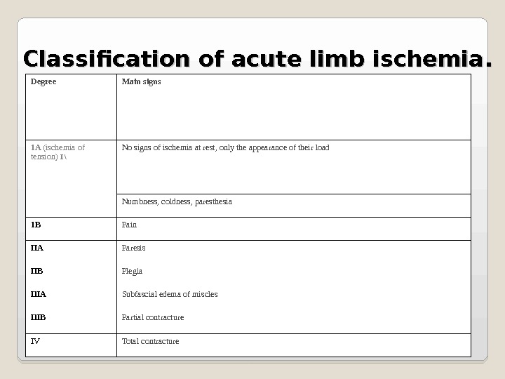
Classification of acute limb ischemia. . Degree Main signs 1 A (ischemia of tension) I \ No signs of ischemia at rest, only the appearance of their load Numbness, coldness, paresthesia 1 B Pain ПА Paresis ПB Plegia ША Subfascial edema of miscles ШB Partial contracture IV Total contracture
Therapeutic tactics and choice of treatment. In acute arterial obstruction of the indications for conservative treatment there may be a contraindication to surgery. Absolute contraindications are agonal state of the patient, total ischemic contracture of the limbs (acute ischemia in the III degree), very severe condition of the patient with pulmonary ischemia (acute ischemia I AI B level).
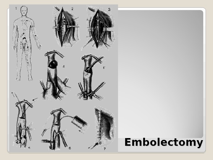
Embolectomy
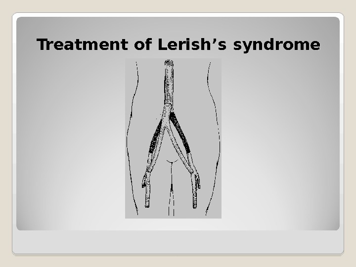
Leriche’s syndrome Leriche’s syndrome is one of the most common diseases of the arterial system due to occlusive lesions (occlusion) of aorto-iliac segment. Poor circulation of the pelvis and lower extremities leads to a characteristic triad of symptoms described in detail Leriche already in 1923: intermittent claudication, absence of pulse in the arteries of the lower limbs and impotence. Accurate data on the incidence of the disease is not present, but his high incidence can be judged by the number of indirect restorative operations on the abdominal aorta. In the USA, the annual production 37 OOO occlusion of the abdominal aorta.
Etiology and pathogenesis The etiology of occlusive lesions of the aorta varies atherosclerosis, nonspecific aortoarteriitis, postembolic occlusion, traumatic thrombosis, fibromuscular dysplasia (mainly affects the iliac arteries), congenital hypoplasia or aplasia of the aorta. Most common atherosclerosis (94%), followed by non-specific aortoarteriitis (5%) and 1% falls on the rest of the pathology. The natural course of the disease is associated with progressive deterioration. Ischemic symptoms usually increase is slow, but this is true only so long until a thrombosis. Then, the clinical course could rapidly deteriorate. Conservative treatment does not stop the progression of the disease and approximately 25% of patients amputation of limbs. The peak of the disease is on 6 decades of life, with the most common in men than women.
Signs and symptoms Is reported as the pain of the lower limbs due to chronic ischemia. Depending on the severity of ischemia of the lower limbs can be divided into four stages of the disease (classification AVPokrovsky): Stage 1 — Pain in the lower extremities appear only at high physical activity, such as walking more than 1 km, Stage 2 — Leg Pain walking in a shorter distance. If the patient is at a normal pace without pain 2 oo m, this Article 2 A. , If pain occurs earlier — Article 2 B, 3 stage — the appearance of pain in the legs at rest, 4 stage — previous complaints join gross tissue trophic disorders of the lower extremities (ulcers , necrosis, gangrene). Occlusion of the aorta pain occurs in the gluteal muscles, lower back and hip muscles — the so-called high intermittent claudication. Some patients the so-called intermittent claudication anal sphincter that results from ischemia of the pelvic floor. Characteristic numbness, cooling of the lower extremities (feet), hair loss, brittle nails and slow growth. Fairly common symptom is impotence. It develops not only due to ischemia of the pelvic organs, and spinal cord.
Laboratory-instrumental diagnostics Additional survey includes a set of methods aimed at establishing the location and extent of occlusion (and isotopic angiography, computed tomography) and hemodynamic (USDG, duplex scanning). Currently, functional hemodynamic changes in aortoiliac occlusive lesion segment quite accurately detected using non-invasive ultrasound examination. When Doppler ultrasound (USDG) the nature of the blood flow (main or collateral) can be defined cross the great arteries, the approximate level of occlusion, to measure the speed of blood flow and pressure in the various segments. Most use the ankle pressure index (LEAD) — the ratio of the pressure in the ankle to brachial pressure. Normally it is just above 1, O. The lower it is, the greater hemodynamic disturbances and severe ischemia. The task is also asking aortography state distal vascular bed (outflow tract). With combined occlusion of the superficial femoral artery is important to determine the condition of the deep femoral artery. Her stenosis, especially the mouth, poorly detected on the pictures in the anteroposterior projection, since in this area the femoral artery are superimposed on one another. Expansion of the information given in the pictures side or oblique.
When occlusive lesions of the aorta may, though rarely, meet syndrome «blue fingers. » He called ateroemboly of distal arteries and shows the typical skin changes as purpur-like rash speckled or reticulated pattern of red, very tender to palpation. This maintains the ripple on the arteries of the foot and ankle pressure is normal. Source of embolism syndrome «blue fingers» usual infrarenal segment of the aorta, and in disseminated ateroembolii (simultaneous failure of internal organs) — thoracoabdominal aorta.
Treatment The main indication for surgery is chronic lower limb ischemia 2 B — 4 tbsp. Reconstructive vascular surgery is contraindicated: with fresh myocardial infarction or stroke, brain (up to 3 months. ), Decompensated cardiac or respiratory failure, renal failure, hepatic, cancer, irreversible contraction of large joints, gangrene of the foot with the transition to the shin. The basic condition for the operation is the availability of satisfactory outflow tract.
Treatment When combined brachiocephalic artery occlusive lesions under transient ischemic initially made a correction of cerebral blood flow, and the second stage of reconstructive surgery of the aorta. In concurrent renovascular hypertension is justified-stage surgical repair of blood flow in the renal arteries and the abdominal aorta. The same applies to the occlusion of the visceral arteries. If shown coronary artery bypass grafting with occlusive coronary artery disease, and Leriche’s syndrome, it is performed in the first place, except for cases in which there is limb ischemia 1 V Art. merger with superinfection. Then the first readjustment is necrotic foci, which is best achieved by means of atypical benign vascular reconstructions type femoral-femoral crossover bypass.