
Молекулярная генетика развития Вельков В В 2013


- Размер: 8.3 Mегабайта
- Количество слайдов: 107
Описание презентации Молекулярная генетика развития Вельков В В 2013 по слайдам
Молекулярная генетика развития Вельков В В 2013 Gene Regulation during Development
Morphogenesis — How do you get from a spherical egg to say a frog?
Dolly and Bonnie Gilbert, SF (2003) Developmental Biology, 7 th ed. The nucleus from an differentiated adult cell (from the mammary gland) was able to replace the nucleus of a fertilized egg and produce an adult complete with functional gametes: Dolly had a daughter – Bonnie. SO: during development genes are just turned on and off in a carefully orchestrated fashion. SO, THE DOGMA OF DEV. BIOL. Is differential gene expression controls development
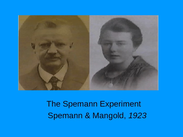
The Spemann Experiment Spemann & Mangold,
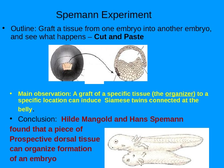
Spemann Experiment • Outline: Graft a tissue from one embryo into another embryo, and see what happens – Cut and Paste • Main observation: A graft of a specific tissue (the organizer ) to a specific location can induce Siamese twins connected at the belly. • Conclusion: Hilde Mangold and Hans Spemann found that a piece of Prospective dorsal tissue can organize formation of an embryo
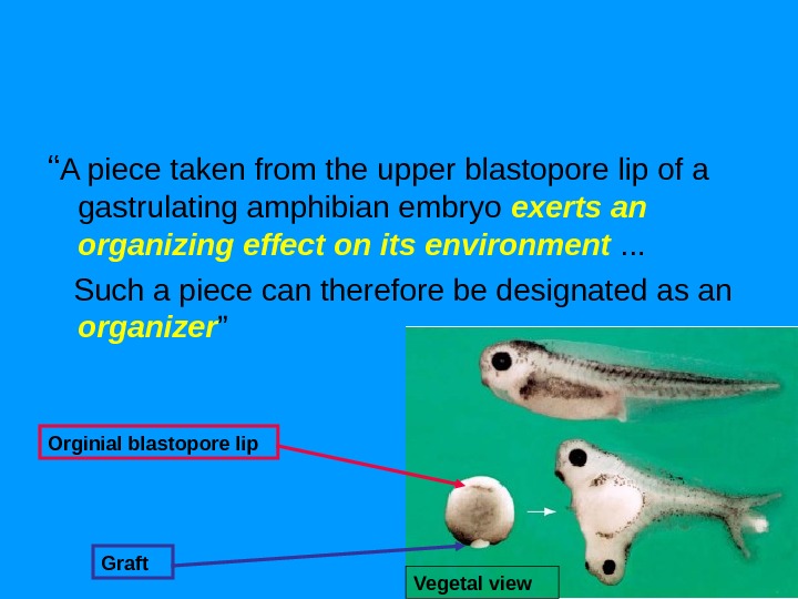
“ A piece taken from the upper blastopore lip of a gastrulating amphibian embryo exerts an organizing effect on its environment . . . Such a piece can therefore be designated as an organizer ” Orginial blastopore lip Graft Vegetal view
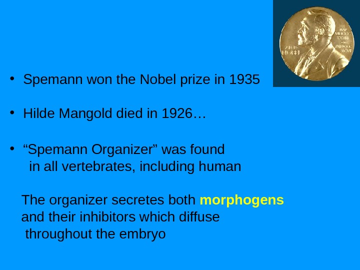
• Spemann won the Nobel prize in 1935 • Hilde Mangold died in 1926… • “ Spemann Organizer” was found in all vertebrates, including human The organizer secretes both morphogens and their inhibitors which diffuse throughout the embryo
Universal mechanism of animal development Gene expression controls 4 essential process. Short- long-range diffusible signaling molecule Changes in cell adhesion
Development is progressive • Specification of cell fate: determination – All cells still ‘look the same’ – Can be tested by transplantation experiments • Interactions can make cells different from each other: induction
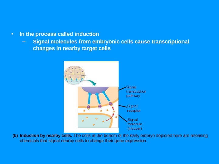
• In the process called induction – Signal molecules from embryonic cells cause transcriptional changes in nearby target cells Early embryo (32 cells) NUCLEUS Signal transduction pathway Signal receptor Signal molecule (inducer) Induction by nearby cells. The cells at the bottom of the early embryo depicted here are releasing chemicals that signal nearby cells to change their gene expression. (b)
From single cell to organism – a life cycle The use of a model organism • Fertilisation followed by cell division • Pattern formation – instructions for – Body plan (Axes: A-P, D-V) – Germ layers (ecto-, meso-, endoderm) • Cell movement — form – gastrulation • Cell differentiation • Cell growth, cell death (apoptosis)
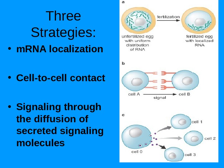
Three Strategies: • m. RNA localization • Cell-to-cell contact • Signaling through the diffusion of secreted signaling molecules
Морфогены и рецепторы морфогенов
• Morphogen – substances that define different cell fates in a concentration-dependent manner • Interaction of two signaling centers located in the anterior and posterior poles of the egg pattern insect body axis
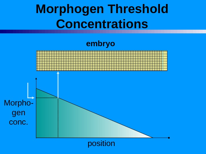
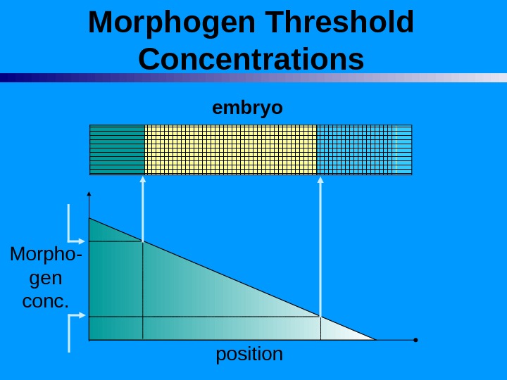
Morphogen = Soluble molecule that causes cellular commitment but is secreted some distance from the target cells. Morphogen Gradient = concentration gradient of a morphogen.
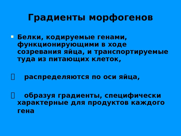
Градиенты морфогенов Белки, кодируемые генами, функционирующими в ходе созревания яйца, и транспортируемые туда из питающих клеток, распределяются по оси яйца, образуя градиенты, специфически характерные для продуктов каждого гена
Morphogen Threshold Concentrations embryo Morpho- gen conc. position
Morphogen Threshold Concentrations embryo Morpho- gen conc. position
Morphogen Threshold Concentrations embryo Morpho- gen conc. position
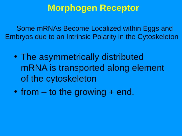
Morphogen Receptor Some m. RNAs Become Localized within Eggs and Embryos due to an Intrinsic Polarity in the Cytoskeleton • The asymmetrically distributed m. RNA is transported along element of the cytoskeleton • from – to the growing + end.
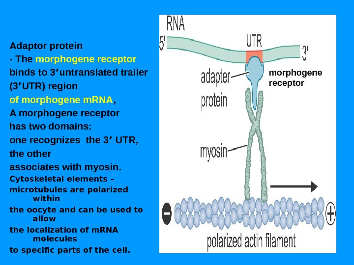
Adaptor protein — The morphogene receptor binds to 3 ’ untranslated trailer (3 ’ UTR) region of morphogene m. RNA , A morphogene receptor has two domains: one recognizes the 3 ’ UTR, the other associates with myosin. Cytoskeletal elements – microtubules are polarized within the oocyte and can be used to allow the localization of m. RNA molecules to specific parts of the cell. morphogene receptor
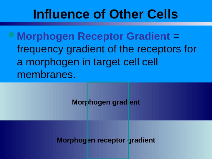
Influence of Other Cells Morphogen Receptor Gradient = frequency gradient of the receptors for a morphogen in target cell membranes. Morphogen gradient Morphogen receptor gradient
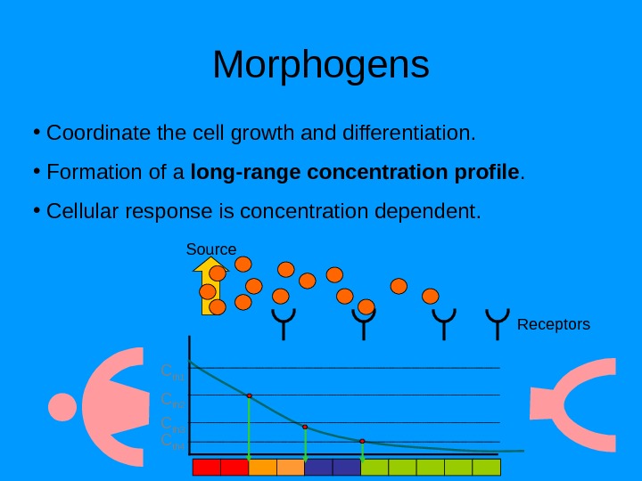
Morphogens • Coordinate the cell growth and differentiation. • Formation of a long-range concentration profile. • Cellular response is concentration dependent. C th 1 C th 2 C th 3 C th 4 Source Receptors
Copyright © The Mc. Graw-Hill Companies, Inc. Permission required to reproduce or display
Life cycle
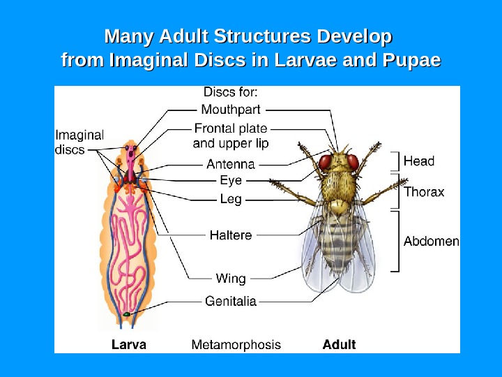
Many Adult Structures Develop from Imaginal Discs in Larvae and Pupae
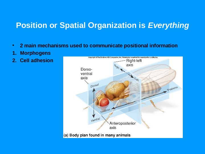
Position or Spatial Organization is Everything • 2 main mechanisms used to communicate positional information 1. Morphogens 2. Cell adhesion
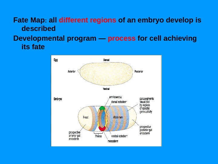
Fate Map : all different regions of an embryo develop is described Developmental program — process for cell achieving its fate
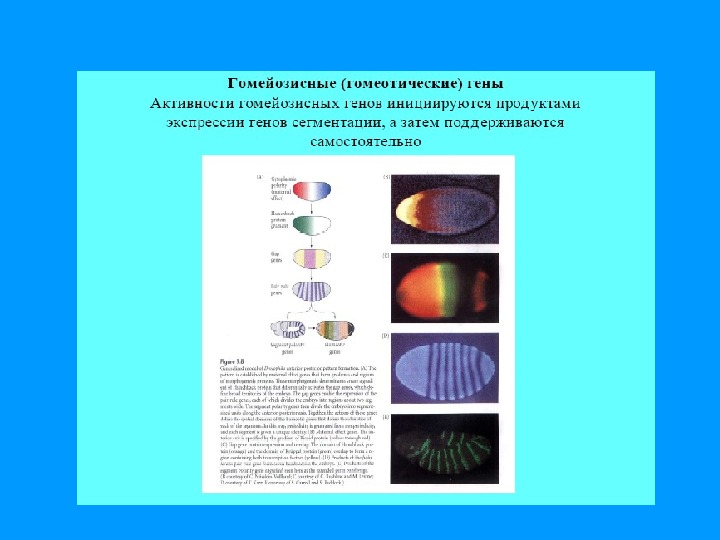
• Морфогены активируют • Гены сегментации активируют • Гомеозисные гены активируют…
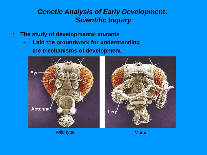
Genetic Analysis of Early Development: Scientific Inquiry • The study of developmental mutants – Laid the groundwork for understanding the mechanisms of development Eye Antenna Leg Wild type Mutant
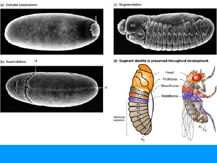
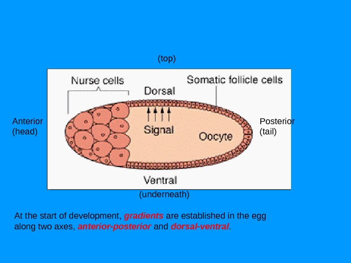
37 Drosophila Embryogenesis First phase is establishment of body axes Before fertilization, specialized nurse cells move maternal m. RNAs (morphogenes) into maturing oocyte Then morphogens are distributed in the oocyte prior to fertilization • The Morphogens organize the structure of the egg These positioned m. RNA will initiate a cascade of gene activations following fertilization Яйцо созревает в особой камере — фолликуле. В фолликулах — гены с материнским эффектом , которые функционируют еще до оплодотворения яйца сперматозоидом В фолликуле ооцит и 15 огромных питающих клеток, которые синтезируют «РНК морфогены» и перекачивают их в ооцит.
At the start of development, gradients are established in the egg along two axes, anterior-posterior and dorsal-ventral. Anterior (head) Posterior (tail) (underneath) (top)
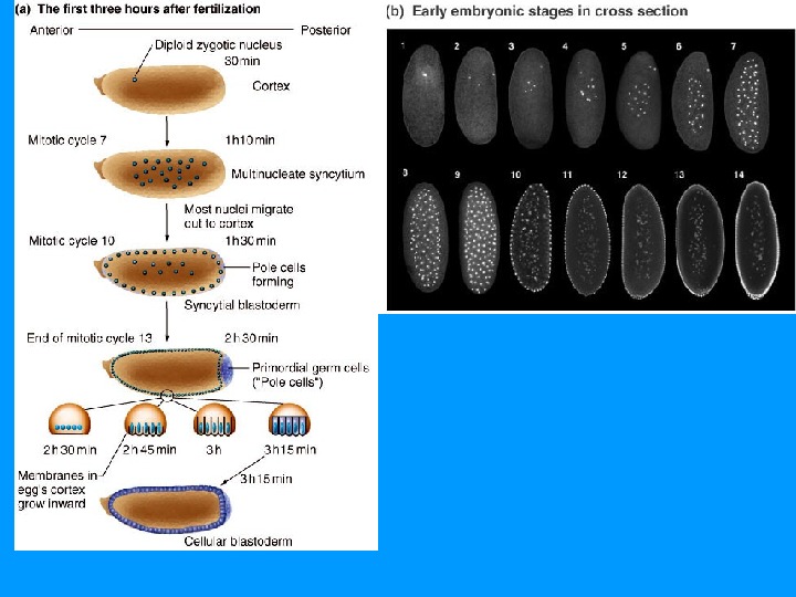
c. b. Nurse cells Anterior. Posterior Movement of maternal m. RNA Oocyte. Follicle cells Fertilized egg a. d. e. Three larval stages Syncytial blastoderm Cellular blastoderm Nuclei line up along surface, and membranes grow between them to form a cellular blastoderm. Segmented embryo prior to hatching Metamorphosis Abdomen Thorax Head. Hatching larva Nucleus Embryo
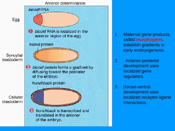
1. Maternal gene products, called morphogens , establish gradients in early embryogenesis. 2. Anterior-posterior development uses localized gene regulators. 3. Dorsal-ventral development uses localized receptor-ligand interactions.
Градиент морфогенов активирует зиготные гены После того как градиенты в яйце созданы, происходит оплодотворение и начинается дробление зародыша образуется однослойная бластодерма. Каждая клетка в ней занимает определенное положение по отношению к сформировавшимся градиентам, — обладает определенной позиционной информацией. Морфогены взаимодействуют с регуляторными участками генов, активирующихся у зигот (зиготических генов).
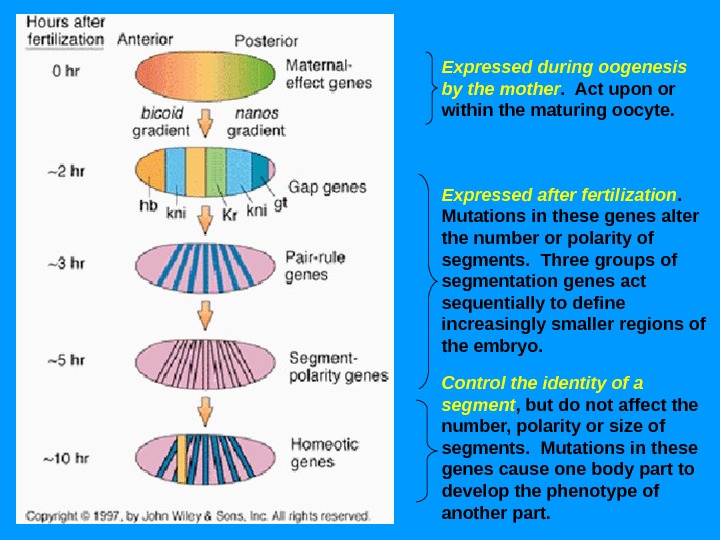
Expressed during oogenesis by the mother. Act upon or within the maturing oocyte. Expressed after fertilization. Mutations in these genes alter the number or polarity of segments. Three groups of segmentation genes act sequentially to define increasingly smaller regions of the embryo. Control the identity of a segment , but do not affect the number, polarity or size of segments. Mutations in these genes cause one body part to develop the phenotype of another part.
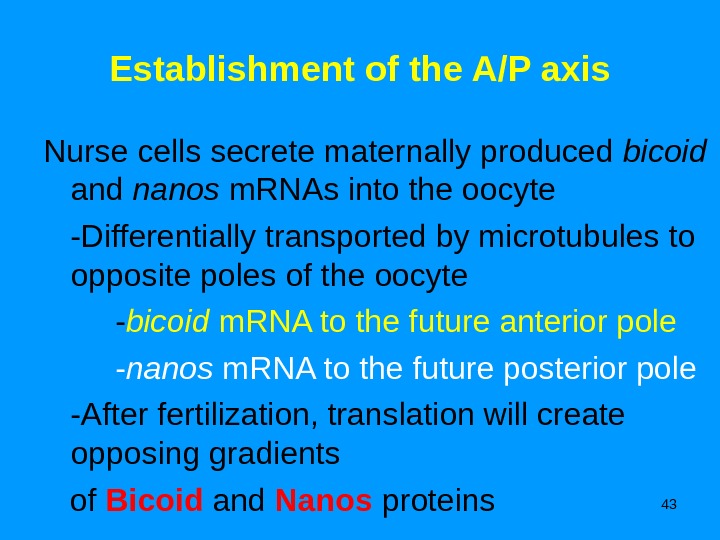
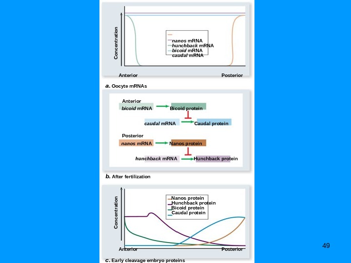
43 Establishment of the A/P axis Nurse cells secrete maternally produced bicoid and nanos m. RNAs into the oocyte -Differentially transported by microtubules to opposite poles of the oocyte — bicoid m. RNA to the future anterior pole — nanos m. RNA to the future posterior pole -After fertilization, translation will create opposing gradients of Bicoid and Nanos proteins
44 a. b. bicoid m. RNA moves toward anterior end bicoid m. RNAMovement of maternal m. RNA Nucleus. Microtubules. Nurse cells Follicle cells nanos m. RNA moves toward posterior end Posterior. Anterior nanos m. RNACopyright © The Mc. Graw-Hill Companies, Inc. Permission required for reproduction or display.
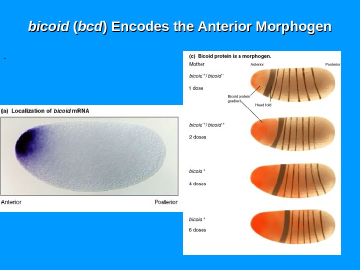
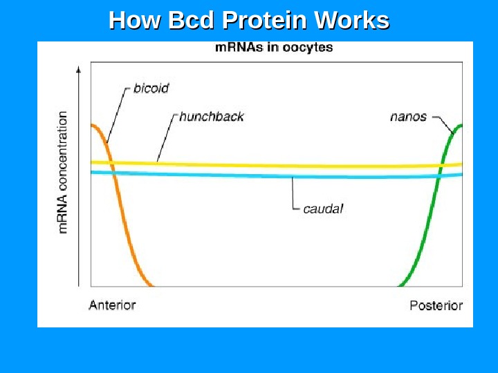
bicoid ( ( bcdbcd ) Encodes the Anterior Morphogen.
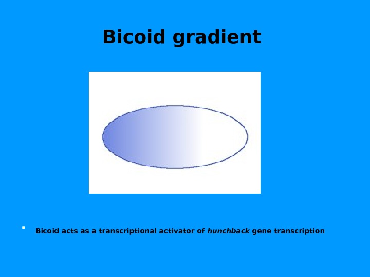
Bicoid gradient Bicoid acts as a transcriptional activator of hunchback gene transcription
Nanos gradient
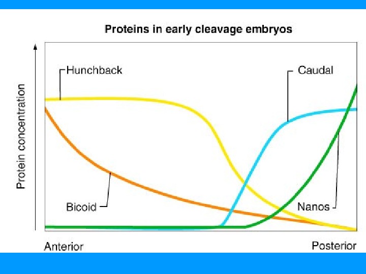
48 Establishment of the A/P axis Bicoid and Nanos control translation of two other maternal m. RNAs, hunchback and caudal , that encode transcription factors — Hunchback activates anterior structures — Caudal activates posterior structures The two m. RNAs are not evenly distributed — Bicoid protein inhibits caudal m. RNA translation — Nanos protein inhibits hunchback m. RNA translation
49 Concentration. Anteriora. Oocyte m. RNAs c. Early cleavage embryo proteinsb. After fertilization Posterior nanos m. RNA hunchback m. RNAbicoid m. RNA caudal m. RNA Nanos protein Hunchback protein. Bicoid protein Caudal protein Concentration Anterior Posteriornanos m. RNA hunchback m. RNA bicoid m. RNA caudal m. RNA Nanos protein Hunchback protein Bicoid protein Caudal protein
How Bcd Protein Works
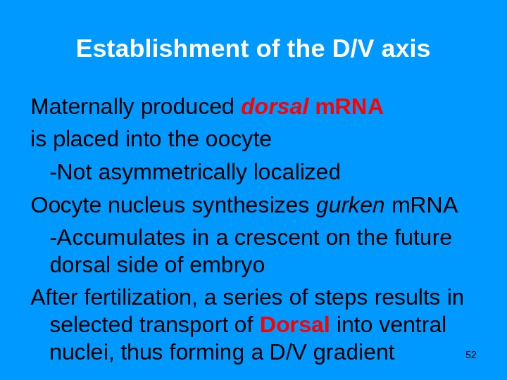
52 Establishment of the D/V axis Maternally produced dorsal m. RNA is placed into the oocyte -Not asymmetrically localized Oocyte nucleus synthesizes gurken m. RNA -Accumulates in a crescent on the future dorsal side of embryo After fertilization, a series of steps results in selected transport of Dorsal into ventral nuclei, thus forming a D/V gradient
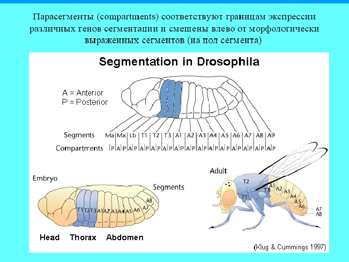
Segmentation genes act sequentially to divide the embryo into segments • Normal Drosophila embryo divided into 15 segments – 3 head , 3 thoracic and 9 abdominal – Each will give rise to unique morphological features in adult
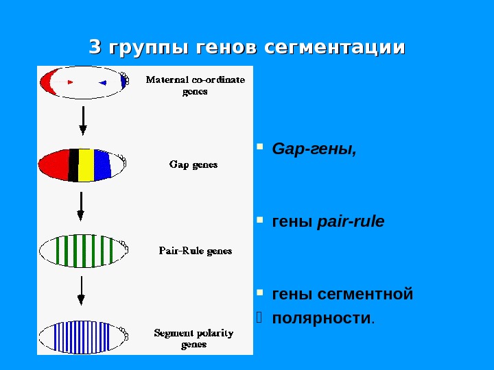
55 Production of Body Plan The body plan is produced by sequential activation of three classes of segmentation genes Segmentation Is Initiated by Localized RNAs at the Anterior and Posterior Poles of the Unfertilized Egg 1. Gap genes -Map out the coarsest subdivision along the A/P axis -All 9 genes encode transcription factors that activate the next gene class 2. Pair-rule genes -Divide the embryo into seven zones -The 8 or more genes encode transcription factors that regulate each other, and activate the next gene class 3. Segment polarity genes -Finish defining the embryonic segments
3 группы генов сегментации Gap -гены, гены pair — rule гены сегментной K полярности.
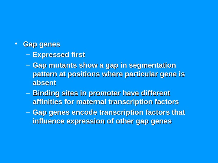
• Gap genes – Expressed first – Gap mutants show a gap in segmentation pattern at positions where particular gene is absent – Binding sites in promoter have different affinities for maternal transcription factors – Gap genes encode transcription factors that influence expression of other gap genes
Gap genes Hunchback , krüppel , giant , tailless and knirps. Their expression patterns in the early embryo are determined by the maternal effect gene products These genes establish the segmented body plan of the embryo along the anterior-posterior axis.
The A-P axis is divided into broad regions by by gapgap gene expression • The first zygotic genes • Respond to maternally-derived instructions • Short-lived proteins, • gives bell-shaped distribution from source
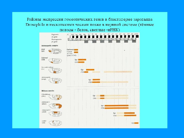
Zones of Expression of Four Gap Genes: Hunchback , , Kruppel , , Knirps , and Giant in Late Syncytial Blastoderm Embryos
Defects in Segmentation from Mutations in Gap Genes A) B) C) D)
Mutations in Gap Gene Result in Loss of Segments Corresponding to Zone of Expression
Transcription factors in cascade GAP genes • Hunchback (hb) , a gap gene, responds to the dose of bicoid protein • A concentration above threshold of bicoid activates the expression of hb • The more bicoid transcripts, the further back hb expression goes
Krüppel reads two values • Krüppel (Kr) , a gap gene, responds to the dose of hb protein • A concentration above minimum threshold of hb activates the expression of Kr • A concentration above maximum threshold of hb inactivates the expression of Kr. GAP genes
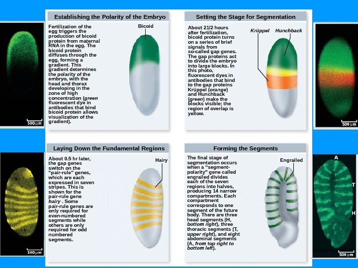
About 21/2 hours after fertilization, bicoid protein turns on a series of brief signals from so-called gap genes. The gap proteins act to divide the embryo into large blocks. In this photo, fluorescent dyes in antibodies that bind to the gap proteins Krüppel ( orange ) and Hunchback ( green ) make the blocks visible; the region of overlap is yellow. Forming the Segments. Laying Down the Fundamental Regions Setting the Stage for Segmentation Bicoid Hairy Krüppel Hunchback Engrailed. Establishing the Polarity of the Embryo Fertilization of the egg triggers the production of bicoid protein from maternal RNA in the egg. The bicoid protein diffuses through the egg, forming a gradient. This gradient determines the polarity of the embryo, with the head and thorax developing in the zone of high concentration ( green fluorescent dye in antibodies that bind bicoid protein allows visualization of the gradient). About 0. 5 hr later, the gap genes switch on the “pair-rule” genes, which are each expressed in seven stripes. This is shown for the pair-rule gene hairy . Some pair-rule genes are only required for even-numbered segments while others are only required for odd numbered segments. The final stage of segmentation occurs when a “segment- polarity” gene called engrailed divides each of the seven regions into halves, producing 14 narrow compartments. Each compartment corresponds to one segment of the future body. There are three head segments (H, bottom right ), three thoracic segments (T, upper right ), and eight abdominal segments (A, from top right to bottom left ).
Homeotic mutations We know some misterious mutations, which generate horroristic monsters
Homeotic-Selector/ HOX Genes
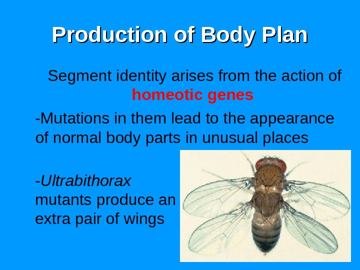
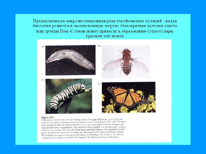
68 Production of Body Plan Segment identity arises from the action of homeotic genes -Mutations in them lead to the appearance of normal body parts in unusual places — Ultrabithorax mutants produce an extra pair of wings
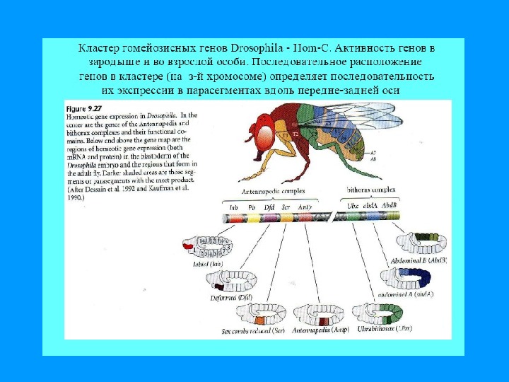
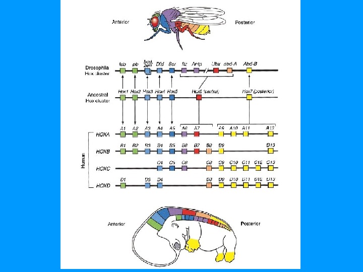
69 Production of Body Plan Homeotic gene complexes -The HOM complex genes of Drosophila are grouped into two clusters — Antennapedia complex , which governs the anterior end of the fly — Bithorax complex , which governs the posterior end of the fly — Interestingly, the order of genes mirrors the order of the body parts they control
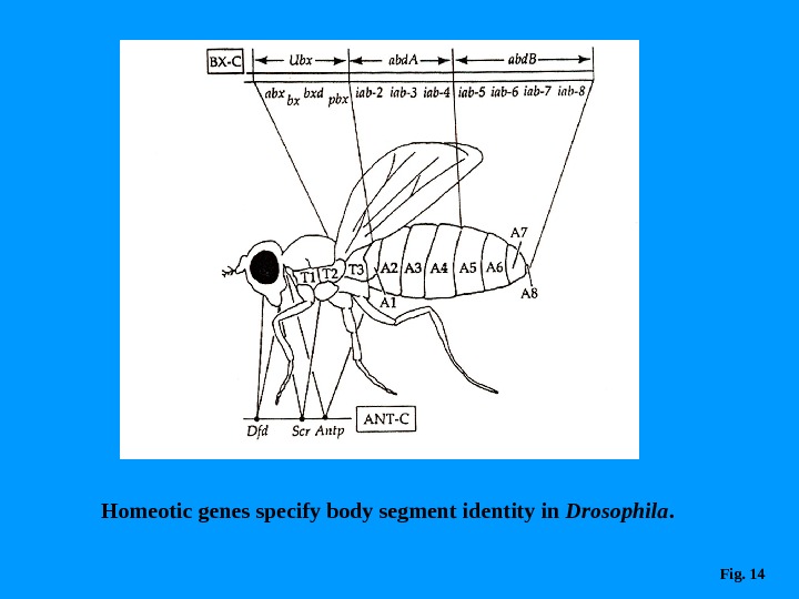
Homeotic genes specify body segment identity in Drosophila. Fig.
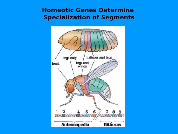
Homeotic Genes Determine Specialization of Segments
Гомеозисные гены Большой класс генов контролируют развитие части тела из определенного сегмента. В результате гомеозисной мутации из данного сегмента развивается другая часть тела Наиболее известны Antennapedia — complex ( Ant — C ). В ithorax — complex ( BX — C ) и
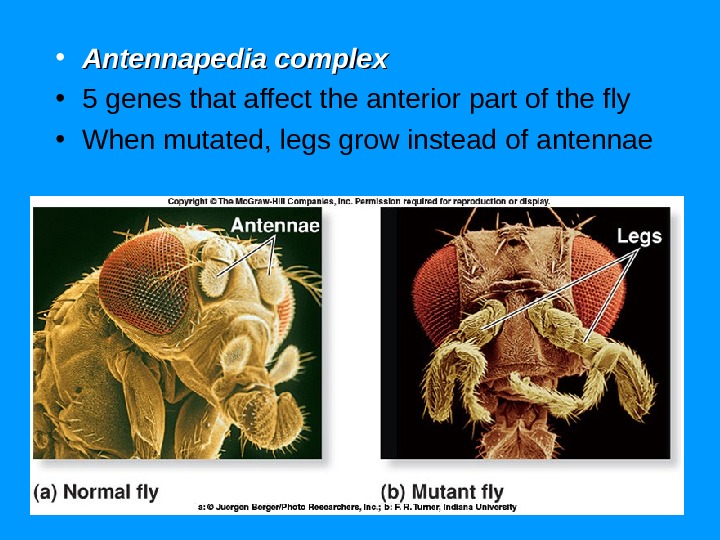
• Antennapedia complex • 5 genes that affect the anterior part of the fly • When mutated, legs grow instead of antennae
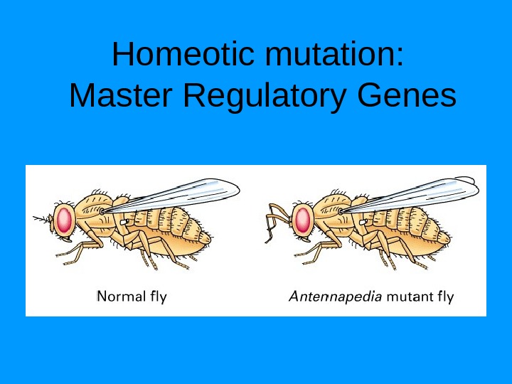
Homeotic Genes: Master Regulatory Genes
Homeotic mutation: Master Regulatory Genes
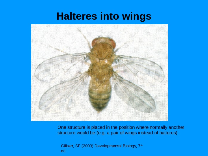
Halteres into wings Gilbert, SF (2003) Developmental Biology, 7 th ed. One structure is placed in the position where normally another structure would be (e. g. a pair of wings instead of halteres)
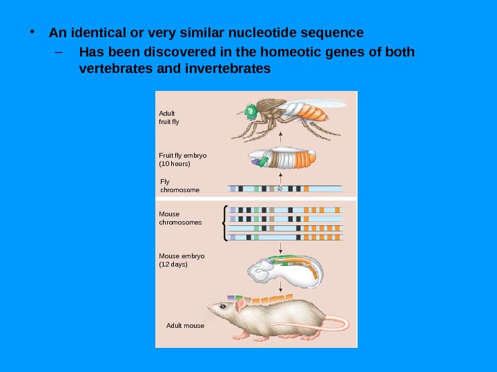
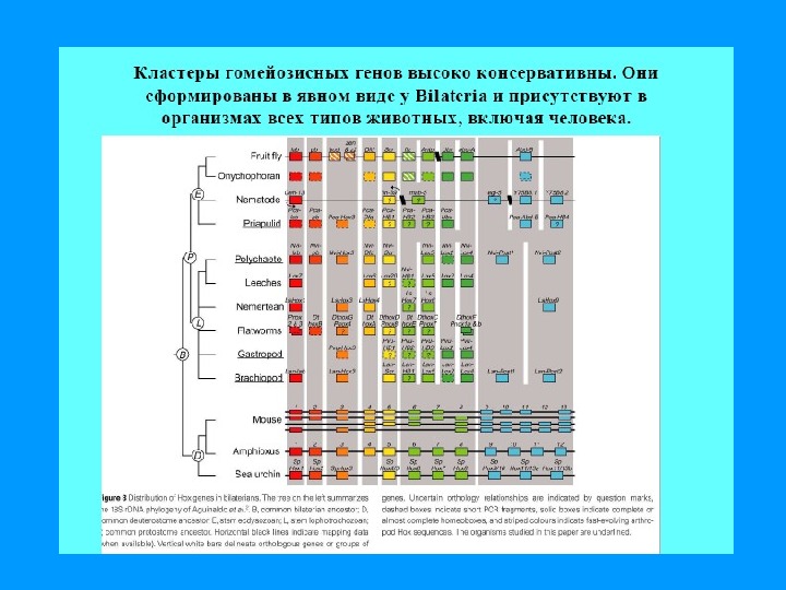
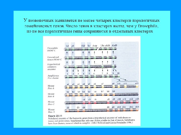
81 Production of Body Plan Homeotic gene complexes -All of these genes contain a conserved 180 -base sequence, the homeobox -Encodes a 60 -amino acid DNA-binding domain, the homeodomain -Homeobox-containing genes are termed Hox genes -Vertebrates have 4 Hox gene clusters
82 Production of Body Plan Drosophila HOM genes Thorax. Antennapedia complex Head Abdomen Bithorax complex Fruit fly embryo Mouse. Hox 1 Hox 2 Hox 3 Hox 4 Mouse embryo a. b. Drosophila HOM Chromosomes Mouse Hox Chromosomes lab pb Dfd Scr Antp Ubxabd-Aabd-
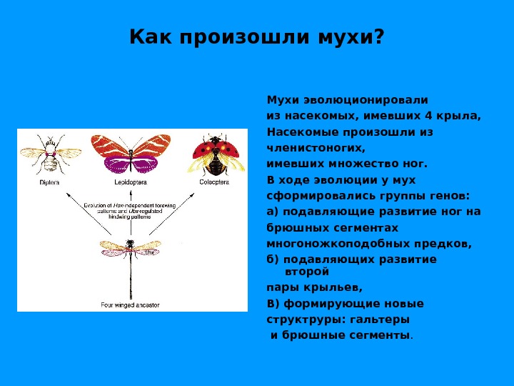
Как произошли мухи? Мухи эволюционировали из насекомых, имевших 4 крыла, Насекомые произошли из членистоногих, имевших множество ног. В ходе эволюции у мух сформировались группы генов: а) подавляющие развитие ног на брюшных сегментах многоножкоподобных предков, б) подавляющих развитие второй пары крыльев, В) формирующие новые структруры: гальтеры и брюшные сегменты.
К чему приводит делеция ВХ-С? Эмбрион развивается до определенной стадии и затем гибнет Эмбрион имел проторакальный сегмент, а все остальные — мезоторакальные Если бы этот организм выжил до взрослой мухи, она бы имела 10 пар крыльев и 10 пар ног K Функция гена ВХ-С — инактивация генов, формирующих ноги и крылья во всех последующих после второго торакального сегментах, — развитии всех структур на брюшных сегментах
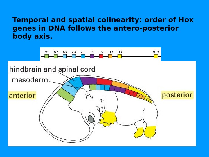
Vertebrates have four Hox complexes, with about 10 genes each. They can be aligned in 13 groups of paralogues. They display colinearity: a) Temporal colinearity: genes on one end of the complex are expressed first, those on the other (posterior) end are turned on last. b) Spatial colinearity: the more anteriorly expressed genes are in one end, the more posterior ones at the other end of the gene complex. c) Anterior Hox genes are activated sequentially by retinoic acid.
Temporal and spatial colinearity: order of Hox genes in DNA follows the antero-posterior body axis.
• An identical or very similar nucleotide sequence – Has been discovered in the homeotic genes of both vertebrates and invertebrates Adult fruit fly Fruit fly embryo (10 hours) Fly chromosome Mouse chromosomes Mouse embryo (12 days) Adult mouse
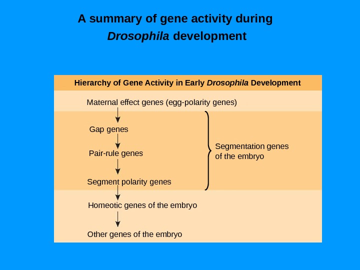
A summary of gene activity during Drosophila development Hierarchy of Gene Activity in Early Drosophila Development Maternal effect genes (egg-polarity genes) Gap genes Pair-rule genes Segment polarity genes Homeotic genes of the embryo Other genes of the embryo Segmentation genes of the embryo
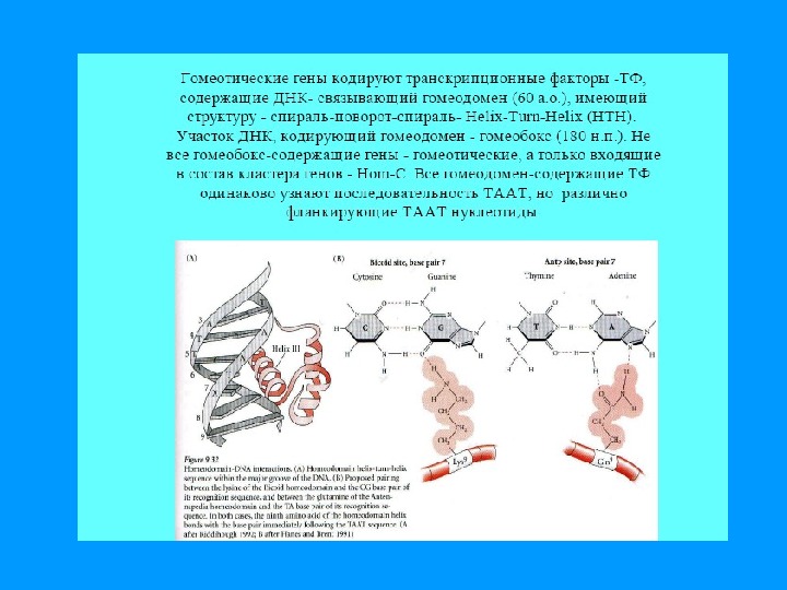
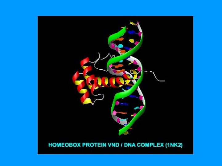
Homeobox & Homeodomain • ДНК связывающий модуль • факторов транскрипции • генов дифференцировки 180 пн 60 аминокислотных остатков
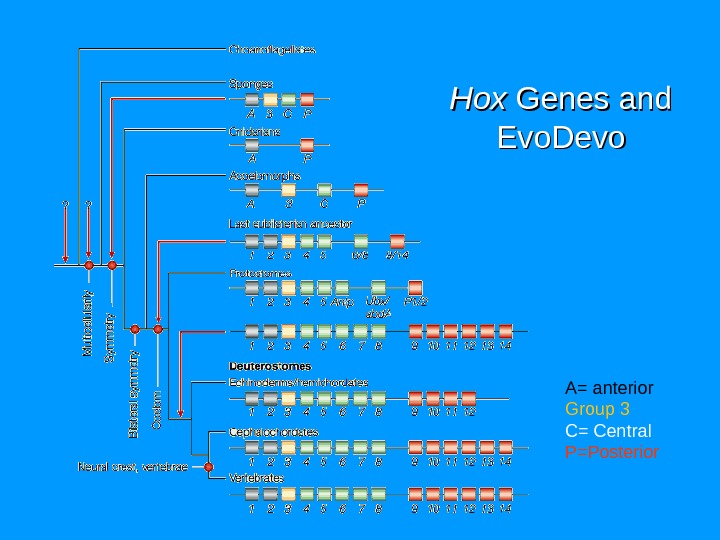
Hox. Hox Genes and Evo. Devo A= anterior Group 3 C= Central P=Posterior
Mutation in Hox. D 13—synpolydactyly Extra digits & interphalangeal webbing (hetero) Similar but more severe & bony malformation of hands, wrists (Homo) Loss of function-mild Gain of function (Polyalanine expansion in Hox. D 13) similar to multiple loss of function-Hox. D 11, 12, 13 Polyalanine expansion- Interfere all three proteins Semidominant effect
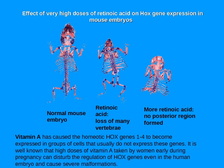
Normal mouse embryo Retinoic acid: loss of many vertebrae More retinoic acid: no posterior region formed Vitamin A has caused the homeotic HOX genes 1 -4 to become expressed in groups of cells that usually do not express these genes. It is well known that high doses of vitamin A taken by women early during pregnancy can disturb the regulation of HOX genes even in the human embryo and cause severe malformations. Effect of very high doses of retinoic acid on Hox gene expression in mouse embryos
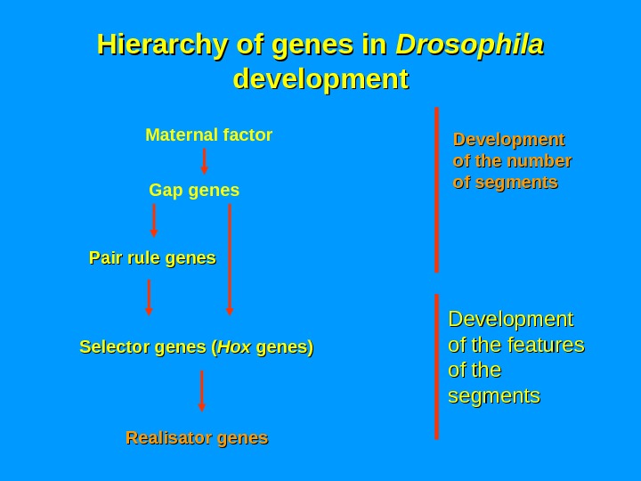
Hierarchy of genes in Drosophila development Realisator genes Maternal factor Pair rule genes Gap genes Development of the number of segments Development of the features of the segments. Selector genes ( Hox genes)
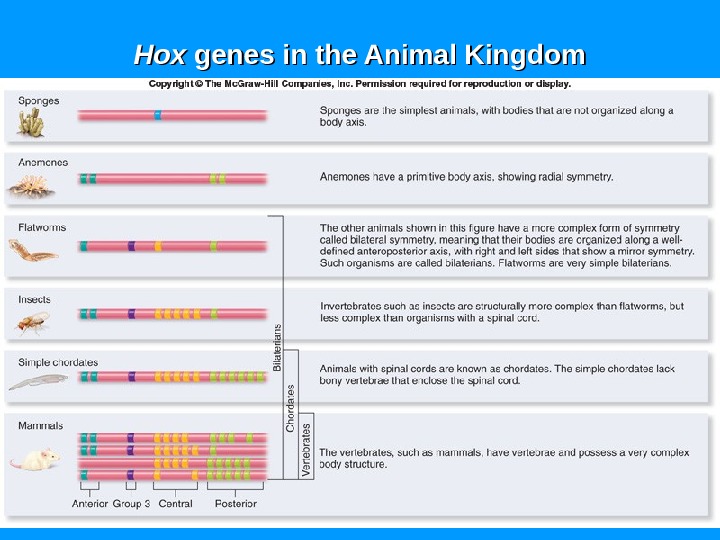
Hox. Hox genes in the Animal Kingdom