
Лимфопролиферативные заболевания . . Проф. М. П. Потапнев


 (2007).")


. Usually monoclonal disease. Lineage committed")
. Mixed phenotype acute leukemia (T or B- myeloid, NK-cell…)")

. Mature B-cell neoplasms - Chronic lymphocytic leukemia/small")
. Mature T-cell and NK-cell neoplasms: - T-cell")


. Chemotherapy,")

. Hodgkin lymphoma: - classical Hodgkin lymphoma, -")


 is mainly found in mantle cell lymphoma,")

 (endemic= EBV positive)")

 in Burkitt’s lymphoma")


")


. . A Bence Jones protein is a monoclonal globulion")
 and serum (down))")


tema_20_limfoproliferativnye_zabolevania.ppt
- Размер: 8.0 Мб
- Автор: Анастасия Медведская
- Количество слайдов: 44
Описание презентации Лимфопролиферативные заболевания . . Проф. М. П. Потапнев по слайдам
Лимфопролиферативные заболевания. . Проф. М. П. Потапнев
Patients with hematological malignancies in Belarus ( ( adults) (2007).
Limphoproliferative diseases
B-cell lymphopoiesis
B-cell malignan- cies
T-cell differen-ti ation stages
Lymphopoiesis in lymph nodes.
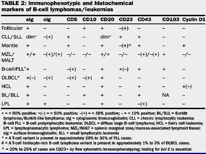
B-cell malignancies
Morphology of leukocytes
Acute leukemia. Originated from bone marrow (>25% blasts). Usually monoclonal disease. Lineage committed morphology (FAB classif. ) B and T or myeloid malignant cells are estimated by immunophenotyping (FAB classif. 1996 classif. ) Cytogenetic abnormalities (WHO classif. 2001, 2008). Fusion genes as markers of disease diagnosis and prognosis.
Acute leukemia (WHO classification, 2008). Mixed phenotype acute leukemia (T or B- myeloid, NK-cell…) B lymphoblastic leukemia/lymphoma with t(9: 22)(q 34; q 11. 2); BCR-ABL 1. B lymphoblastic leukemia/lymphoma with t (v; 11 q 23); MLL rearranged. B lymphoblastic leukemia/lymphoma with t(12; 21)(p 13; q 22) TEL-AML 1 (ETV 6 -RUNX 1) B lymphoblastic leukemia/lymphoma with hyperdiploidy. B lymphoblastic leukemia/lymphoma with hypodiploidy. B lymphoblastic leukemia/lymphoma with t(5; 14)(q 31; q 32); IL-3 -Ig. H B lymphoblastic leukemia/lymphoma with t (1; 19)(q 23; p 13. 3); TCF-PBX 1 T lymphoblastic leukemia/lymphoma.
Cytogenetic and genetic features of ALL. Fusion oncogene Translocation Clinical Frequency Prognosis BCR-ABL t(9; 22)(q 34; q 11) >95% in adult CML, 30% in adult ALL favorable MLL-AF 4 t(4; 11)(q 21; q 23) 5% ALL poor TEL-AML 1 T(12; 21)(p 13; q 22) 25% in pediatric B-ALL favorable E 2 A-PBX 1 T(4; 11)(p 13; q 22) 3 -5% ALL favorable Ig. H, IGL — >95% B-ALL n. d. (diagn. ) TCRδ, TCRγ — >95% T-ALL n. d. (diagn. )
Chronic lymphocytic leukemia (WHO classification, 2008). Mature B-cell neoplasms — Chronic lymphocytic leukemia/small lymphocytic lymphoma , — B-cell prolymphocytic leukemia , — Splenic marginal zone lymphoma , — Hairy cell leukemia, — Lymphoplasmacytic lymphoma, — Waldenstrom macroglobulinemia, — Heavy chain diseases, — Plasma cell myeloma, — -MALT lymphoma, — Follicular lymphoma, — Diffuse large B-cell lymphoma, — Plasmablastic lymphoma, — Burkitt lymphoma.
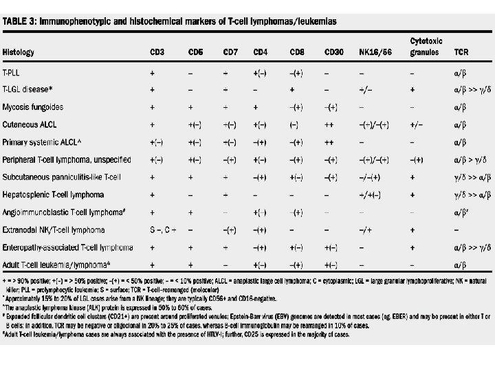
Chronic lymphocytic leukemia (WHO classification, 2008). Mature T-cell and NK-cell neoplasms: — T-cell prolymphocytic leukemia , — T-cell large granular lymphocytic leukemia , — Aggressive NK-cell leukemia, — Adult T-cell leukemia/lymphoma, — Mycosis fungoides, — Sezary syndrome, — Primary cutaneous CD 30+ T cell lymphoproliferative disorders, — Peripheral T-cell lymphoma, — Anaplastic large cell lymphoma… —
Adverse prognostic factors of CLL Diffuse infiltration of bone marrow by lymphocytes; Advanced age; Male gender; Deletions in chr. 17 p (p 53!) or 11 q (ATM !) (5 -10% of pts for each) ; High serum level of beta-2 – microglobulin; Increased fraction of prolymphocytes in PB; >20% of ZAP-70 -positive cells, >30% CD 38+ cells; No rearangement in Ig. H V region. Favorable prognostic factors No diffuse infiltration of bone marrow by lymphocytes; Deletion in chr. 13 q (50% of pts); <20% of ZAP-70 -positive cells, <30% CD 38+ cells; Mutations in Ig. H V region.
Typical B cell phenotype in CLL
Strategy for CLL therapy. First line of therapy : Fludarabine, Cyclophosphamine, Rituximabe (FCR). Chemotherapy, MABs such as alemtuzumab (directed against CD 52) and ofatumumab (directed against CD 20) are also used. Stem cell transplantation – rare. Survival: Subclinical “disease” can be identified in 3, 5% of normal adults and up to 7% of individuals over the age of 70. Survival rate depends on subtypes (6 -8 years to 22 years).
Types of lymphomas.
Hodgkin Lymphoma et al. (WHO, 2008). Hodgkin lymphoma: — classical Hodgkin lymphoma, — Lymphocyte-rich classical Hodgkin lymphoma, … Histiocytic and dendritic cell neoplasms: — histiocytic sarcoma, — Langerhans cell histiocytic, — Follicular dendritic cell sarcoma, … Posttranplantation lymphoproliferative disorders: -plasmacytic hyperplasia, -Infectious mononucleous-like PTLD, -polymorphic PTLD, — monomorphic PTLD (B- and T/NK-cell types), …
Histological diagnosis of HD. The Reed–Sternberg cells are identified as large often bi-nucleated cells with prominent nucleoli and an unusual CD 45 -, CD 30+, CD 15+/- immunophenotype. In approximately 50% of cases, the Reed–Sternberg cells are infected by the Epstein–Barr virus.
The adverse prognostic factors for HD Age ≥ 45 years Stage IV disease Hemoglobin < 105 g/l Lymphocyte count < 600/µl or < 8% Male gender Albumin < 40 g/l White blood count ≥ 15, 000/µl
Stages and Therapy of HD Therapy strategy : radiation therapy +/- chemotherapy. Prognosis : The 5 -year survival rate for those patients with a favorable prognosis was 98%, while that for patients with worse outlooks was at least 85% Stage I is involvement of a single lymph node region (I) (mostly the cervical region) or single extralymphatic site (IIe); Stage II is involvement of two or more lymph node regions on the same side of the diaphragm (II) or of one lymph node region and a contiguous extralymphatic site (IIe); Stage III is involvement of lymph node regions on both sides of the diaphragm, which may include the spleen (IIIs) and/or limited contiguous extralymphatic organ or site (IIIe, IIIes); Stage IV is disseminated involvement of one or more extralymphatic organs
Non-Hodgkin lymphoma Causes The many different forms of lymphoma likely have different causes. These possible causes and associations with at least some forms of NHL include: Infectious agents like Epstein-Barr virus, Human T-cell leukemia virus, Helicobacter pylori, HHV-8 and HIV infection. Chemicals, like diphenylhydantion, dioxin, and phenoxyherbicides. Medical treatments like radiation therapy and chemotherapy. Genetic diseases , like Klinefelter ‘s syndrome, Chediak-Higashi syndrome, ataxia-telangiectasia syndrome Autoimmune diseases , like Sjogren’s syndrome, celiac sprue, rheumatoid arthritis and systemic lupus erythematosis
Cytogenetic analysis for B-cell malignancies t(11; 14) is mainly found in mantle cell lymphoma, but also in B-prolymphocytic leukaemia, in plasma cell leukaemia, in splenic lymphoma with villous lymphocytes, in chronic lymphocytic leukaemia, and in multiple myeloma, herein briefly described; all these diseases involve a B-lineage lymphocyte
Diagnosis of DLBCL by Micro. Array technique: Germinal center B cell DLBCL vs activated (post-germinal center) B cell DLBCL
Burkitt’s lymphoma (rare type of NHL) (endemic= EBV positive)
Immunophenotypic diagnosis of Burkitt’s lymphoma The cells of BL typically express monotypic surface Ig. M, CD 19, CD 20, CD 22, CD 10, Bcl-6, and CD 79 a, and are negative for CD 5, CD 23, Bcl-2, and nuclear terminal deoxyribonucleotide transferase (Td. T). Lack of surface immunoglobulin has been reported in a few cases. The presence of CD 10 and Bcl-6 expression supports the germinal center-cell stage of differentiation. A remarkable feature of BL is the high growth fraction (> 95%) as demonstrated by Ki-67. The leukemic cells of BL express a mature immunophenotype that distinguishes it from precursor B-cell acute lymphoblastic leukemia (ALL).
T (8, 14) in Burkitt’s lymphoma
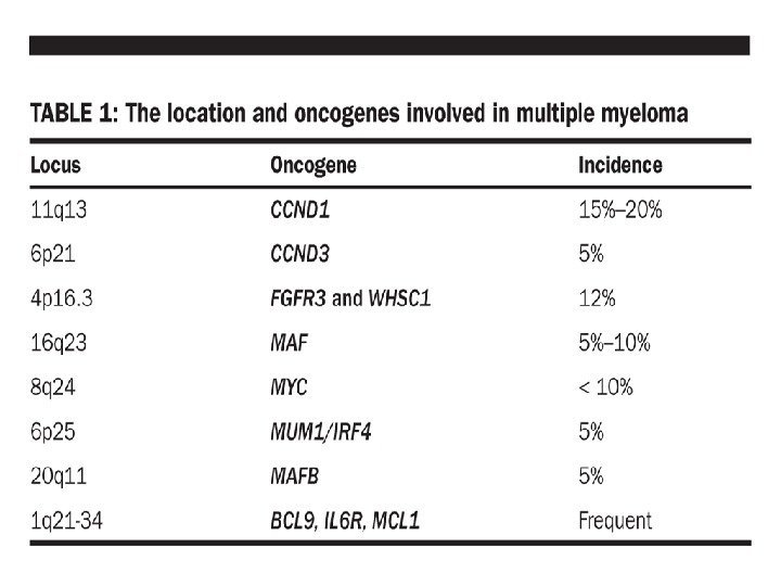
Path from Normal plasma cells through Monoclonal Gammopathy of Undetermined Significance to Multiple Myeloma. .
Plasma cell malignancies
Morphology of malignant plasma cells in blood (H&E staining)
Immunophenotyping of Plasma Cells
Multiple Myeloma diagnosis and therapy. Diagnosis: Roentgen + BM biopsy+. . Therapy: chemotherapy, BMT. Survival: 5 -8 years.
Serum paraprotein detection
M-protein and diseases. More than 50% of patients with serum M protein have an initial clinical diagnosis of MGUS ( M protein <30 g/l in serum, +10% plasma cells in BM). The prevalence of MGUS increases with age, from approximately 1% in patients 50 to 60 years old to greater than 5% in those older than 70 years. The age-adjusted prevalence is higher in males than in females and is twice as high in patients of African descent as in patients of European descent
Waldenstrom macroglobulinemia: pathogenesis Immunophenotype of BM cells in WM Ig light chain — Positive CD 19 — Positive CD 20 — Positive CD 52 — Positive Surface Ig. M — Positive CD 79 b — Positive CD 11 c — Usually negative CD 25 — Positive CD 23 — Usually negative CD 38 — Dim positive FMC 7 — Usually dim positive CD 22 — May be positive CD 5 — Negative CD 10 — Negative CD 27 — Dim positive CD 75 — Usually negative CD 138 — Usually negative Bcl 2 — Dim positive Bcl 6 — Usually absent PAX 5+ — Dim positive CD 45 (RA) — Usually positive
Diagnosis and Therapy of WM.
Light chain Disease (Bence-Jones proteins). . A Bence Jones protein is a monoclonal globulion protein or immunoglobulin light chain found in the urine, with a molecular weight of 22 -24 k. Da. Detection of Bence Jones protein may be suggestive of Multiple Myeloma or Waldenstrom’s macroglobulinemia.
(Bence-Jones protein in serum/urine (up) and serum (down))
HEAVY CHAIN DISEASE Heavy chain disease is a form of paraproteinemia with a proliferation of cells producing immunoglobulin heavy chains There are four forms : alpha chain disease (Seligmann’s disease) gamma chain disease (Franklin’s disease) mu chain disease delta chain disease
Secondary immunodeficiency in lymphoproliferative diseases. 1. Lymphoadenopathy (decreased lymphocyte proliferation to mitogens, T cell subpopulation imbalance). 2. Autoimmunity (autoantibodies, amyloidosis, renal and liver failure, coagulopathy, vasculitis).