
LE 12 -1 Dendrites Perikaryon Nucleus Cell body


- Размер: 21 Mегабайта
- Количество слайдов: 80
Описание презентации LE 12 -1 Dendrites Perikaryon Nucleus Cell body по слайдам
LE 12 -1 Dendrites Perikaryon Nucleus Cell body Axon Telodendria. Axon Regions of a neuron. Dendritic branches Nissl bodies (RER and free ribosomes) Mitochondrion Axon hillock Initial segment of axon Axolemma Golgi apparatus Neurofilament Nucleus Nucleolus Synaptic terminals Structural components of a neuron. Dendrite See Figure 12 -2 POSTSYNAPTIC CELLPRESYNAPTIC CELL
LE 12 -1 a Dendrites Perikaryon Nucleus Cell body Axon Telodendria Regions of a neuron
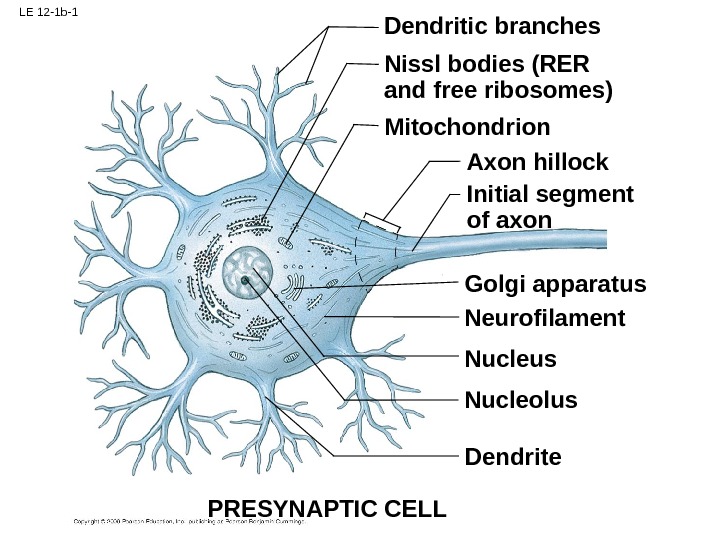
LE 12 -1 b-1 Dendritic branches Nissl bodies (RER and free ribosomes) Mitochondrion Axon hillock Initial segment of axon Golgi apparatus Neurofilament Nucleus Nucleolus Dendrite PRESYNAPTIC CELL
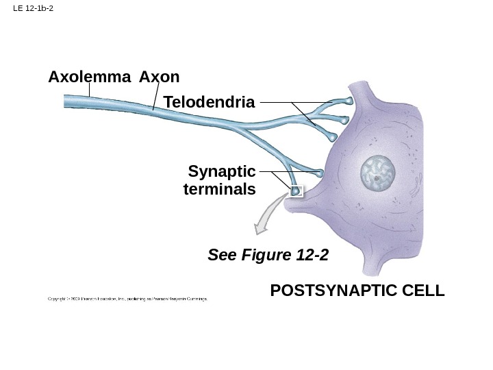
LE 12 -1 b-2 Telodendria. Axon Axolemma Synaptic terminals See Figure 12 -2 POSTSYNAPTIC CELL
LE 12 -2 Telodendrion Synaptic vesicles Presynaptic membrane Postsynaptic membrane Synaptic cleft. Endoplasmic reticulum Synaptic knob Mitochondrion
LE 12 -2 -1 Telodendrion Synaptic vesicles Presynaptic membrane Postsynaptic membrane Synaptic cleft. Endoplasmic reticulum Synaptic knob Mitochondrion
LE 12 -2 —
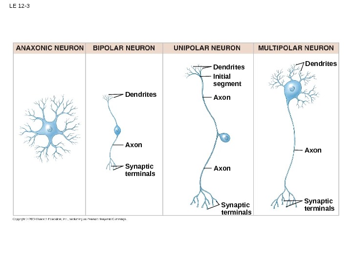
LE 12 -3 Dendrites Axon Synaptic terminals. Axon. Dendrites Initial segment
LE 12 -3 -2 Dendrites Axon Synaptic terminals
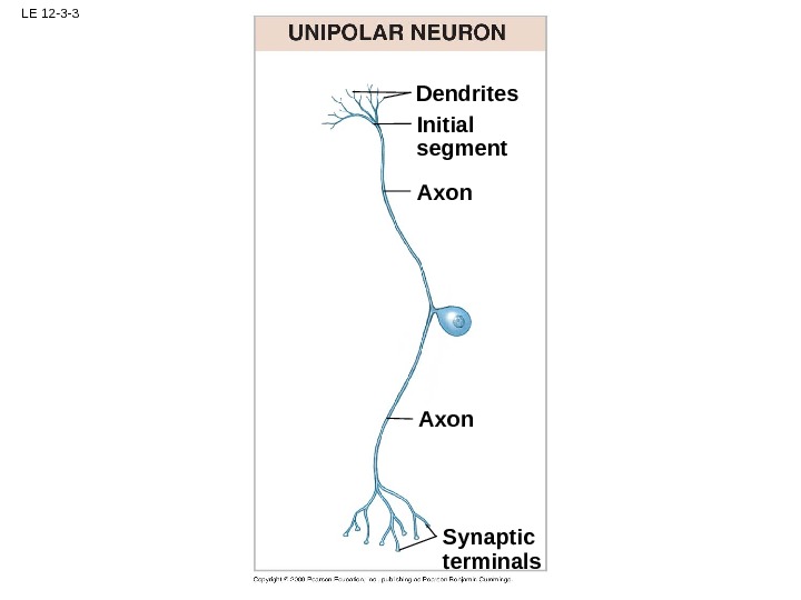
LE 12 -3 -3 Synaptic terminals. Axon. Dendrites Initial segment
LE 12 -3 -4 Synaptic terminals Axon. Dendrites
LE 12 -4 Peripheral Nervous System Central Nervous System containsare found in Satellite cells Oligodendrocytes. Schwann cells Astrocytes Microglia Surround all axons in PNS; responsible for myelination of peripheral axons; participate in repair process after injury Maintain blood–brain barrier; provide structural support; regulate ion, nutrient, and dissolved- gas concentrations; absorb and recycle neurotransmitters; form scar tissue after injury. Myelinate CNS axons; provide structural framework Remove cell debris, wastes, and pathogens by phagocytosis. Line ventricles (brain) and central canal (spinal cord); assist in producing, circulating, and monitoring cerebrospinal fluid Ependymal cells Surround neuron cell bodies in ganglia; regulate O 2 , CO 2 , nutrient, and neurotransmitter levels aound neurons in ganglia
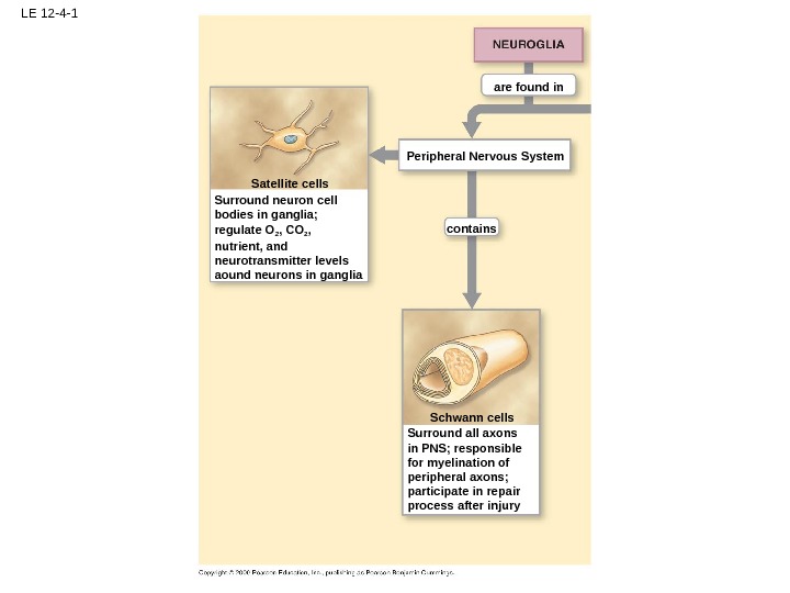
LE 12 -4 -1 Peripheral Nervous System contains are found in Satellite cells Schwann cells Surround all axons in PNS; responsible for myelination of peripheral axons; participate in repair process after injury. Surround neuron cell bodies in ganglia; regulate O 2 , CO 2 , nutrient, and neurotransmitter levels aound neurons in ganglia
LE 12 -4 -2 Central Nervous System containsare found in Oligodendrocytes Astrocytes Microglia Maintain blood–brain barrier; provide structural support; regulate ion, nutrient, and dissolved- gas concentrations; absorb and recycle neurotransmitters; form scar tissue after injury. Myelinate CNS axons; provide structural framework Remove cell debris, wastes, and pathogens by phagocytosis. Line ventricles (brain) and central canal (spinal cord); assist in producing, circulating, and monitoring cerebrospinal fluid Ependymal cells
LE 12 -5 Ependymal cells Gray matter Ependymal cells. Central canal CENTRAL CANAL Microglial cell Astrocyte Unmyelinated axon Basal lamina Capillary. Oligodendrocyte Axolemma. Nodes Myelinated axons Axon Myelin (cut) Neurons. Gray matter White matter Internode
LE 12 -5 a Ependymal cells Gray matter. Central canal
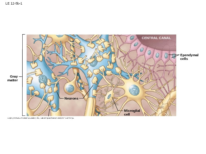
LE 12 -5 b-1 CENTRAL CANAL Microglial cell Ependymal cells Neurons. Gray matter
LE 12 -5 b-2 Astrocyte Unmyelinated axon Basal lamina Capillary. Oligodendrocyte Axolemma Nodes Myelinated axons Axon Myelin (cut) White matter Internode
LE 12 -6 Nucleus Axon hillock Axon Myelinated internode Initial segment (unmyelinated) Nodes Schwann cell nucleus Axon Axolemma Myelin covering internode Myelin. Schwann cell Axons. Neurilemma Myelinated axon Unmyelinated axon Schwann cell nucleus Axons. Schwann cell nucleus Neurilemma Axons Node. Dendrite Neurilemma
LE 12 -6 a Nucleus Axon hillock Axon Myelinated internode Initial segment (unmyelinated) Nodes Schwann cell nucleus Axon Axolemma Myelin covering internode Myelin. Axons. Neurilemma Myelinated axon. Neurilemma Dendrite
LE 12 -6 a-1 Nucleus Axon hillock Axon Myelinated internode Initial segment (unmyelinated) Nodes Schwann cell nucleus Axon Axolemma Myelin covering internode Dendrite Neurilemma
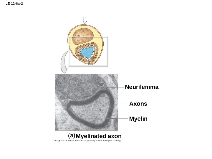
LE 12 -6 a-2 Myelin Axons. Neurilemma Myelinated axon
LE 12 -6 b Schwann cell Axons. Neurilemma Unmyelinated axon Schwann cell nucleus Axons. Schwann cell nucleus Neurilemma Axons Node
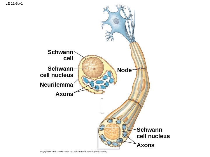
LE 12 -6 b-1 Schwann cell nucleus Axons. Schwann cell nucleus Neurilemma Axons Node
LE 12 -6 b-2 Axons. Neurilemma Unmyelinated axon Schwann cell nucleus Axons
LE 12 -7 Axon continues to grow into distal stump and is enclosed by Schwann cells. Axon sends buds into network of Schwann cells and then starts growing along cord of Schwann cells form cord, grow into cut, and unite stumps. Macrophages engulf degenerating axon and myelin. Schwann cell Macrophage. Fragmentation of axon and myelin occurs in distal stump. Myelin Axon Proximal stump Distal stump Site of injury
LE 12 -7_1 Fragmentation of axon and myelin occurs in distal stump. Myelin Axon Proximal stump Distal stump Site of injury
LE 12 -7_2 Schwann cells form cord, grow into cut, and unite stumps. Macrophages engulf degenerating axon and myelin. Schwann cell Macrophage
LE 12 -7_3 Axon sends buds into network of Schwann cells and then starts growing along cord of Schwann cells.
LE 12 -7_4 Axon continues to grow into distal stump and is enclosed by Schwann cells.
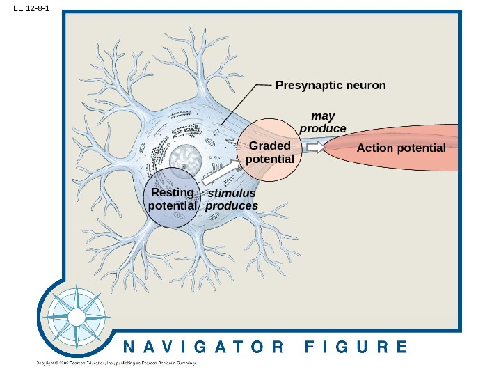
LE 12 -8 Action potential Postsynaptic cell Information processingtriggers. Presynaptic neuron Graded potential may produce stimulus produces. Resting potential. Synaptic activity
LE 12 -8 -1 Action potential. Presynaptic neuron Graded potential may produce stimulus produces. Resting potential
LE 12 -8 -2 Action potential Postsynaptic cell Information processing triggers. Synaptic activity
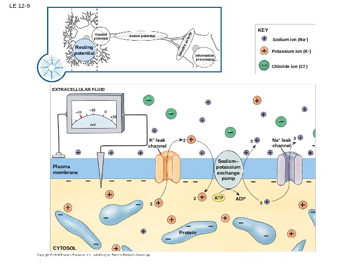
LE 12 -9 EXTRACELLULAR FLUID Resting potential Graded potential Action potential. Synaptic activity Information processing Sodium ion (Na + ) Potassium ion (K + ) Chloride ion (Cl – )KEY Plasma membrane CYTOSOL 2 K + leak channel 2 2 Protein 3 Sodium– potassium exchange pump Na + leak channel
LE 12 -9 -1 EXTRACELLULAR FLUID Sodium ion (Na + ) Potassium ion (K + ) Chloride ion (Cl – )KEY Plasma membrane CYTOSOL K + leak channel 2 2 Protein 3 Sodium– potassium exchange pump Na + leak channel
LE 12 -10 EXTRACELLULAR FLUID Potassium chemical gradient EXTRACELLULAR FLUID Potassium electrochemical gradient (net)Potassium electrical gradient K +– 70 m. V CYTOSOL Na +K + K +– 90 m. V +66 m. V – 70 m. V Potassium chemical gradient Potassium electrical gradient Sodium chemical gradient Sodium electrochemical gradient (net) Sodium chemical gradient Sodium electrical gradient At the normal resting potential, an electrical gradient opposes the chemical gradient for potassium ions (K + ). The net electrochemical gradient tends to force potassium ions out of the cell. If the plasma membrane were freely permeable to sodium ions, the influx of Na + would continue until the equilibrium potential was reached. The chemical and electrical gradients would then be equal and opposite in direction, and no net movement of Na + would occur across the membrane. At the normal resting potential, chemical and electrical gradients combine to drive sodium ions (Na + ) into the cell. If the plasma membrane were freely permeable to potassium ions, the outflow of K + would continue until the equilibrium potential was reached.
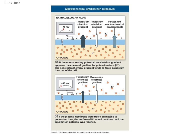
LE 12 -10 ab EXTRACELLULAR FLUID Potassium chemical gradient Potassium electrochemical gradient (net)Potassium electrical gradient K +– 70 m. V CYTOSOL K +– 90 m. V Potassium chemical gradient Potassium electrical gradient At the normal resting potential, an electrical gradient opposes the chemical gradient for potassium ions (K + ). The net electrochemical gradient tends to force potassium ions out of the cell. If the plasma membrane were freely permeable to potassium ions, the outflow of K + would continue until the equilibrium potential was reached.
LE 12 -10 cd EXTRACELLULAR FLUID CYTOSOL Na ++66 m. V – 70 m. V Sodium chemical gradient Sodium electrochemical gradient (net) Sodium chemical gradient Sodium electrical gradient If the plasma membrane were freely permeable to sodium ions, the influx of Na + would continue until the equilibrium potential was reached. The chemical and electrical gradients would then be equal and opposite in direction, and no net movement of Na + would occur across the membrane. At the normal resting potential, chemical and electrical gradients combine to drive sodium ions (Na + ) into the cell.
LE 12 -11 Chemically gated channel Resting state ACh +30 m. VGated channel Activation gate Applied pressure. Mechanically gated channel. Voltage-gated channel – 60 m. V– 70 m. V Inactivation gate Pressure removed. Binding site Channel open Channel closed Channel inactivated Channel closed. Channel open. Channel closed Channel open. Presence of ACh
LE 12 -11 a Chemically gated channel Resting state ACh Gated channel. Binding site Channel open. Channel closed Presence of ACh
LE 12 -11 b +30 m. V Activation gate. Voltage-gated channel – 60 m. V– 70 m. V Inactivation gate Channel inactivated. Channel closed Channel open
LE 12 -11 c Applied pressure. Mechanically gated channel Pressure removed Channel closed. Channel open. Channel closed
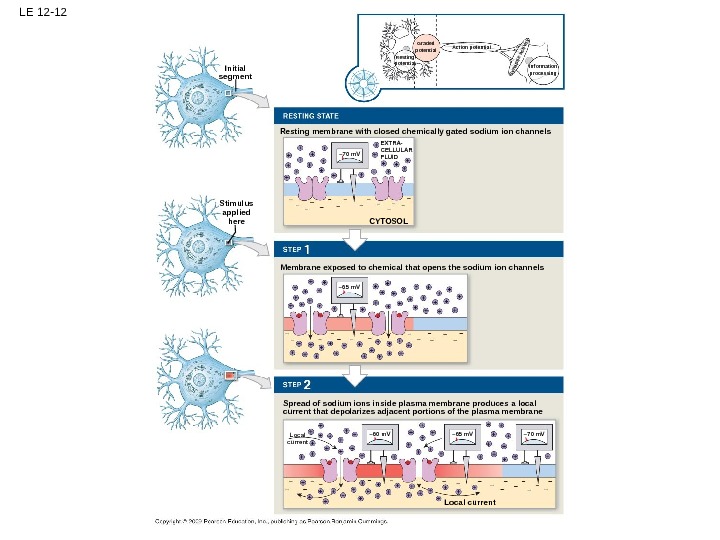
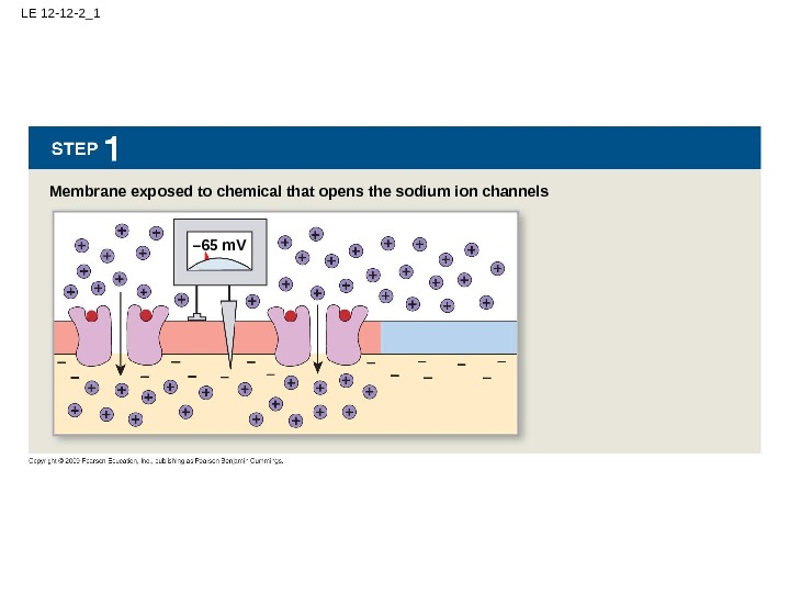
LE 12 -12 Initial segment Stimulus applied here Resting membrane with closed chemically gated sodium ion channels Membrane exposed to chemical that opens the sodium ion channels Spread of sodium ions inside plasma membrane produces a local current that depolarizes adjacent portions of the plasma membrane – 70 m. V CYTOSOL – 65 m. V – 60 m. V – 65 m. V – 70 m. V Local current Local EXTRA- CELLULAR FLUID Resting potential Graded potential Action potential Information processing. Synaptic activity
LE 12 -12 -1 Resting membrane with closed chemically gated sodium ion channels – 70 m. V CYTOSOLEXTRA- CELLULAR FLUI
LE 12 -12 -2_1 Membrane exposed to chemical that opens the sodium ion channels – 65 m. V
LE 12 -12 -3_2 Spread of sodium ions inside plasma membrane produces a local current that depolarizes adjacent portions of the plasma membrane – 60 m. V – 65 m. V – 70 m. V Local current Local
LE 12 -13 ab – 60 – 70 Time. Transmembrane potential (m. V) Chemical stimulus removed. Chemical stimulus applied. Chemical stimulus removed Return to resting potential. Resting potential Hyperpolarization. Repolarization Depolarization –
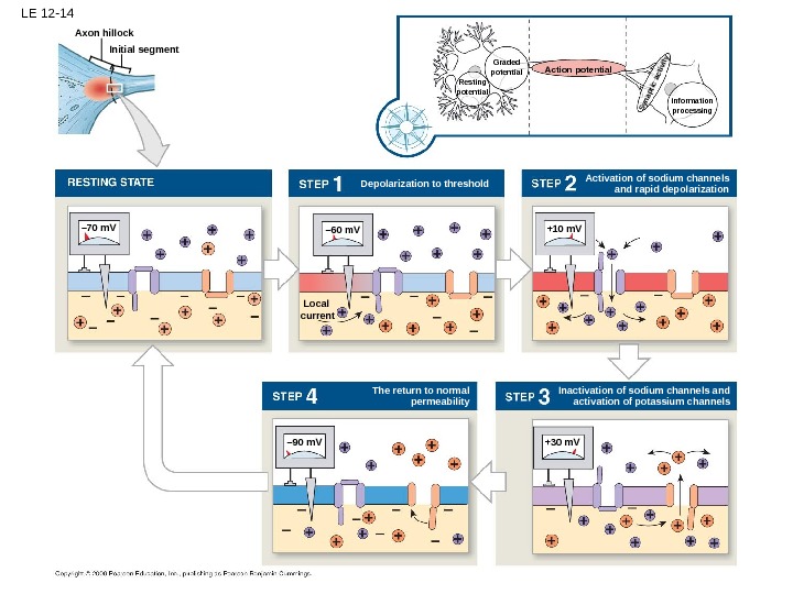
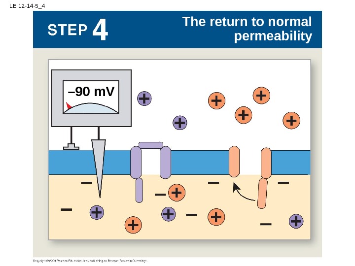
LE 12 -14 Initial segment The return to normal permeability– 60 m. V– 70 m. V Local current Resting potential Graded potential Action potential Information processing. Synaptic activity – 90 m. V +30 m. V +10 m. VAxon hillock Activation of sodium channels and rapid depolarization. Depolarization to threshold Inactivation of sodium channels and activation of potassium channels
LE 12 -14 -1 – 70 m. V
LE 12 -14 -2_1 – 60 m. V Local current Depolarization to threshold
LE 12 -14 -3_2 +10 m. V Activation of sodium channels and rapid depolarization
LE 12 -14 -4_3 +30 m. V Inactivation of sodium channels and activation of potassium channels
LE 12 -14 -5_4 The return to normal permeability – 90 m. V
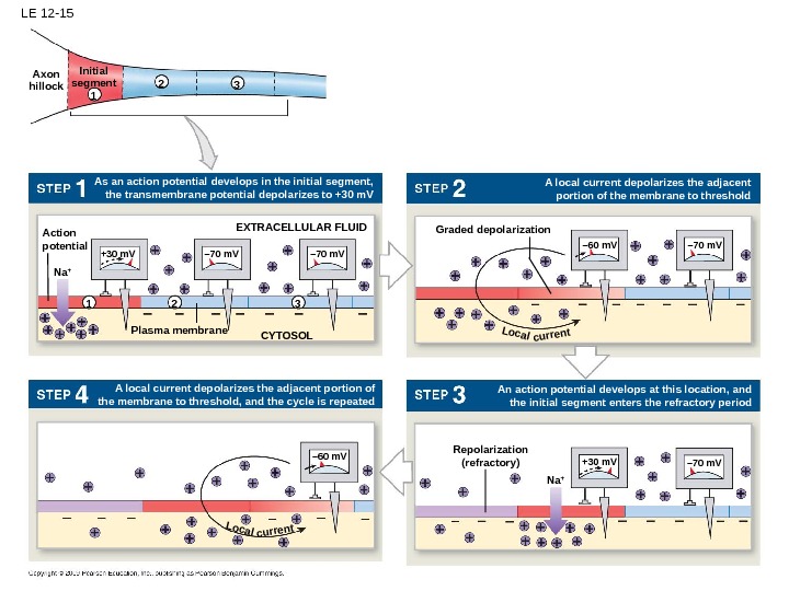
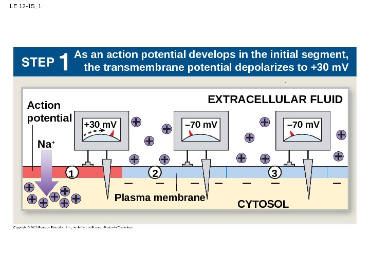
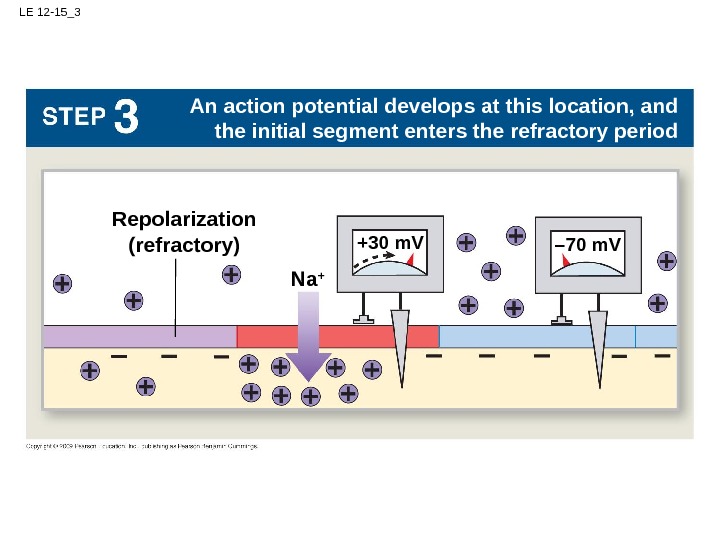
LE 12 -15 EXTRACELLULAR FLUIDInitial segment. Axon hillock – 70 m. V CYTOSOL Na +– 60 m. V +30 m. V– 70 m. V Repolarization (refractory) A local current depolarizes the adjacent portion of the membrane to threshold – 70 m. VLocal. Graded depolarization Plasma membrane. Na +Action potential – 70 m. V – 60 m. V +30 m. V 32 1 As an action potential develops in the initial segment, the transmembrane potential depolarizes to +30 m. V An action potential develops at this location, and the initial segment enters the refractory period. A local current depolarizes the adjacent portion of the membrane to threshold, and the cycle is repeatedcurrent Localcurrent
LE 12 -15_1 EXTRACELLULAR FLUID CYTOSOL– 70 m. V Plasma membrane. Na +Action potential – 70 m. V+30 m. V 3 2 1 As an action potential develops in the initial segment, the transmembrane potential depolarizes to +30 m. V
LE 12 -15_2 – 70 m. V Local current A local current depolarizes the adjacent portion of the membrane to threshold Graded depolarization – 60 m. V
LE 12 -15_3 Na + +30 m. VRepolarization (refractory) – 70 m. V An action potential develops at this location, and the initial segment enters the refractory period
LE 12 -15_4 – 60 m. VLocal. A local current depolarizes the adjacent portion of the membrane to threshold, and the cycle is repeatedcurrent
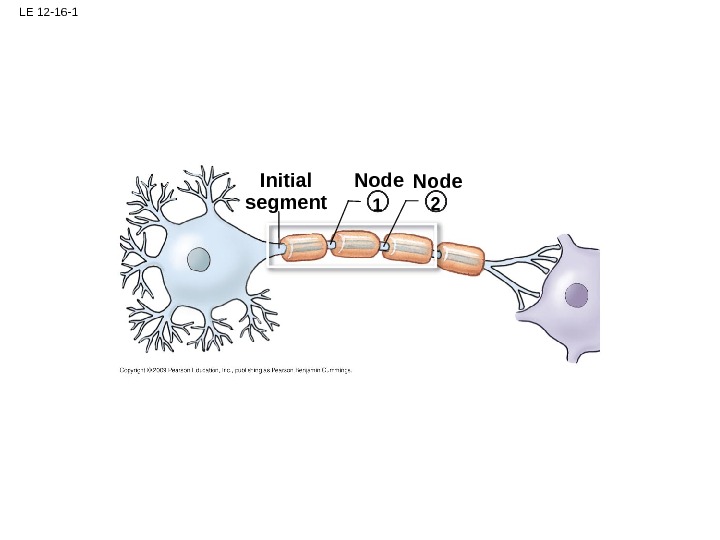
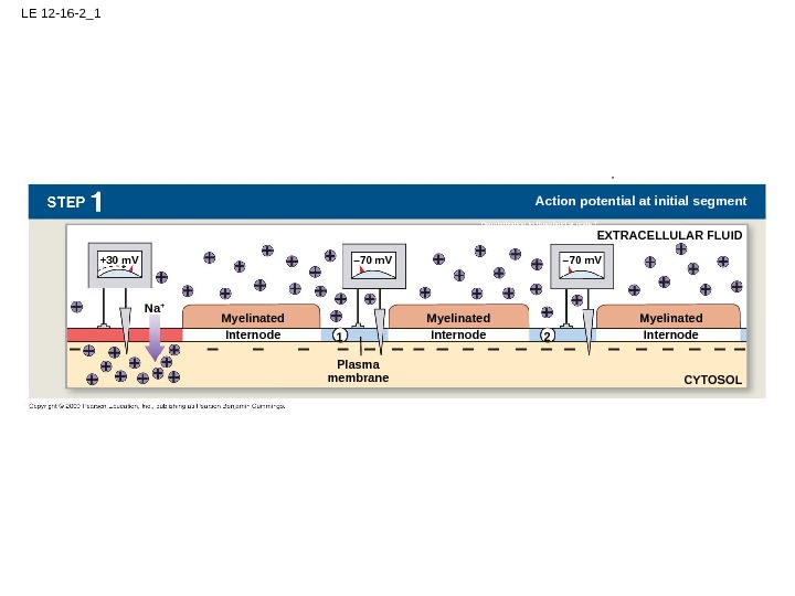
LE 12 -16 Initial segment EXTRACELLULAR FLUID – 70 m. V CYTOSOL Na + – 70 m. V+30 m. V – 70 m. V Repolarization (refractory) – 70 m. V Local current. Plasma membrane. Na + Myelinated Internode – 60 m. V +30 m. V 2 1 21 Action potential at initial segment 1 2 – 60 m. VLocal current Myelinated Internode Action potential at node 1 Depolarization to threshold at node 1 Depolarization to threshold at node 2 Node
LE 12 -16 -1 Initial segment 2 1 Node
LE 12 -16 -2_1 EXTRACELLULAR FLUID CYTOSOL– 70 m. V+30 m. V – 70 m. V Plasma membrane. Na + Myelinated Internode 2 1 Action potential at initial segment Myelinated Internode. Depolarization to threshold at node
LE 12 -16 -3_2 – 70 m. V– 60 m. V Local current Depolarization to threshold at node
LE 12 -16 -4_3 – 70 m. V Na +Repolarization (refractory) +30 m. V Action potential at node
LE 12 -16 -5_4 Local current 1 2 – 60 m. VDepolarization to threshold at node
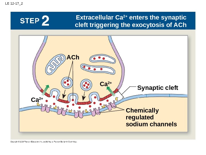
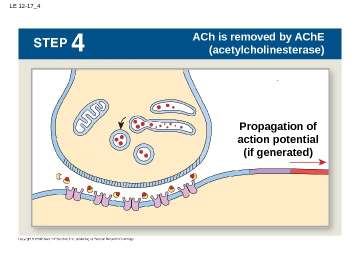
LE 12 -17 Action potential. EXTRACELLULAR FLUIDPRESYNAPTIC NEURON Ca 2+ Synaptic vesicles Na + An action potential arrives and depolarizes the synaptic knob Action potential at initial segment Action potential at node 1 Na + POSTSYNAPTIC NEURON CYTOSOLACh. E ER Action potential Synaptic knob Initial segment Extracellular Ca 2+ enters the synaptic cleft triggering the exocytosis of ACh binds to receptors and depolarizes the postsynaptic membrane ACh is removed by ACh. E (acetylcholinesterase) Information processing. Graded potential Resting potential Ca 2+ACh Chemically regulated sodium channels Synaptic cleft Na + Receptor Propagation of action potential (if generated)Initiation of action potential if threshold is reached. Synaptic activity
LE 12 -17_1 EXTRACELLULAR FLUIDPRESYNAPTIC NEURON Synaptic vesicles An action potential arrives and depolarizes the synaptic knob POSTSYNAPTIC NEURON CYTOSOLACh. E ER Action potential Synaptic knob Initial segment
LE 12 -17_2 Ca 2+ Extracellular Ca 2+ enters the synaptic cleft triggering the exocytosis of ACh Ca 2+ACh Chemically regulated sodium channels Synaptic cleft
LE 12 -17_3 Na + ACh binds to receptors and depolarizes the postsynaptic membrane Na + Receptor Initiation of action potential if threshold is reached
LE 12 -17_4 Action potential at node 1 ACh is removed by ACh. E (acetylcholinesterase) Propagation of action potential (if generated)
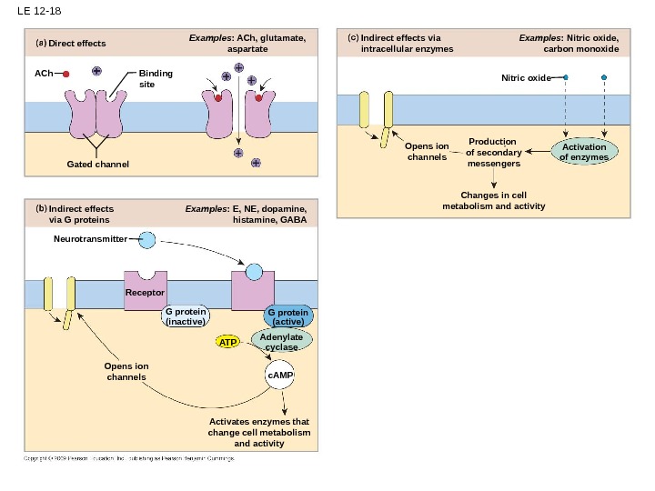
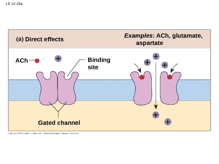
LE 12 -18 Changes in cell metabolism and activity Examples : Nitric oxide, carbon monoxide Opens ion channels Activation of enzymes. Nitric oxide Production of secondary messengers. Indirect effects via intracellular enzymes. Examples : ACh, glutamate, aspartate Examples : E, NE, dopamine, histamine, GABAIndirect effects via G proteins Opens ion channels Activates enzymes that change cell metabolism and activity. Receptor. Neurotransmitter G protein (inactive) G protein (active) Adenylate cyclase. ATP c. AMPGated channel Binding site. ACh Direct effects
LE 12 -18 a Examples : ACh, glutamate, aspartate Gated channel Binding site. ACh Direct effects
LE 12 -18 b Examples : E, NE, dopamine, histamine, GABAIndirect effects via G proteins Opens ion channels Activates enzymes that change cell metabolism and activity. Receptor. Neurotransmitter G protein (inactive) G protein (active) Adenylate cyclase. ATP c. AMP
LE 12 -18 c Changes in cell metabolism and activity Examples : Nitric oxide, carbon monoxide Opens ion channels Activation of enzymes. Nitric oxide Production of secondary messengers. Indirect effects via intracellular enzymes
LE 12 -19 ACTION POTENTIAL PROPAGATIONResting potential Graded potential Action potential Information processing. Synaptic activity ACTION POTENTIAL PROPAGATION TWO SIMULTANEOUS STIMULIFIRST STIMULUS SECOND STIMULUS Threshold reached. Initial segment
LE 12 -19 a ACTION POTENTIAL PROPAGATION FIRST STIMULUS SECOND STIMULUS Threshold reached. Initial segment
LE 12 -19 b ACTION POTENTIAL PROPAGATION TWO SIMULTANEOUS STIMULI Threshold reached
LE 12 -20 Time IPSPTime 2: Hyperpolarizing stimulus applied EPSP Time 1: Depolarizing stimulus applied Time 3: Hyperpolarizing stimulus applied Time 3: Depolarizing stimulus applied. Stimulus removed Stimuli removed. Resting potential– 70 m. V (resting potential)
LE 12 -21 Action potential arrives GABA release Ca 2+ 1. Action potential arrives 2. Less calcium enters 3. Less neurotransmitter released Inactivation of calcium channels 4. Reduced effect on postsynaptic membrane Presynaptic inhibition Presynaptic facilitation. Action potential arrives Activation of calcium channels Ca 2+ 1. Action potential arrives 2. More calcium enters 3. More neurotransmitter released 4. Increased effect on postsynaptic membrane. Serotonin release
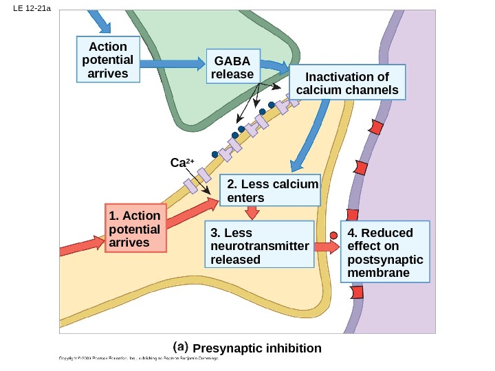
LE 12 -21 a Action potential arrives GABA release Ca 2+ 1. Action potential arrives 2. Less calcium enters 3. Less neurotransmitter released Inactivation of calcium channels 4. Reduced effect on postsynaptic membrane Presynaptic inhibition
LE 12 -21 b Presynaptic facilitation. Action potential arrives Activation of calcium channels Ca 2+ 1. Action potential arrives 2. More calcium enters 3. More neurotransmitter released 4. Increased effect on postsynaptic membrane. Serotonin release