

anemia in children.ppt
- Количество слайдов: 123
 Karazin Kharkiv National University Department of Pediatrics Anemia in children of different age. Assistant Tatyana Golovko
 Аnemia – pathologic state, accompanied by decrease in the level of hemoglobin and the quantity of erythrocytes per unit of volume of the blood.
, hemoglobin (Hb), red blood cells (RBC) concentration >") Anemia is defined as hematocrit (Hct), hemoglobin (Hb), red blood cells (RBC) concentration > 2 SD below age index. 4
Anemia is defined as hematocrit (Hct), hemoglobin (Hb), red blood cells (RBC) concentration > 2 SD below age index. 4
 Anemia is a condition in which a person’s blood has a lower number of red blood cells than it must be in normal condition, or red blood cells don’t have enough hemoglobin. Hemoglobin is an iron-rich protein that gives blood its red color and carries oxygen from the lungs to the rest of the body.
 Normal red blood cells live about 120 days in the bloodstream and then die. Their main role is to carry oxygen, but they also remove carbon dioxide (a waste product) from cells and carry it to the lungs to be exhaled.
 Erythrocytes - less informative index of anemia than the level of hemoglobin. In the general practice the basic criterion of severity is precisely Hb: • Light degree of anemia - Hb 110 -90 g / l, • The average degree of severity - Hb 90 -70 g / l, • Severe anemia - Hb below 70 g / l.
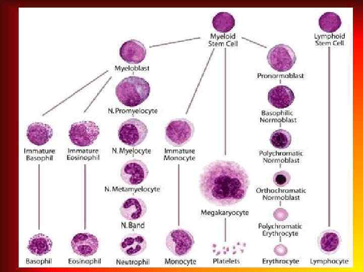
 RETICULOCYTE Reticulocytes are immature red blood cells, typically composing about 1% of the red cells in the human body. Reticulocytes develop and mature in the red bone marrow and then circulate for about a day in the blood stream before developing into mature red blood cells. Normal index: • The relative number of reticulocyte is 0, 5 -1, 2%; • The absolute number of reticulocyte is 30 -70 x 109 / L; • In cord blood of newborns is 20 -60%.
 The clinical index of blood. (Institute of Pediatric Hematology, the drafters of the A. G. Rumyantsev, E. B. Vladimirskaya), Moscow, 1999 Automatical counting Units measurement Normal Level Short Form HGB- Hemoglobin G/L 120 -160 Hb RBC - erythrocyte * 1012 /L 3, 9 -5, 9 Er HCT Hematocryte % 36, 0 -48, 0 Ht
 MCH –") MCV- average volume 1 micron = of erythrocyte 1 - femtoliter (fl) MCH – the average content of Hb in erythrocyte 80 - 95 Pikogram 1 г =1012 pikograms 27, 0 -31, 02 MCHC – average G/dl или concentration of Hb in g %, erythrocyte less g/l 32, 0 -36, 0 RDW – width of the distribution curve of erythrocyte by volume % 11, 5 -14, 5 Colour Index (0, 85 -1, 0) anisocytosis
MCV- average volume 1 micron = of erythrocyte 1 - femtoliter (fl) MCH – the average content of Hb in erythrocyte 80 - 95 Pikogram 1 г =1012 pikograms 27, 0 -31, 02 MCHC – average G/dl или concentration of Hb in g %, erythrocyte less g/l 32, 0 -36, 0 RDW – width of the distribution curve of erythrocyte by volume % 11, 5 -14, 5 Colour Index (0, 85 -1, 0) anisocytosis
 Normal levels of index in CBT in children different ages. Index IU age Neonatal period Early Later neonatal period Erythrocytes (RBC) T/l 7, 2 -5, 4 4, 7 Haemoglobin (Hb) g/l 220 -180 150 Color index - 0, 9 -1, 2 0, 9 -1, 1 Reticulocytes ‰ Thrombocytes g/l Leukocytes (WBC) g/l Early infancy 10 -30 1 -5 years 4, 2 -4, 8 Before 5 month 120150; Older 120 -140 (but not less 110) 130 -150 (but not less 120) 0, 8 -1, 0 5 -10 6 -8 (allowable 4 -9) Older by 14 years 5, 2 (male); 4, 8 (female). 150 -300. By WHO 140 -400 30 -10 5 -12 years 2, 5 -5, 0
 in newborns In newborns of the first and the second") Anemia criteria (capillary blood) in newborns In newborns of the first and the second weeks of life: • Hb < 145 g/L; • Hct < 45 %; • RBC < 4, 5 * 1012/L. The third week and the fourth weeks of life: • Hb < 120 g/L; • Hct < 40 %; • RBC < 4, 0* 1012/L.
Anemia criteria (capillary blood) in newborns In newborns of the first and the second weeks of life: • Hb < 145 g/L; • Hct < 45 %; • RBC < 4, 5 * 1012/L. The third week and the fourth weeks of life: • Hb < 120 g/L; • Hct < 40 %; • RBC < 4, 0* 1012/L.
 : • In infants") Normal index of Hb according WHO criteria (capillary blood) : • In infants from 2 to 6 months > 95 g/L; • In children from 6 months to 5 years > 110 g/L; • Elder than 5 years > 120 g/L.
Normal index of Hb according WHO criteria (capillary blood) : • In infants from 2 to 6 months > 95 g/L; • In children from 6 months to 5 years > 110 g/L; • Elder than 5 years > 120 g/L.
 the commonest cause of anemia, including:") CAUSES OF NEONATAL ANEMIA: 1. BLOOD LOSS (POSTHEMORRHAGIC) the commonest cause of anemia, including: A. Obstetrical causes: placental abruption, placenta previa, trauma to placenta or umbilical cord during delivery and rupture of anomalous placental vessels. B. Feto-maternal transfusion: 8% of normal pregnancies have some blood admixture.
CAUSES OF NEONATAL ANEMIA: 1. BLOOD LOSS (POSTHEMORRHAGIC) the commonest cause of anemia, including: A. Obstetrical causes: placental abruption, placenta previa, trauma to placenta or umbilical cord during delivery and rupture of anomalous placental vessels. B. Feto-maternal transfusion: 8% of normal pregnancies have some blood admixture.
 twins and if") C. Feto-placental transfusion Occurs only with monochorionic (i. e. , monozygotic) twins and if placental vessels allow shunting of blood from one twin to the other. Donor will have anemia of variable severity. Recipient will have polycythemia of variable severity. D. Internal hemorrhage such as intracranial hemorrhage, subgaleal hemorrhage, cephalohematoma, adrenal hemorrhage, subcapsular hematoma of liver etc. E. Iatrogenic blood loss is secondary in frequent sampling of blood for laboratory tests. This is the commonest cause of anemia in newborns.
C. Feto-placental transfusion Occurs only with monochorionic (i. e. , monozygotic) twins and if placental vessels allow shunting of blood from one twin to the other. Donor will have anemia of variable severity. Recipient will have polycythemia of variable severity. D. Internal hemorrhage such as intracranial hemorrhage, subgaleal hemorrhage, cephalohematoma, adrenal hemorrhage, subcapsular hematoma of liver etc. E. Iatrogenic blood loss is secondary in frequent sampling of blood for laboratory tests. This is the commonest cause of anemia in newborns.
 Blood loss Gastrointestinal: gastro-oesophageal reflux, Meckel’s diverticulum, cow’s milk protein intolerance; Parasites – hookworm; Menstruation in adolescent females; Epistaxis (nasal bleeding); Iatrogenic – excessive venepuncture in infants; Bleeding disorders – haemophilia, Willebrand’s disease (Epistaxis; Menorrhagia).
: A. Endogenic causes: Hereditary RBC disorders (rare), including: •") 2. INCREASED RBC DESTRUCTION (HAEMOLYTIC): A. Endogenic causes: Hereditary RBC disorders (rare), including: • RBC Enzyme defects (e. g. , G 6 PD deficiency); • RBC membrane defects (e. g. , hereditary spherocytosis); • Hemoglobinopathies (e. g. , α-thalassemia).
2. INCREASED RBC DESTRUCTION (HAEMOLYTIC): A. Endogenic causes: Hereditary RBC disorders (rare), including: • RBC Enzyme defects (e. g. , G 6 PD deficiency); • RBC membrane defects (e. g. , hereditary spherocytosis); • Hemoglobinopathies (e. g. , α-thalassemia).
 B. Exogenic causes: • Immune hemolysis: -Rh incompatibility, - ABO incompitability, - Minor blood group incompatibility (e. g. , Kell, Duffy), -Hemangiomas (Kasabach-Merritt syndrome). • Setting up hemolysis: - Infection, -Vitamin E deficiency (more often in earlier times but is very rare nowadays), - Drugs.
: A. Anemia of prematurity due to transient deficiency") 3. DECREASED RBC PRODUCTION (HYPOPLASTIC, DEFICIENCY): A. Anemia of prematurity due to transient deficiency of erythropoietin; B. Aplastic or hypoplastic anemia (e. g. , Diamond. Blackfan); C. Bone marrow suppression (e. g. , with Rubella or Parvovirus B 19 infection); D. Nutritional anemia (e. g. , iron deficiency), usually after neonatal period; E. Megaloblastic anemia (B 12 and/or folic acid deficiency).
3. DECREASED RBC PRODUCTION (HYPOPLASTIC, DEFICIENCY): A. Anemia of prematurity due to transient deficiency of erythropoietin; B. Aplastic or hypoplastic anemia (e. g. , Diamond. Blackfan); C. Bone marrow suppression (e. g. , with Rubella or Parvovirus B 19 infection); D. Nutritional anemia (e. g. , iron deficiency), usually after neonatal period; E. Megaloblastic anemia (B 12 and/or folic acid deficiency).
 Physiological anemia of infancy • Normal newborn: - High Hb level progressively declines by 8 -12 week of life up to 9 -11 g/dl. • - Hypoxia stimulates Renal and Hepatic oxygen sensors – erythropoietin production increases. • Preterm: - Hb level declines extremely by 3 -6 week of life to 7 -9 g/dl. There are relatively insensitive Hepatic oxygen sensors; as Renal Oxygen sensors switch on at 40 week of gestation.
 Classification of Anemia according to the leading mechanism of genesis I. Anemia resulting from blood loss: A) Acute; B) Chronical. II. Anemia resulting from a insufficiency of erythropoesis: A) Inheritable aplastic anemia (atrophic anemia, anemia gravis): a) Pancytopenia (Fanconi's anemia, Estren-Dameshek anemia): b) With partial damage of erythroid lineage (Blackfan. Diamond syndrome);
 Acquired aplastic anemia (atrophic anemia, anemia gravis): a) With Pancytopenia (acute, subacute, chronic);") B) Acquired aplastic anemia (atrophic anemia, anemia gravis): a) With Pancytopenia (acute, subacute, chronic); b) With partial damage of erythroid lineage. Which includes transitory erythroblastopenia in infants; C) Dyserythropoietic anemia (Inheritable, Acquired); D) Sideroblastic anemia( Inheritable, Acquired); E) Deficiency anemia: a) megaloblastic anemia: - folic acid deficiency anemia; - B 12 Deficiency Anemia; - orotic aciduria.
B) Acquired aplastic anemia (atrophic anemia, anemia gravis): a) With Pancytopenia (acute, subacute, chronic); b) With partial damage of erythroid lineage. Which includes transitory erythroblastopenia in infants; C) Dyserythropoietic anemia (Inheritable, Acquired); D) Sideroblastic anemia( Inheritable, Acquired); E) Deficiency anemia: a) megaloblastic anemia: - folic acid deficiency anemia; - B 12 Deficiency Anemia; - orotic aciduria.
 microcytic anemia: - iron deficiency (asiderotic anemia); - copper deficiency; - Lead poisoning;") b) microcytic anemia: - iron deficiency (asiderotic anemia); - copper deficiency; - Lead poisoning; - Thalassaemia. F) physiologic anemia in infant: G) Early anemia in preterm. III. Hemolytic anemia: A) Inherited: a) Membranopathy (spherocytosis, elliptocytosis); b) Enzymopathy (Glucoze-6 -Phosphate-Dehydrogenaze); c) Hemoglobinopathies (sickle cell anemia, unstable hemoglobins).
b) microcytic anemia: - iron deficiency (asiderotic anemia); - copper deficiency; - Lead poisoning; - Thalassaemia. F) physiologic anemia in infant: G) Early anemia in preterm. III. Hemolytic anemia: A) Inherited: a) Membranopathy (spherocytosis, elliptocytosis); b) Enzymopathy (Glucoze-6 -Phosphate-Dehydrogenaze); c) Hemoglobinopathies (sickle cell anemia, unstable hemoglobins).
 Aquired: A. Immunopathologic; Б. Infectious; B. Vitamin deficiency; Г. Toxic; Д. Marchiafava-Micheli") A) Aquired: A. Immunopathologic; Б. Infectious; B. Vitamin deficiency; Г. Toxic; Д. Marchiafava-Micheli anemia; Е. Disseminated intravascular coagulation - the syndrom of different etymology and mechanical damage of erythrocytes.
A) Aquired: A. Immunopathologic; Б. Infectious; B. Vitamin deficiency; Г. Toxic; Д. Marchiafava-Micheli anemia; Е. Disseminated intravascular coagulation - the syndrom of different etymology and mechanical damage of erythrocytes.
 IV. Mixed genesis anemia: A. In acute infections, sepsis. Б. In burns. B. In tumors and leucosis. Г. In endocrinopathy.
 Anemia are divided in according to number of reticulocyte : • Regenerative- reticulocyte from 1. 5 to 5% (or 15 to 50 ppm) • Hyperregenerative - reticulocyte more than 5% (or more than 50 ppm) • Aregenerative - Low reticulocyte (less than 0. 5%), not according to the severity of anemia or lack of reticulocyte.
 Anemia are divided in according erythrocytic indexes Index Normal Level MCV- average volume 80 – 95 fl of erythrocyte MCH – the average content of Hb in erythrocyte Ø Ø Ø Increase microcytic anemia Decrease macrocytic anemia hypochromic hyperchromic 27, 0 -31, 02 pg anemia microcytic anemia (MCV less than 75 fl); normocytic anemia (MCV 75 -100 fl); macrocytic anemia (MCV more than 100 fl).
 Classification of anemia Reticulocyte count Microcytic anemia Normocytic anemia Macrocytic anemia Aregenerative Iron deficiency, Lead poisoning, Chronic disease, Aluminum toxicity, Copper deficiency, Protein malnutrition Chronic disease, RBC aplasia, Malignancy, Juvenile rheumatoid arthritis, Endocrinopathies, Renal faile Folate deficiency, Vitamine B 12 deficiency, Aplastic anemia, Congenital bone marrow dysfunction, Drug induced, Trisomy 21, Hypothyroidism. Regenerative Thalassemia trait, Sideroblastic anemia Acute bleeding, Hypersplenism, Dyserythropoietic anemia II. Hyperregenerative Thalassemia syndromes, Hemoglobin C disorders. Antibody-mediated hemolysis, Hypersplenism, Microangiopathy, Membranopathies, Enzyme disorders, Hemoglobinopathies. Dyserythropoietic anemia I, III. Active hemolysis.
 Anemia are divided in order of severity Mild Hb 91 -110 g/l Erythrocytes (for children under 6 years old); 3 -3, 5 х 1012/l Hb 91 -120 g/l (for children older 6 years old) Hb 71 -90 g/l Erythrocytes 2, 5 -3 х 1012/l Hb менее 70 g/l Erythrocytes less than 2, 5 х 1012/l Moderate Severe
 Morphology Nomenclature of Erythrocytes Acanthocyte Dacrocyte Drepanocyte Echinocyte Elliptocyte Megalocyte Schizocyte Spherocyte Stomatocyte Spicule: Liver disease Tear: Thalassemia, Heinz body Sickle: Sickle cell anemia Sea urchin: Senescent/metabolic Oval: Hereditary elliptocytosis Large: Megaloblastic anemia Cut: Microangiopathic hemolytic Sphere: Hereditary Spherocytosis Mouth: Hereditary or acquired
 Anemia are divided in according Colour Index Ø Normochromal anemia – colour index = 0, 85 - 1, 05; Ø Hypochromal anemia – colour index < 0, 85; Ø Hyperchromal anemia – colour index > 1, 05.
 • IDА is recorded in 20% of the world's population.") IRON DEFICIENCY ANEMIA (IDA) • IDА is recorded in 20% of the world's population. • 83 -90% of all anemia constitute IDA. • In children the first 2 years of life the frequency of iron deficiency anemia is 73%. • The second peak of IDA development is adolescence.
IRON DEFICIENCY ANEMIA (IDA) • IDА is recorded in 20% of the world's population. • 83 -90% of all anemia constitute IDA. • In children the first 2 years of life the frequency of iron deficiency anemia is 73%. • The second peak of IDA development is adolescence.
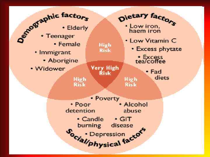
 CAUSES FOR DEVELOPMENT of IDA • Alimentary iron deficiency as a consequence of an unbalanced diet; • Increasing demand in iron and the reduction of the deposit (more often in multiple pregnancies, in prematurity, while lactation and rapid growth periods, also in sportmen); • Chronic blood loss (nasal bleeding, diaphragmatic hernia, and bleeding from GIT and diverticulitis, menorrhagia, renal hemorrhage, idiopathic lung hemosiderosis); • Reduction of iron absorption (malabsorption, chronic inflammatory diseases of GIT, gastrectomy).
 The total content of iron in the body is about 4. 2 g. They are: - 75 -80% belongs to the hemoglobin; - 20 - 25% reserve; - 5 -10% part of the myoglobin; -1% is part of the enzyme for the tissue respiration.
 Anemic Syndrome - Decrease amount of Hemoglobin Complaints: General weakness, reduction in the appetite, physical and mental fatigue, dispnae, vertigo, noise in the ears, flashing “flies” before the eyes, unconscious states, in heavy cases – leads to coma. Symptoms: the pallor of the skin and mucosa, tachycardia, hypotonia, the expansion of the boundaries of heart, muting tones and systolic murmur. Laboratory signs: a decrease in the level Hb and a drop in hematocrit (lower than 35% in children, 37% in girls and 42% in young men)
 • dystrophic changes in the skin and its appendages") Sideropenic Syndrome (Deficit of Iron) • dystrophic changes in the skin and its appendages (shedding of hair, the brittleness of nails), the atrophy of the mucous membranes of nose, esophagitis and gastritis, gingivitis, glossitis, angular stomatitis; • the distortion of taste and sense of smell • muscular pain (deficit of myoglobin) • muscular hypotonia • alteration in the nervous system: slowing down of conditioned reflexes, decrease attentiveness, worsening of memory, delay of intellectual development.
Sideropenic Syndrome (Deficit of Iron) • dystrophic changes in the skin and its appendages (shedding of hair, the brittleness of nails), the atrophy of the mucous membranes of nose, esophagitis and gastritis, gingivitis, glossitis, angular stomatitis; • the distortion of taste and sense of smell • muscular pain (deficit of myoglobin) • muscular hypotonia • alteration in the nervous system: slowing down of conditioned reflexes, decrease attentiveness, worsening of memory, delay of intellectual development.
 Laboratory Signs of Iron Deficit Anemia v A decrease MCV - less than 75; v Reduction in the color index - less than 0, 85; v Increase RDW; v A decrease MCHC - less than 30. v Morphology of the erythrocytes - hypochromic, anisocytosis and poikilocytosis. § § Biochemical - decrease level of serum ferritin; Decrease level of serum iron; Increase Total Iron Binding Capacity (TIBC); Increase level of serum transferrin.
 Ø Pre-latent (exhaustion of iron reserve") Developmental Stages of Iron Deficiency Anemia (WHO, 1977) Ø Pre-latent (exhaustion of iron reserve in tissues; index of the blood within the standard; there are no clinical manifestations); Ø Latent (deficit of iron in the tissues and the decrease of its transport; index of the blood within the standard; clinical picture is caused by the sideropenic syndrome); Ø Iron Deficiency Anemia (deviation of the standard index of the blood in dependence on the degree of severity; the clinical manifestations in the form of sideropenic syndrome and general anemic symptoms).
Developmental Stages of Iron Deficiency Anemia (WHO, 1977) Ø Pre-latent (exhaustion of iron reserve in tissues; index of the blood within the standard; there are no clinical manifestations); Ø Latent (deficit of iron in the tissues and the decrease of its transport; index of the blood within the standard; clinical picture is caused by the sideropenic syndrome); Ø Iron Deficiency Anemia (deviation of the standard index of the blood in dependence on the degree of severity; the clinical manifestations in the form of sideropenic syndrome and general anemic symptoms).
 Differential Diagnosis of Iron Deficiency Anemia it is carried out with other forms of the hypochromic anemia: • Thalassemia - there are no signs of deficit of iron. Synthesis of polypeptide chains reduction which include the structure of normal haemoglobin. • Sideroblastic Anemia – can be diagnosed by the puncture specimen of bone marrow. • Chronic poisoning by lead - the erythrocytes destruction because their membrane damages. It is followed by the increasing of iron level in blood. • Against the background chronic infectious and inflammatory diseases - hypochromic normocytic anemia, normal or increased level of ferritin in combination with the lowered content of serum iron and transferrin.
 Ferritin • Water-soluble complex of iron hydroxide with the protein apoferritin. • It is located in cells of the liver, spleen, bone marrow, in the reticulocytes. • Ferritin is the basic protein in human which deposits iron and concentration of ferritin in the serum which reflects the reserve of iron in the organism. • Normal level of ferritin in blood in male 20 -250 mkg/l, in female 10 -120 mkg/l.
. • Main function to transport of absorbed iron in the depot") Serum Transferrin (Beta-globulin). • Main function to transport of absorbed iron in the depot (liver, spleen), into the medullary erythroid predecessors and into the reticulocytes. • Basic place of synthesis is liver. • An increase in the content of transferrin with lowering in the level of iron of serum is the characteristic for the iron-deficiency state. • In the loss of protein (for example, in nephrotic syndrome) and damage of the liver (different genesis) a decrease in the level of transferrin may occur. • The level of transferrin is increased in the last term of pregnancy. • Normal level of transferrin in serum 2, 0 -4, 0 g/l.
Serum Transferrin (Beta-globulin). • Main function to transport of absorbed iron in the depot (liver, spleen), into the medullary erythroid predecessors and into the reticulocytes. • Basic place of synthesis is liver. • An increase in the content of transferrin with lowering in the level of iron of serum is the characteristic for the iron-deficiency state. • In the loss of protein (for example, in nephrotic syndrome) and damage of the liver (different genesis) a decrease in the level of transferrin may occur. • The level of transferrin is increased in the last term of pregnancy. • Normal level of transferrin in serum 2, 0 -4, 0 g/l.
 Transferrin •
 Iron absorption: – We take in 10 -30 mg Fe/day; – We absorb 0. 5 -1. 0 mg/day (5. 5 -10%); – Absorption may increase to ~20% in deficiency states; – Meat iron (heme) is much better absorbed than vegetable iron; – Poorest Fe absorption with grains; – Vitamin C promotes the increased Fe uptake.
 Treatment of Iron Deficiency Anemia • Diet: meat, liver, yeast, fish. • Oral preparations: the rate of absorbtion of the iron does not differ from parenteral introduction, side effects are less, excessive introduction does not lead to hemosiderosis. - Dosage: 1 hour before meat with verjuice (apple juice) in the evening time (absorption increase in the second-half of a day). If child have pain in abdomen take a drugs with meal.
 During first 3 days - half dose of the selected active substance. Possibilities: dark colour of stool and transitory dyspeptic disorders (nausea, diarrhea or watery stool) can be observed. Colour of urine may be red. Check analysis of the blood: in 7 -10 days – reticulocyte reaction; in 4 weeks - increase Hb and Ht. During the normalization of the indices of the blood – reduce the dose of active substance but not discontinue. Treatment must continue during 8 -9 weeks.
 Therapeutic dose of Iron drug 0 – 3 years – 5 – 8 mg/kg/day; 3 – 7 years – 100 mg/day; > 7 years – 200 mg/day.
 Indication for Parenteral Introduction of Iron • in exceptional cases; • in severe iron deficiency anemia; • intolerance of oral drugs (after repeated replacement and reduction in the dose); • diseases of gastro-intestinal tract; • syndrome of the disrupted intestinal absorbtion; • after the extensive resection of the small intestine; • continuous blood loss;
; • General reactions (anaphylaxis, fever,") Complications of Parenteral Introduction • Local reactions (pains, phlebitis); • General reactions (anaphylaxis, fever, head and articulate pains, vomiting, rash, bronchospasm). Drugs for parenteral introduction: Venofer - for the intravenous introduction, Maltofer, Ferrum-Lek – for the intramuscular introduction.
Complications of Parenteral Introduction • Local reactions (pains, phlebitis); • General reactions (anaphylaxis, fever, head and articulate pains, vomiting, rash, bronchospasm). Drugs for parenteral introduction: Venofer - for the intravenous introduction, Maltofer, Ferrum-Lek – for the intramuscular introduction.
 Signs of overdose of Iron In the first 6 -8 hours - epigastral pains, nausea, vomiting (including with the blood), diarrhea, pallor, sleepiness, acrocyanosis). For 12 -24 hours - metabolic acidosis, leukocytosis, there can be spasms, coma, after 2 -4 days - necroses of the liver and kidneys. Treatment: emetic means, stomach lavage, the method of milk with the egg white, Deferoksamin, Desferal, symptomatic therapy.
 Iron Overload Syndrome • ! Human does not have special mechanism of the excretion of iron! Its excessive introduction leads to hemosiderosis. Clinical manifestations: Gradual increase of the liver and spleen sizes, cardiopathy, adrenal insufficiency, diabetes mellitus, eunuchoidism. Laboratory signs: • Increase of serum iron (more than 30 mmol/liter), percentage of saturation transferrin of iron is more than 45%, ferritin of serum is more than 1000 ng/ml; Test with desferalom; + the specific signs of the defect of internal organs (ECG, level biochemical index of functions of the liver, the level of hormones and others).
 Megaloblastic anemia it is inheritable and acquired anemia when in bone marrow the megaloblasts are present. Megaloblastic anemia is an uncommon problem in childhood that is most frequently associated with vitamin deficiency or gastrointestinal disease. The megaloblastic effect is characterized by an aregenerative macrocytic anemia with nuclear dysmaturity B 12 and Folic acid Deficiency more often are causes this disease in children. Megaloblastic anemia is most often caused by an acquired lack of vitamin B 12 or lack of folic acid. Inherited abnormalities of the metabolism of these nutrients may be the cause.
 Causes of B Deficiency Anemia 12 • Decreased ingestion (e. g. , poor dietary intake); • Atrophy of the mucous membrane of stomach as the most frequent reason, gastroectomy; • Inflammatory or autoimmune diseases of small intestine, the removal of its certain sections; • Helminthic invasion (tapeworms); • Impaired absorption (e. g. , failure to release B-12 from protein, IF deficiency, chronic pancreatic disease, competitive parasites, intrinsic intestinal disease); • Impaired use (eg, congenital enzyme deficiencies, lack of transcobalamin II, administration of nitrous oxide). Inadequate vitamin B-12 dietary intake is rare in children, though it may be seen in breastfed infants whose mothers are themselves deficient.
 Causes of Folic Acid Deficiency • Alimentary; • Increased need (prematurity birth, rapid growth rate, pregnancy); • Feeding by the goat milk; • Disease of the small intestine; • Comsumtion of folate antagonist with Methotrexate, Anti-convulsant (diphenine), oral contraceptives; • Chronic hemolysis.
 Clinical Anemic syndrome; Skin is pale with the lemon shade; Slight jaundice of the scleras; Glossitis, stomatitis, hyperpigmentation, and weight loss; • Disturbance of the proliferation of the epithelium of gastro-intestinal tract: dry-red tongue, loss of appetite, achylia (an absence or severe deficiency of hydrochloric acid and pepsinogen (that is, pepsin) in the stomach), diarrhea, erosive and ulcerous changes in the mucous membranes. • •
 Signs only for B -Deficiency Anemia 12 Damage of CNS - funicular myelosis (degeneration and the sclerosis of the posterior and lateral horns of spinal cord), paresthesia, paralyses with the disorder of the function of pelvic organs.
 Diagnosis of B and Folic acid Deficiency Anemia 12 •
 Bone marrow: • Irritation in erythroid lineage, megaloblasts, the disintegration of erithrokaryocytes. Biochemical Analysis of Blood • an increase in unconjugated bilirubin; • an increase in serum iron. • В 12 vitamin and Folic acid in blood– decrease
 Laboratory Criteria of B 12 and Folic acid Deficiency Anemia Form of disease Latent form Folic acid Deficiency B 12 Deficiency Concentration of folates in serum Concentration of vitamin B 12 in serum decrease less than 5 ng/ml (normal decrease less than 200 pg/ml (normal level 5 -20 ng/ml) level 200 -800 pg/ml) Normal concentration of folic acid in erythrocytes. Manifest form Normal concentration of folic acid in erythrocytes. Concentration of folates in serum decrease less than 3 ng/ml Concentration of vitamin B 12 in serum decrease less than 100 pg/ml Decrease concentration of folic acid in erythrocytes less than 100 ng/ml (normal level 74 -640 ng/ml) Decrease concentration of folic acid in erythrocytes (above 150 ng/ml). Increase renal excretion of formiminoglutamic acid Increase renal excretion of methylmalonic acid
 Treatment of megaloblastic anemia • Treatment of megaloblastic anemia depends on the underlying cause. Folate deficiency due to dietary deficiency or increased demands is best treated with folate supplements. In addition, a diet rich in green, leafy vegetables is essential for normal intake of folic acid. • Folate deficiency caused by the use of sulfa drugs or other antifolate medications may be addressed by folate supplementation or by reducing or eliminating the drug. Folate deficiency due to celiac sprue requires treatment of the underlying disorder and folate supplements.
 • Oral B-12 supplementation is as effective as parenteral supplementation in patients with nutritional deficiency. Even in patients with intrinsic factor (IF) deficiency, oral supplements may be effective, using higher doses. • Often, however, high-dose oral B-12 supplements are unsuccessful in patients with IF deficiency or who have undergone intestinal surgery. These patients may require parenteral supplementation because of impaired secretion or absorption of IF. • Because vitamin B-12 is contained exclusively in animal products (meat), vitamin supplementation is the only means of appropriate vitamin B-12 intake in individuals choosing vegetarian diets.
 Criteria of Effective Treatment • Subjective improvement of patient condition during the first days of treatment; • Reticulocytosis, maximally expressed (to 20%) on the 5 -7 th day of treatment; • Increase in hemoglobin and number of erythrocytes, beginning from the 2 nd week of treatment; • The normalization of the blood index (number of leukocytes and thrombocytes ) in 3 -4 weeks of treatment.
 Folic acid 0 -6 months: 65 mcg/day PO 7 -12 months: 80 mcg/day PO 1 -4 years: 150 mcg/day PO 4 -9 years: 200 mcg/day PO 9 -14 years: 300 mcg/day PO 14 -18 years: 400 mcg/day PO Upper limit: 1 -4 years, 300 mcg/day PO; 4 -8 years, 400 mcg/day PO. Folic Acid Deficiency Infants: 15 mcg/kg/day or 50 mcg/day IV/PO/IM/SC 1 -10 years: 1 mg/day IV/PO/IM/SC initially, then 0. 1 -0. 4 mg/day Cyanocobalamin 0 -6 months: 0. 4 mcg 7 -12 months: 0. 5 mcg 1 -3 years: 0. 9 mcg 4 -8 years: 1. 2 mcg 9 -13 years: 1. 8 mcg >14 years: 2. 4 mcg. Pernicious Anemia 30 -50 mcg IM/SC once daily for 2 weeks for total dose of 1, 000 mcg to 5, 000 mcg administer concomitantly with 1 mg/day of folic acid for 1 month. Maintenance: 100 mcg IM/SC monthly. B 12 Deficiency 0. 2 mcg/kg for 2 days; follow by 1, 000 mcg/day for 2 -7 days; follow by 100 mcg/day for 2 -7 days; then 100 mcg/week for 1 month. Maintenance: 100 mcg IM/SC monthly
 Hemolytic anemia = reduced red-cell life span. Hemolysis is the premature destruction of erythrocytes. A hemolytic anemia will develop if bone marrow activity cannot compensate for the erythrocyte loss. Mild hemolysis can be asymptomatic while the anemia in severe hemolysis can be life threatening and cause angina and cardiopulmonary decompensation. Hemolysis can be due to hereditary and acquired disorders. Hemolytic anemia represents approximately 5% of all anemias. Mechanism of hemolysis: - intravascular; - extravascular.
 • Intravascular hemolysis occurs in hemolytic anemia due to the following: - Artificial cardiac valves; - Glucose-6 -phosphate dehydrogenase (G 6 PD) deficiency; - Thrombotic thrombocytopenic purpura; - Disseminated intravascular coagulation; - Transfusion of ABO incompatible blood; - Paroxysmal nocturnal hemoglobinuria (PNH). Red cells destruction occurs in vascular space.
 Laboratory sings of Intravascular hemolysis: Indirect hyperbilirubinemia; Erythroid hyperplasia; Hemoglobinemia; Reticulocytosis; Hemoglobinuria; Absence or reduced of free serum haptoglobin; Hemosiderynuria.
 Extravascular hemolysis: - Red cells destruction occurs in reticuloendothelial system; - Clinical states associated with extravascular hemolysis: autoimmune hemolysis; delayed hemolytic transfusion reactions; ; hemoglobinopathies; hereditary spherocytosis; hypersplenism; hemolysis with liver disease.
 Laboratory sings of Extravascular hemolysis: Indirect hyperbilirubinemia; Erythroid hyperplasia; Inareased excretion of bilirubin by bile; Hemosiderosis.
 Etiology Hereditary disorders may cause hemolysis as a result of erythrocyte membrane abnormalities, enzymatic defects, and hemoglobin abnormalities. Hereditary disorders include the following: - Glucose-6 -phosphate dehydrogenase (G 6 PD) deficiency; Hereditary spherocytosis; - Sickle cell anemia. Acquired causes of hemolysis include the following: Immune disorders; - Toxic chemicals and drugs; - Antiviral agents (e. g. , ribavirin); - Physical damage; Infections.
 Clinical features of hemolytic anemia: Patients with minimal or long-standing hemolytic anemia may be asymptomatic, and hemolysis is often found incidentally during routine laboratory testing. • In intravascular hemolysis, iron deficiency due to chronic hemoglobinuria can exacerbate anemia and weakness; • Tachycardia, dyspnea, angina, and weakness occur in patients with severe anemia, as cardiac function is sensitive to anoxia; • Persistent hemolysis may result in the development of bilirubin gallstones; these patients may present with abdominal pain;
 • Bronze skin color and diabetes occur in hematosiderosis; iron overload may occur in patients who have received multiple transfusions or those who have been administered iron therapy erroneously; • Dark urine may be due to hemoglobinuria; • In addition to hemolysis, patients with thrombotic thrombocytopenic purpura (TTP) may experience fever, neurologic signs, renal failure, and thrombocytopenia; • Leg ulcers may develop in patients with sickle cell anemia and other hemolytic disorders, as a result of decreased red blood cell (RBC) deformability and endothelial changes; • Splenomegaly occurs in hereditary spherocytosis and other hemolytic anemias, but it is not present in all hemolytic disorders.
 Laboratory sings of hemolytic anemia: - normocytic/macrocytic, hyperchromatic anemia; - reticulocytosis; - Increased serum iron; - Antiglobin Coomb’ test is possitive; - Anisopoikilocytosis, spherocytosis; - Erythroblasts; - Shistocytes. Bone marrow smear: erythroid hyperplasia.
 Autoimmune hemolytic anemia it is status when in organism development antibodies against own erythrocytes with unchanged antigenic structure. can be due to warm or cold autoantibody types and, rarely, mixed types. These antibodies can be detected by a direct Coombs test, which also is known as a direct antiglobulin test (DAT). Causes of Autoimmune hemolytic anemia
 Autoimmune hemolytic anemia caused by warm-reactive antibodies: I. Primary. II. Secondary: 1. acute - viral infections; - drugs (α-Methyldopa, Penicillin) 2. chronic - rheumatoid arthritis, systemic lupus erythematosus; - lymphoproliferative disorders (leukemia, lymphomas); - miscellaneous (thyroid disease, malignancy).
 Autoimmune hemolytic anemia caused by cold-reactive antibodies: I. Primary. II. Secondary: - mycoplasma infections; - viral infections; - lymphoproliferative disorders (leukemia, lymphomas); III. Paroxysmal cold hemoglobinuria.
 Autoimmune hemolytic anemia - diagnosis - Possitive direct Coombs’ test. Treatment: - Steroids; Splenectomy; Immunosupressive agents; Transfusion.
 resulting cytoskeleton") Hereditary microspherocytosis 1. Pathophysiology: - red cell membrane protein defects (spectrin deficiency) resulting cytoskeleton instability. 2. Family history. 3. Clinical features - splenomegaly. 4. Laboratory features: - Hemolytic anemia; - Blood smear-microspherocytes; - Abnormal osmotic fragility test; - Positive autohemolysis test; - Prevention of increased autohemolysis by including glucose in incubation medium. 5. Treatment: - splenectomy.
Hereditary microspherocytosis 1. Pathophysiology: - red cell membrane protein defects (spectrin deficiency) resulting cytoskeleton instability. 2. Family history. 3. Clinical features - splenomegaly. 4. Laboratory features: - Hemolytic anemia; - Blood smear-microspherocytes; - Abnormal osmotic fragility test; - Positive autohemolysis test; - Prevention of increased autohemolysis by including glucose in incubation medium. 5. Treatment: - splenectomy.
 Thalassemia syndromes a heterogeneous group of inherited anemia characterized by reduced or absent synthesis of either alpha or beta globin chains. The thalassemias are inherited disorders of hemoglobin (Hb) synthesis. Their clinical severity widely varies, ranging from asymptomatic forms to severe or even fatal entities. Most common single gene disorder.
 Alpha Thalassemia • The alpha thalassemia patient's hemoglobin does not produce enough alpha protein. This type is commonly found in southern China, Southeast Asia, India, the Middle East, and Africa. • To make alpha globin protein chains we need four genes, two on each chromosome 16. We get two from each parent. If at least one of these genes is missing, it produces alpha thalassemia. The severity of thalassemia depends on how many genes are faulty.
 gene - there are either no symptoms at all,") • One faulty (mutated) gene - there are either no symptoms at all, or they are very mild. A person who is apparently "healthy" and has a child with symptoms of thalassemia is known as a Silent Carrier. This type is also known as alpha thalassemia minima, or 2 trait. • Two mutated genes - the patient will have mild anemia. Also known as alpha thalassemia minor, or 1 trait.
• One faulty (mutated) gene - there are either no symptoms at all, or they are very mild. A person who is apparently "healthy" and has a child with symptoms of thalassemia is known as a Silent Carrier. This type is also known as alpha thalassemia minima, or 2 trait. • Two mutated genes - the patient will have mild anemia. Also known as alpha thalassemia minor, or 1 trait.
 • Three mutated genes - the patient will have hemoglobin H disease, i. e. chronic anemia. A person with hemoglobin H disease needs regular blood transfusions throughout his/her life. • Four genes are mutated - the patient has alpha thalassemia major, the severest form of this type of thalassemia. Fetuses with four mutated genes cannot produce normal hemoglobin and do not survive. Blood transfusions given to the fetus have a low success rate. This type of thalassemia is also known as hemoglobin Bart hydrops fetalis.
 Mortality/Morbidity • α thalassemia major is a mortal disease, and virtually all affected fetuses are born with hydrops fetalis as a result of severe anemia. Such patients require extensive medical care thereafter, including regular blood transfusions and chelation therapy. Morbidity and mortality remain high among such patients. • Patients with Hb H disease also require close monitoring. They may require frequent or only occasional blood transfusions, depending on the severity of the condition. Some patients may require splenectomy. Morbidity is usually related to the anemia, complications of blood transfusions, massive splenomegaly in some patients, or the complications of splenectomy in others.
 Beta Thalassemia • We need two globin genes to make beta globin chains. We get one from each parent. If one or two of these genes are faulty, it produces beta thalassemia. • Beta thalassemia is much more common among people of Mediterranean ancestry, hence its other name, Mediterranean anemia. It is also more prevalent in North Africa and West Asia. Sixteen percent of the people in the Maldives, some islands in the Indian Ocean, are carriers.
 Severity of beta thalassemia also depends on how many genes are mutated: • If one globin gene is mutated - the patient may have Beta thalassemia minor. • If both globin genes are mutated - the patient may have either moderate or severe symptoms (Colley's anemia).
 Signs And Symptoms Of Beta Thalassemia The majority of infants with beta thalassemia will not have symptoms until they reach six months, because they start off with a different type of hemoglobin called fetal hemoglobin. After the age of six months "normal" hemoglobin starts replacing the fetal one. In patients with Cooley's anemia, the stigmata of severe untreated β thalassemia major included the following: • Severe anemia, with an Hb level of 3 -7 g/d. L; • Massive hepatosplenomegaly; • Severe growth retardation; • Bony deformities.
 Mortality/Morbidity • In patients with various types of β thalassemia, mortality and morbidity vary according to the severity of the disease and the quality of care provided. Severe cases of β thalassemia major are fatal if not treated. Heart failure due to severe anemia or iron overload is a common cause of death in affected persons. Liver disease, fulminating infection, or other complications precipitated by the disease or by its treatment are some of the causes of morbidity and mortality in the severe forms of thalassemia.
 Age • Despite thalassemia's inherited nature, age at onset of symptoms varies significantly. In α thalassemia, clinical abnormalities in patients with severe cases and hematologic findings in carriers are evident at birth. Unexplained hypochromia and microcytosis in a neonate, depicted below, are highly suggestive of the diagnosis. • Milder forms of thalassemia are frequently discovered by chance and at various ages. Many patients with an apparent homozygous β thalassemia condition (ie, hypochromasia, microcytosis, electrophoresis negative for Hb A, evidence that both parents are affected) may show no significant symptoms or anemia for several years.
 People with thalassemia mainly have anemia-like symptoms. • Jaundice; • Fatigue; • Pale skin; • Cold hands and feet; • Dyspnea; • Poor feeding; • Delayed growth; • Skeletal deformities; • Too much iron - the body will try to absorb more iron to compensate. Iron may also accumulate from blood transfusions. Excessive iron can harm the spleen, heart and liver; • Greater susceptibility to infections; • Delayed puberty.
 Signs And Symptoms Of Alpha Thalassemia The majority of children with hemoglobin H are generally healthy. Symptoms will range from mild to moderate anemia. • Fatigue; • Drowsiness; • Chest pain; • Pale skin; • Cold hands and feet; • Headaches; • Dizziness and feeling faint; • Dyspnea.
 - to measure hemoglobin levels, quantities") Diagnosing of thalassemia A complete blood count (CBC) - to measure hemoglobin levels, quantities of red blood cells and their size. The red blood cells may be particularly small (target cell, poikilocyte). A reticulocyte count - this blood test measures how fast red blood cells (reticulocytes) are being made by the bone marrow and released into the blood. The high level of reticulocytes is present (about 50‰). In the severe forms of thalassemia, the Hb level ranges from 2 -8 g/d. L. Mean corpuscular volume (MCV) and mean corpuscular Hb (MCH) are significantly low, but, unlike thalassemia trait, thalassemia major is associated with a markedly elevated RDW, reflecting the extreme anisocytosis.
Diagnosing of thalassemia A complete blood count (CBC) - to measure hemoglobin levels, quantities of red blood cells and their size. The red blood cells may be particularly small (target cell, poikilocyte). A reticulocyte count - this blood test measures how fast red blood cells (reticulocytes) are being made by the bone marrow and released into the blood. The high level of reticulocytes is present (about 50‰). In the severe forms of thalassemia, the Hb level ranges from 2 -8 g/d. L. Mean corpuscular volume (MCV) and mean corpuscular Hb (MCH) are significantly low, but, unlike thalassemia trait, thalassemia major is associated with a markedly elevated RDW, reflecting the extreme anisocytosis.
 The WBC count is usually elevated in β thalassemia major; this is due, in part, to miscounting the many nucleated RBCs as leukocytes. Leukocytosis is usually present, even after excluding the nucleated RBCs. A shift to the left is also encountered, reflecting the hemolytic process. Platelet count is usually normal, unless the spleen is markedly enlarged. Serum iron level is elevated, with saturation reaching as high as 80%. The serum ferritin level, which is frequently used to monitor the status of iron overload, is also elevated. Genetic testing - DNA analysis will help either diagnose thalassemia or tell whether a person is carrying faulty hemoglobin genes.
 Treatment of thalassemia Blood transfusions - this is done to replenish hemoglobin and red blood cell levels. Patients with moderate to severe thalassemia will have repeat transfusions every 4 months, while those with more severe disease may require transfusions every two to four weeks. Patients with mild symptoms may require occasional transfusions when they are ill or have an infection. Splenectomy is the principal surgical procedure used for many patients with thalassemia. Bone marrow transplant - also called a stem cell transplant - is the most effective treatment. However, there are significant risks.
; Infection; Bone deformities.") Complications of thalassemia Iron overload; Enlarged spleen (splenomegaly); Infection; Bone deformities.
Complications of thalassemia Iron overload; Enlarged spleen (splenomegaly); Infection; Bone deformities.
 Congenital sideroblastic anemias • Congenital sideroblastic anemias generally present with lower hemoglobin and more microcytosis than myelodysplastic syndrome and are usually associated with higher serum iron levels than myelodysplastic syndrome. • Of the congenital sideroblastic anemias, X-linked sideroblastic anemias are further divided into pyridoxine-responsive (>50%) and pyridoxineresistant subtypes. • X-linked recessive types of sideroblastic anemia occur more commonly in males.
 Etiology Congenital causes of sideroblastic anemia include the following: • δ-ALAS mutation; • ABC 7 mutation; • PSU 1 mutation; • Pearson syndrome (mitochondrial protein defects); • DIDMOAD syndrome; • Mitochondrial SLC 25 A 38 usually presenting in children; • SCL 19 A 2 (thiamine transporter) gene defects; • Glutaredoxin 5 defects; • Erythropoietic protoporphyria (ferrochelatase deficiency).
 • Major causes of death in cases of sideroblastic anemia are secondary hemochromatosis from transfusions and leukemia. The patients who die of acute leukemia tend to have a more severe anemia, a lower reticulocyte count, an increased transfusion requirement, and thrombocytopenia.
; Failure of growth; Diarrhea (malabsorption); Polyuria, blindness, deafness (associated") Clinical Presentation Incoordination (cerebellar symptoms); Failure of growth; Diarrhea (malabsorption); Polyuria, blindness, deafness (associated with DIDMOAD syndrome); • History of exposure to cold for prolonged periods; • Family history of mitochondrial disease and anemia; • •
Clinical Presentation Incoordination (cerebellar symptoms); Failure of growth; Diarrhea (malabsorption); Polyuria, blindness, deafness (associated with DIDMOAD syndrome); • History of exposure to cold for prolonged periods; • Family history of mitochondrial disease and anemia; • •
 • Medication history of antibiotics, antituberculous agents, chelators, or chemotherapy; • Ingestion of supplements, especially zinc; • Prolonged dependence on parenteral nutrition, with insufficient replacement of copper; • Chronic dialysis with higher than normal zinc levels in dialysis fluid; • Psychiatric disease with possible coin ingestion; • History of myelodysplastic syndrome; • General symptoms of anemia, including malaise, fatigue, and dyspnea on exertion.
 Physical Examination • • • General - Growth retardation in children; Vital signs – Hypothermia; Oral - Lead line on teeth margins; Skin - Photosensitivity (porphyria), petechiae (myelodysplastic syndrome); Eyes - Optic atrophy (associated with DIDMOAD syndrome); Neurologic - Ataxia, diminished deep-tendon reflexes, incoordination; Cardiovascular – Fatigue; Respiratory – Dyspnea; Abdominal – Splenomegaly; Musculoskeletal - Muscular weakness; Genitourinary - Pink staining of diapers from porphyrins in urine.
 Complete Blood Cell Count and Peripheral Smear • CBC count usually reveals anemia. • The mean corpuscular volume (MCV) is usually low, with a microcytic picture. • Siderocytes with Pappenheimer bodies (hypochromic erythrocytes with basophilic iron deposits) are sideroblasts that have matured enough to make it to peripheral blood.
 • Sideroblastic anemia also seen in combined vitamin B-12 deficiency with iron deficiency. But iron studies may show increased an iron level with decreased TIBC. A very low ferritin level strongly favors iron deficiency as the primary cause of anemia. • Sideroblastic anemia that is associated with myelodysplastic syndrome (MDS), leukopenia, thrombocytopenia, or even thrombocytosis may be observed. • The peripheral smear may exhibit basophilic stippling in cases of lead poisoning.
 Treatment • Treatment of sideroblastic anemia may include removal of toxic agents; • administration of pyridoxine, thiamine, or folic acid; • transfusion (along with antidotes if iron overload develops from transfusion); other medical measures; • bone marrow or liver transplantation. Bone marrow transplantation is a treatment of last resort and is best saved for young patients whose conditions are pyridoxine resistant and transfusion dependent and who have a human leukocyte antigen (HLA)-matched sibling.
 SICKLE CELL ANEMIA A serious condition in which red blood cells can become sickle-shaped. In sickle cell anemia, a lower-than-normal number of red blood cells occurs because sickle cells don’t last very long. Sickle cells die faster than normal red blood cells, usually within 10 to 20 days. The bone marrow can’t make new red blood cells fast enough to replace the dying ones. The result is anemia.
 Sickle-shaped cells don’t move easily through blood. They’re stiff and sticky and tend to form clumps and get stuck in blood vessels. The clumps of sickle cells block blood flow in the blood vessels that lead to the limbs and organs. Blocked blood vessel can cause pain, serious infection, and organ damage.
 Normal and Sickled Red Blood Cells in Blood Vessels Figure B shows abnormal, sickled red blood cells clumping and blocking the blood flow in a blood vessel. The inset image shows a cross-section of a sickled red blood cell with abnormal strands of hemoglobin. Figure A shows normal red blood cells flowing freely in a blood vessel. The inset image shows a cross-section of a normal red blood cell with normal hemoglobin. Source from http: //www. nhlbi. nih. gov/health/dci/Diseases/Sca/SCA_What. Is. html
 • People who have sickle cell anemia are born with it; means inherited, lifelong condition. • They inherit two copies of sickle cell genes, one from each parent. • Sickle cell trait is different from sickle cell anemia. People with sickle cell trait don’t have the disease signs, but they have only one of the pathologic genes. • People with sickle cell anemia and sickle cell trait can pass the gene to their children.
 Inheritance of Sickle Cell Anemia • If one parent has sickle cell anaemia (Hb. SS) and the other is completely unaffected (Hb. AA) then all the children will have sickle cell trait. • None will have sickle cell anemia. • The parent who has sickle cell anemia (Hb. SS) can pass the sickle hemoglobin gene to each of their children. Source from http: //www. sicklecellsociety. org/education/inherit. htm#anchor 298279
 Inheritance of Sickle Cell Anemia • If both parents have sickle cell trait (Hb. AS) there is a one in four (25%) chance that any child could be born with sickle cell anemia. • There is also a one in four chance that any child could be completely unaffected. • There is a one in two (50%) chance that any child will get the sickle cell trait.
 Signs and Symptoms Individual signs and symptoms varies. Some have mild symptoms, others have very severe symptoms and may be hospitalized for treatment. Present at birth, many infants don’t show signs until 4 months of life. Anemia: fatigue (tiredness), pale skin and nail beds, jaundice, and shortness of breath. Pain (Sickle Cell Crisis): Sudden episode of pain throughout the body. Common sites: bones, lungs, abdomen, and joints. Lack of blood flow can cause pain and organ damage.
 Differential Diagnoses • • • Acute Anemia; Carotid Cavernous Fistula; Hands Rheumatoid Arthritis; Hemoglobin C Disease; Hemolytic Anemia; Legg-Calve-Perthes Disease Imaging; Leukemias; Osteomyelitis; Pulmonary Embolism; Septic Arthritis.
 Complication of Sickle Cell Anemia Splenic Crisis Infections Acute Chest Syndrome Delayed growth and puberty in children • Stroke • Eye problem • • • Gallstone • Ulcers on the legs • Pulmonary Arterial Hypertension • Multiple Organ Failure
 Treatment Effective treatment is available to help relieve the symptoms and complications of sickle cell anemia. no cure. The goal is to relieve the pain; prevent infections, eye damage, strokes and control complications if they occur. Pain medicine: Acetaminophen, nonsteroidal antiinflammatory drugs (NSAIDs), and narcotics such as Meperidine, Morphine, Oxycodone, and etc. Heating pads Hydroxyurea, Folic Acid Blood Transfusions
 Prevention Identify what can trigger the “Crisis” such as stress, avoid extremes of heat and cold weather, don’t travel by airplane that is not cabin pressurized Maintain healthy lifestyle habits Eating healthy Avoid dehydration Exercise regularly Get enough sleep and rest Avoid alcohol and don’t smoke Regular medical checkups and treatment are important
 Aplastic Anemia is a syndrome of bone marrow failure characterized by peripheral pancytopenia and marrow hypoplasia. Although often normocytic, mild macrocytosis can also be observed in association with stress erythropoiesis and elevated fetal hemoglobin levels. Congenital or inherited causes of aplastic anemia are responsible for at least 25% of children with this condition and for perhaps up to 10% of adults.
 Fanconi Anemia • Fanconi anemia is the most frequently reported of the rare inherited bone marrow failure syndromes. Fanconi anemia has been reported in persons of all races. • The male-to-female ratio in the literature cases is 1. 2: 1. • Fanconi anemia has been diagnosed in patients from birth to age 49 years, with a median age of 7 years. Individuals with birth defects are diagnosed at younger ages than are persons without birth defects. • Treatment of aplastic anemia with medications, supportive use of blood products, and stem cell transplantation increases the life expectancy beyond the projected median of approximately age 30 years.
: Short stature, petechiae and bruises, abnormal skin") • Multiple congenital anomalies (60 -75%): Short stature, petechiae and bruises, abnormal skin pigmentation, café au lait spots, malformations of the thumbs with or without dysplastic or absent radii, microphthalmos, pallor, fatigue, and infections, malformations of the heart, kidneys, intestines, and ears.
• Multiple congenital anomalies (60 -75%): Short stature, petechiae and bruises, abnormal skin pigmentation, café au lait spots, malformations of the thumbs with or without dysplastic or absent radii, microphthalmos, pallor, fatigue, and infections, malformations of the heart, kidneys, intestines, and ears.
 A 3 -year-old patient with Fanconi anemia. Note the multiple birth defects, including short stature, microcephaly, microphthalmia, epicanthal folds, dangling thumbs, site of ureteral reimplantation, congenital dislocated hips, and rocker bottom feet. Café au lait spot and hypopigmented area in a 3 -year-old patient with Fanconi anemia. Thumbs attached by threads on a 3 -year-old patient with Fanconi anemia
 • Bone marrow failure: Thrombocytopenia, leukopenia, or aplastic anemia; most patients with Fanconi anemia have bone marrow failure by adulthood. • Predominantly autosomal recessive inheritance pattern: Fifteen genes are now known to be causative for Fanconi anemia; only 1 of these— FANCB (X-lined recessive)—is not inherited in an autosomal recessive manner.
 • Gonads may display the following abnormalities: • Males - Hypogenitalia, undescended testes, hypospadias, abnormal or absent testis, atrophic testes, azoospermia, phimosis, abnormal urethra, micropenis, delayed development • Females - Hypogenitalia; bicornuate uterus; aplasia of uterus and vagina; atresia of uterus, vagina, or ovary/ovaries
") Cancer: Hematologic malignancies are common with Fanconi anemia and myelodysplasia: acute myeloid leukemia (AML) being the most common; solid tumors such as those in squamous cell head and neck cancer, female genital tumors, and liver tumors are also seen; Fanconi anemia is associated with increased chromosomal breakage and abnormal sister chromatid exchange in the presence of agents such as diepoxybutane or mitomycin C.
Cancer: Hematologic malignancies are common with Fanconi anemia and myelodysplasia: acute myeloid leukemia (AML) being the most common; solid tumors such as those in squamous cell head and neck cancer, female genital tumors, and liver tumors are also seen; Fanconi anemia is associated with increased chromosomal breakage and abnormal sister chromatid exchange in the presence of agents such as diepoxybutane or mitomycin C.
 Supportive Care • Treatment is recommended for significant cytopenias, such as hemoglobin less than 8 g/d. L, platelets fewer than 30, 000/µL, or neutrophils fewer than 500/µL. • Transfusions of packed RBCs; • Symptomatic thrombocytopenia can be treated with similarly treated platelets; singledonor platelets are preferred to reduce the frequency of antibody formation.
 Thank you for attention!