 Ischmic Heart Disease Myocardial Infarction
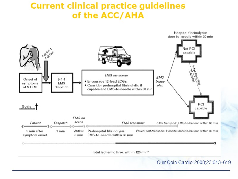 Current clinical practice guidelines of the ACC/AHA Curr Opin Cardiol 2008;23:613–619
 Acute Coronary Syndrome Electrocardiographic changes are often nonspecific in more than 50% of patients with chest pain. Cardiac biomarkers take hours from symptoms onset to exceed the normal range.
 Role of Echocardiography in AMI Anatomical, functional and hemodynamic information Diagnosis and exclusion of AMI in patients with chest pain and non-diagnostic ECG findings Estimation of the amount of myocardium at risk and final infarct size after reperfusion therapy
 Role of Echocardiography in AMI 3. Evaluation of unstable hemodynamics 4. Detection of infarct complications 5. Evaluation of myocardial viability 6. Risk stratification
 Wall motion abnormality in AMI Compensatory hyperkinesis RWMA at other area : Multi vessel involvement Wall Motion Score Index
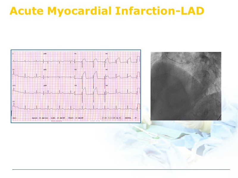 Acute Myocardial Infarction-LAD
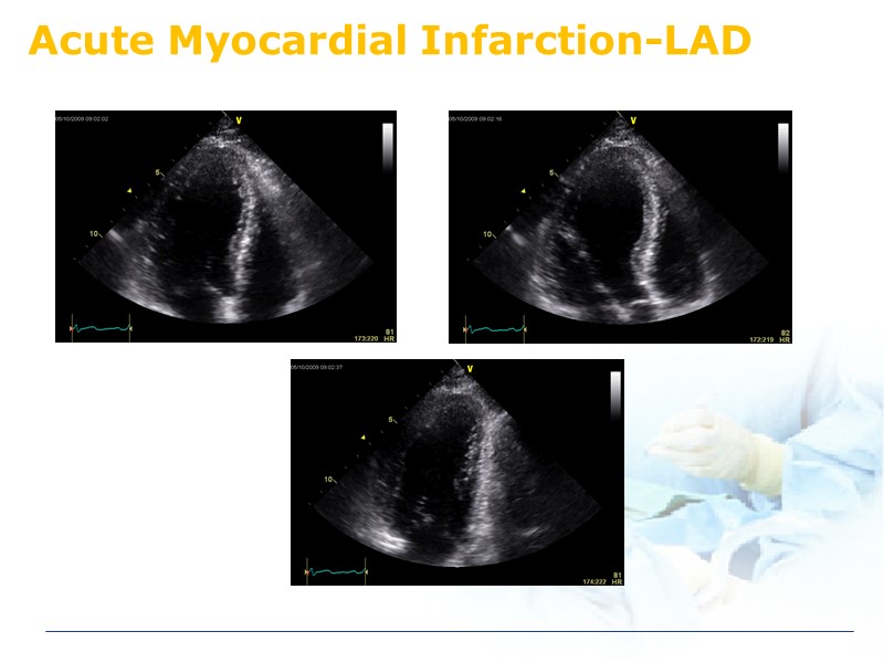 Acute Myocardial Infarction-LAD
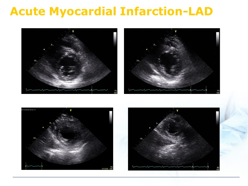 Acute Myocardial Infarction-LAD
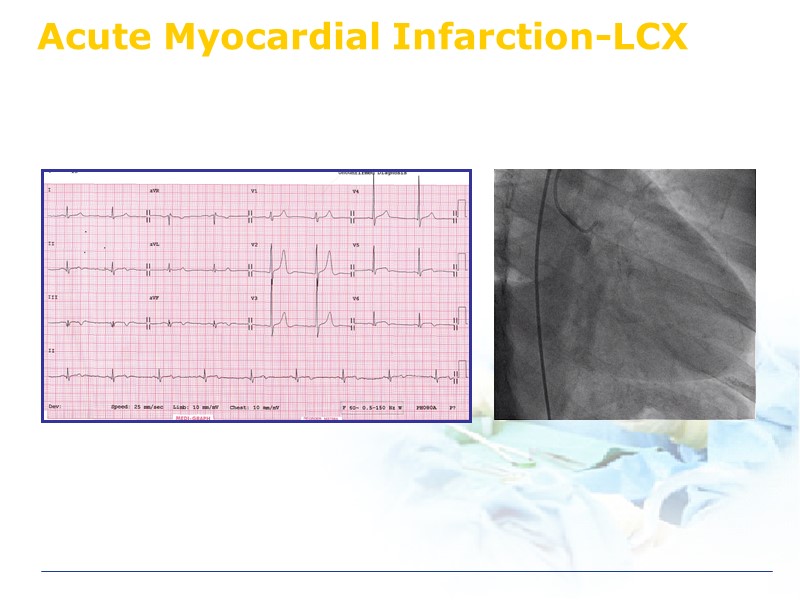 Acute Myocardial Infarction-LCX
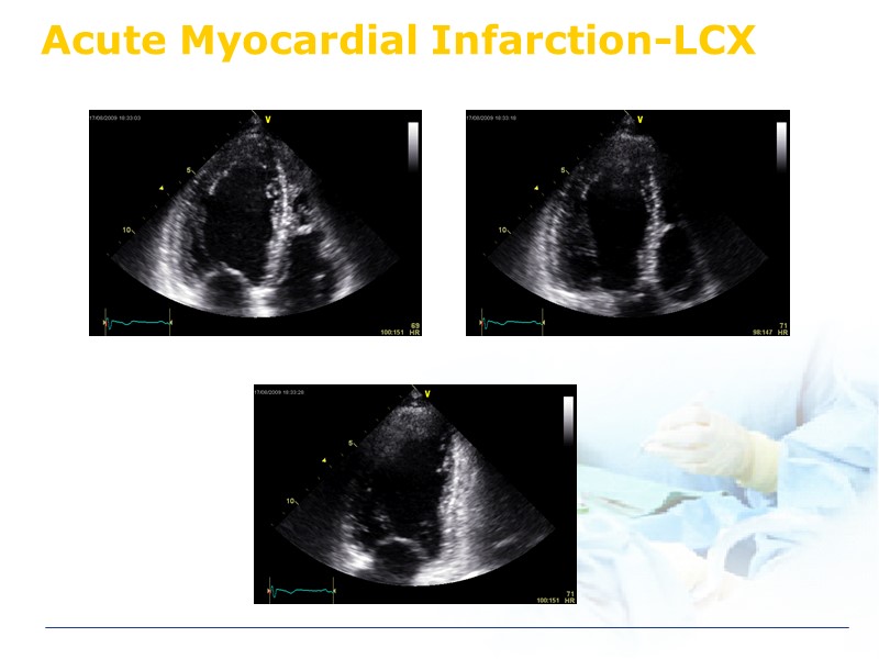 Acute Myocardial Infarction-LCX
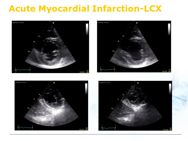 Acute Myocardial Infarction-LCX
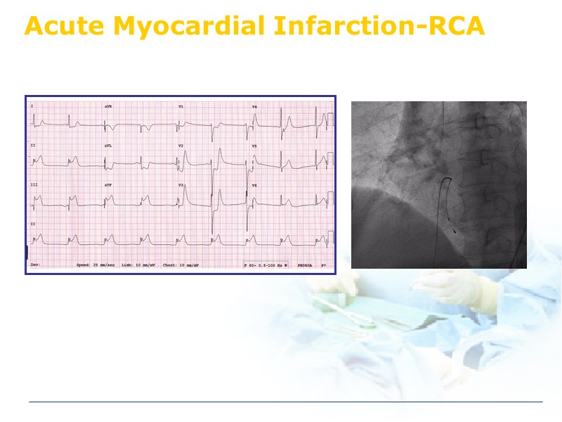 Acute Myocardial Infarction-RCA
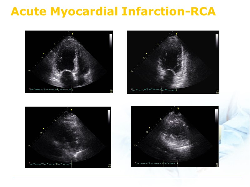 Acute Myocardial Infarction-RCA
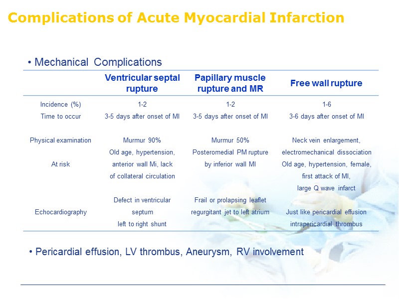 Complications of Acute Myocardial Infarction Pericardial effusion, LV thrombus, Aneurysm, RV involvement
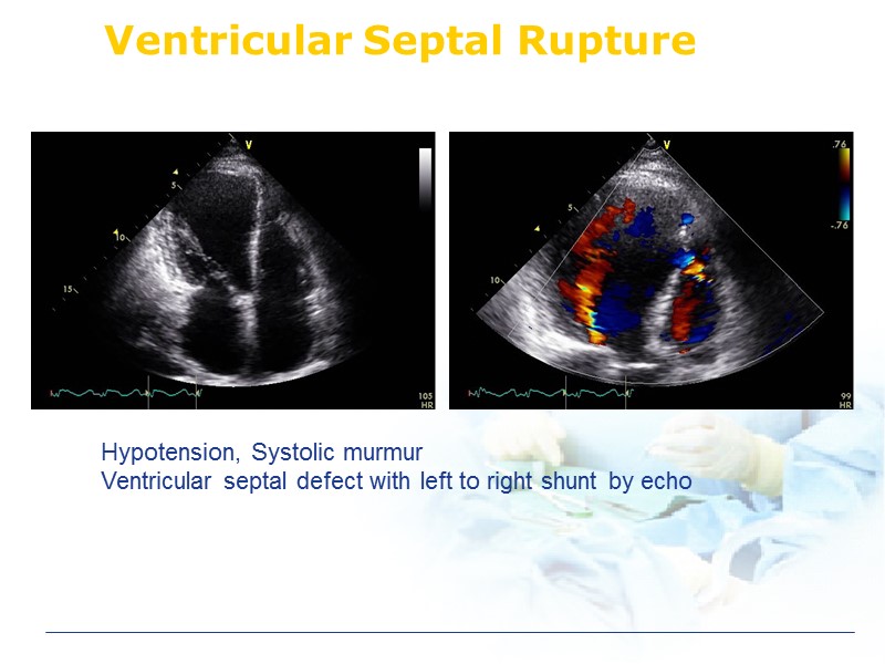 Ventricular Septal Rupture Hypotension, Systolic murmur Ventricular septal defect with left to right shunt by echo
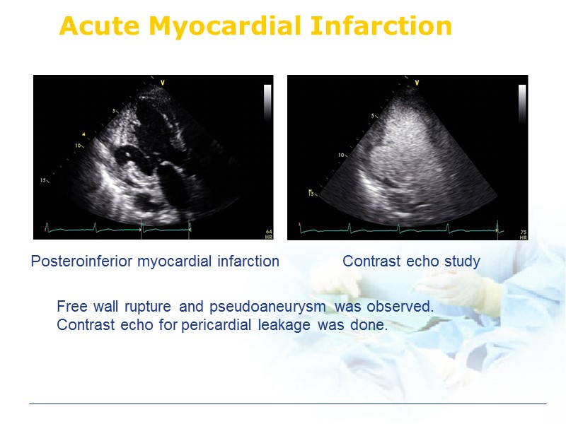 Acute Myocardial Infarction Posteroinferior myocardial infarction Contrast echo study Free wall rupture and pseudoaneurysm was observed. Contrast echo for pericardial leakage was done.
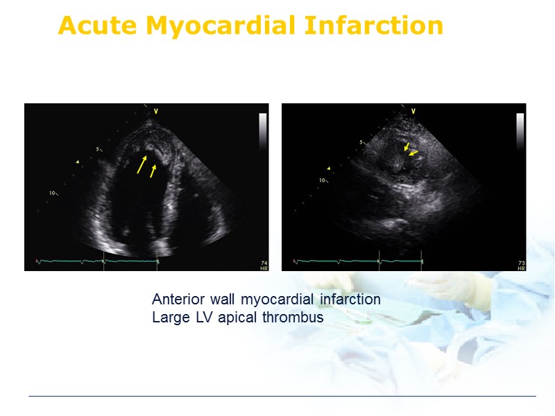 Acute Myocardial Infarction Anterior wall myocardial infarction Large LV apical thrombus
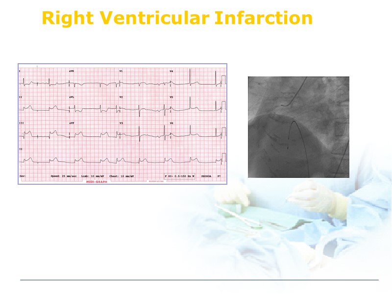 Right Ventricular Infarction
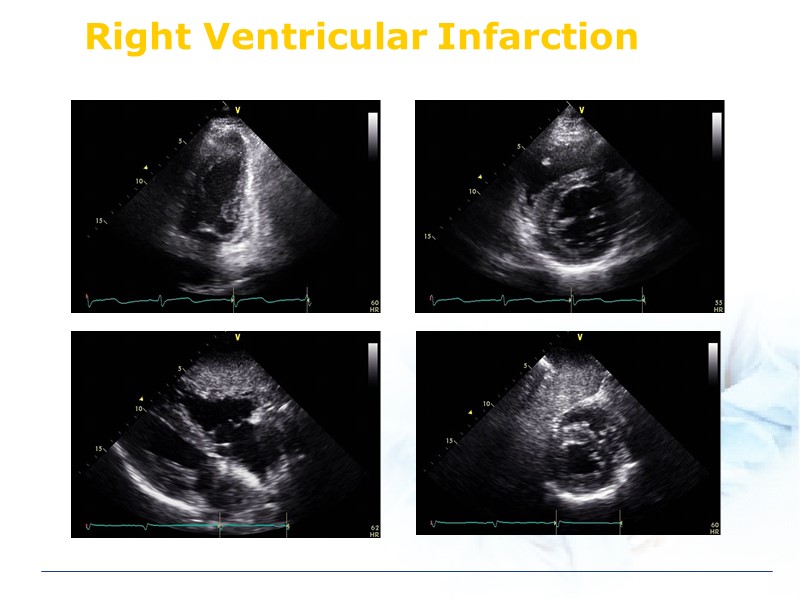 Right Ventricular Infarction
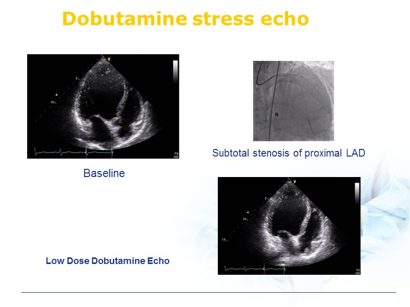 Dobutamine stress echo Low Dose Dobutamine Echo Baseline Subtotal stenosis of proximal LAD
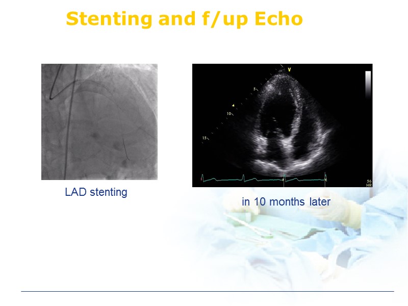 Stenting and f/up Echo in 10 months later LAD stenting
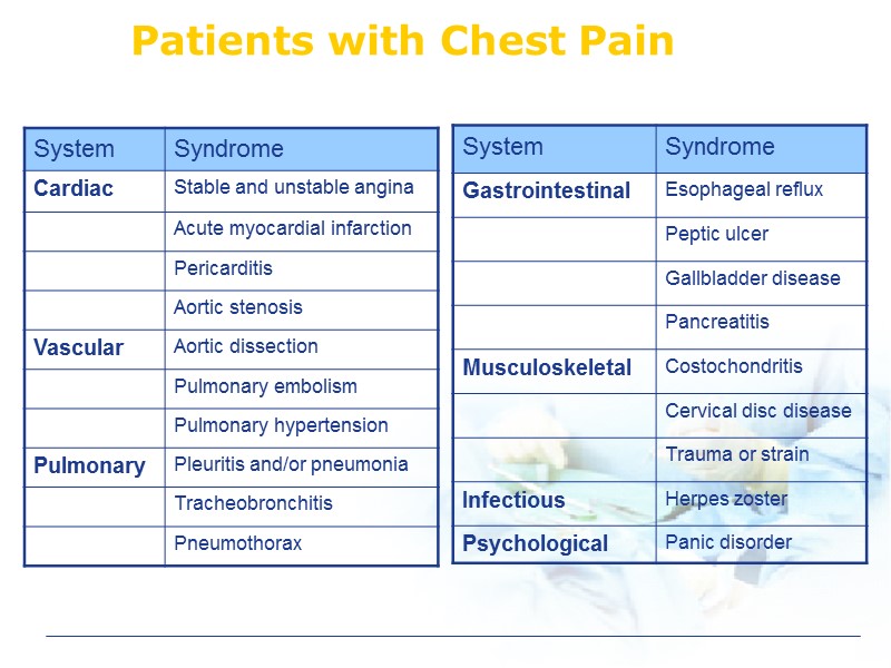 Patients with Chest Pain
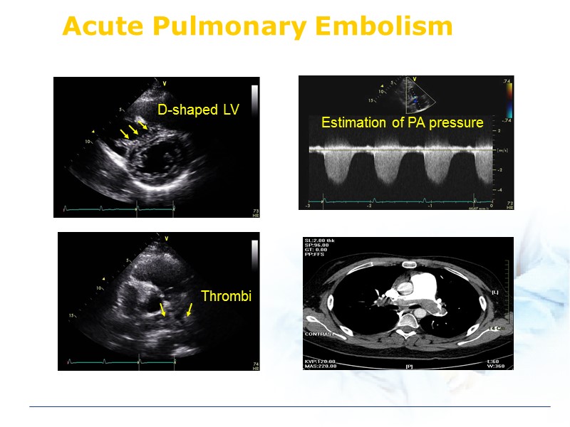 Acute Pulmonary Embolism D-shaped LV Thrombi
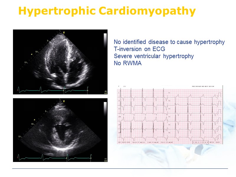 Hypertrophic Cardiomyopathy No identified disease to cause hypertrophy T-inversion on ECG Severe ventricular hypertrophy No RWMA
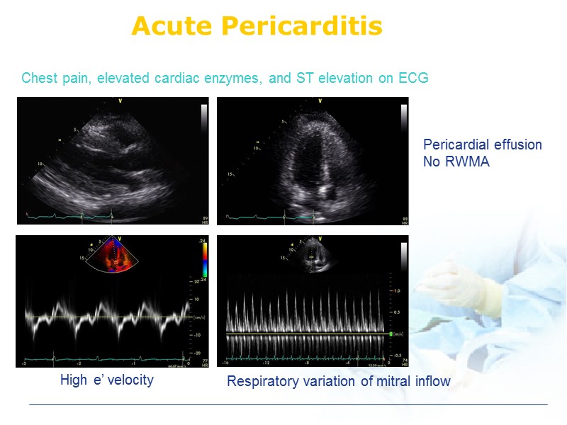 Acute Pericarditis Pericardial effusion No RWMA Chest pain, elevated cardiac enzymes, and ST elevation on ECG Respiratory variation of mitral inflow High e’ velocity
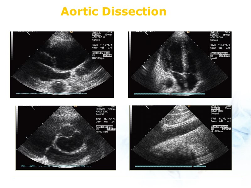 Aortic Dissection
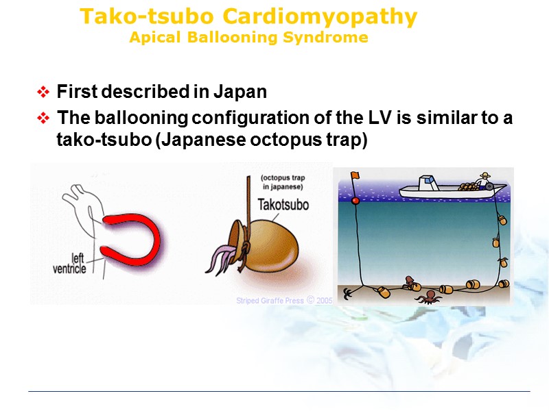 Tako-tsubo Cardiomyopathy Apical Ballooning Syndrome First described in Japan The ballooning configuration of the LV is similar to a tako-tsubo (Japanese octopus trap)
 Modified Mayo Clinic Criteria of stress induced cardiomyopathy transient hypokinesis, akinesis, or dyskinesis in the LV mid segments with or without apical involvement; RWMA that extend beyond a single epicardial vascular distribution; and frequently, but not always, a stressful trigger (2) the absence of obstructive coronary disease or angiographic evidence of acute plaque rupture
 Modified Mayo Clinic Criteria of stress induced cardiomyopathy (3) new ECG abnormalities (ST-segment elevation and/or T-wave inversion) or modest elevation in cardiac troponin (4) the absence of pheochromocytoma and myocarditis ***Patients were assigned this diagnosis when they satisfied all these criteria.
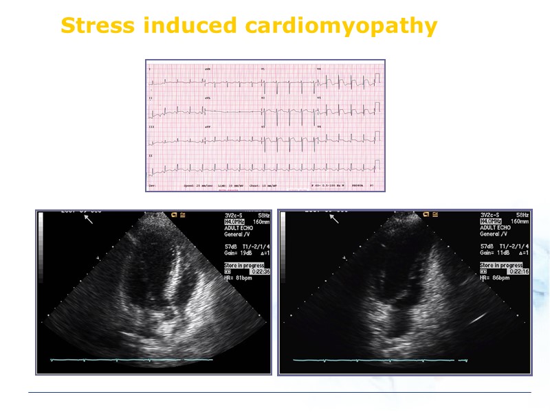 Stress induced cardiomyopathy
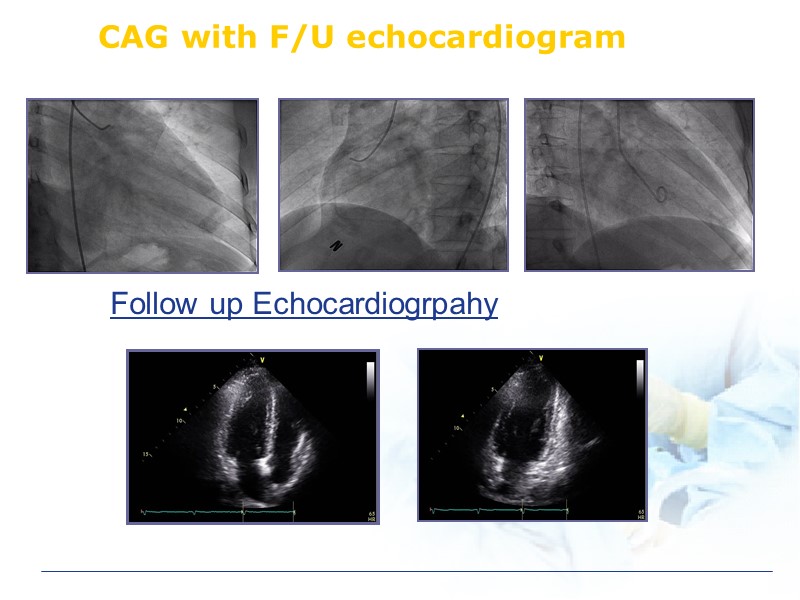 CAG with F/U echocardiogram Follow up Echocardiogrpahy


