Infectious Diseases Выполнила: Шаймерденова Ш. Проверила: Кыдырмолдина Э.
shyrynay_angl.ppt
- Размер: 1.8 Мб
- Автор:
- Количество слайдов: 29
Описание презентации Infectious Diseases Выполнила: Шаймерденова Ш. Проверила: Кыдырмолдина Э. по слайдам
 Infectious Diseases Выполнила: Шаймерденова Ш. Проверила: Кыдырмолдина Э. Курс: 5 Группа: 59 —
 Infectious Disease Terms 1. Epidemiology 2. Epidemic 3. Endemic 4. Pandemic 5. Pathogen 6. Opportunist 7. Nosocomial 8. virulence
 Normal Micro flora & its importance 1. Prevent the growth of pathogens 2. Stimulate the immune system to produce antibodies that cross-react with invading pathogens 3. Aid in digestion of cellulose in ruminants. 4. Produce essential nutrients
 Koch’s Postulates 1. The same pathogen must be present in every case of the disease; 2. The pathogen must be isolated from the diseased host and grown in pure culture; 3. The pathogen from the pure culture must cause the disease when it is introduced into a healthy but susceptible organism. 4. The pathogen must be isolated from the inoculated animal and be shown to be the original organism.
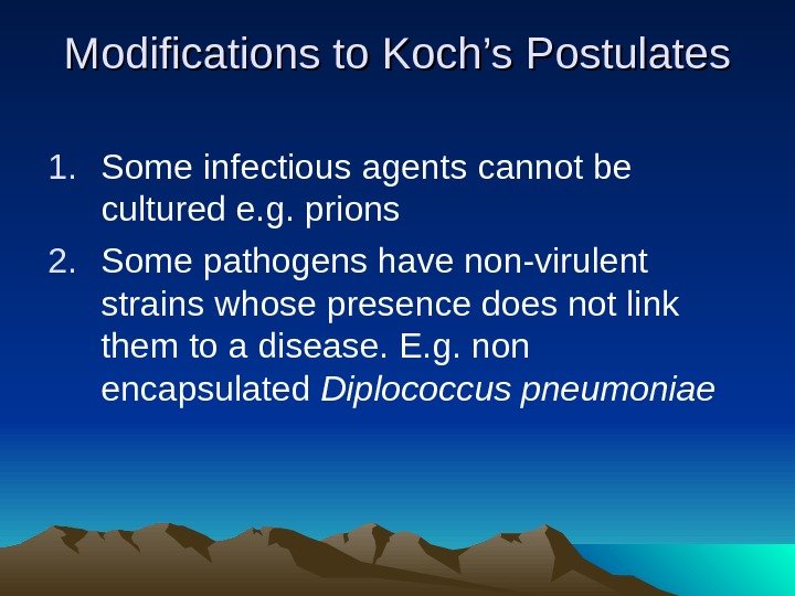
 Modifications to Koch’s Postulates 1. Some infectious agents cannot be cultured e. g. prions 2. Some pathogens have non-virulent strains whose presence does not link them to a disease. E. g. non encapsulated Diplococcus pneumoniae
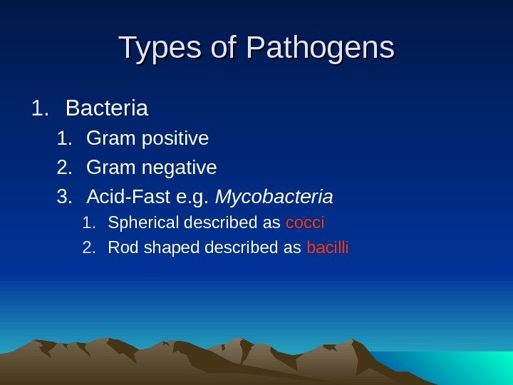
 Types of Pathogens 1. Bacteria 1. Gram positive 2. Gram negative 3. Acid-Fast e. g. Mycobacteria 1. Spherical described as cocci 2. Rod shaped described as bacilli
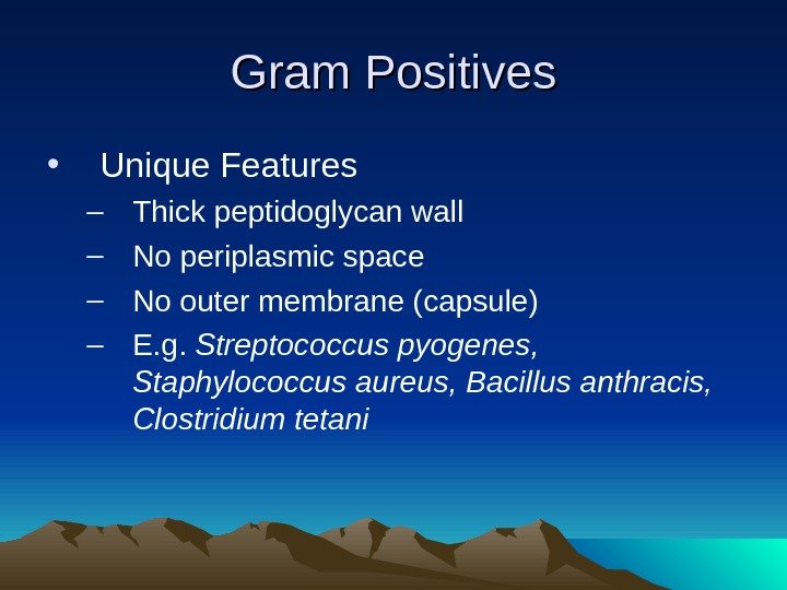
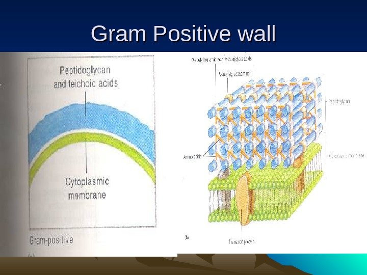
 Gram Positives • Unique Features – Thick peptidoglycan wall – No periplasmic space – No outer membrane (capsule) – E. g. Streptococcus pyogenes, Staphylococcus aureus, Bacillus anthracis, Clostridium tetani
 Gram Positive wall
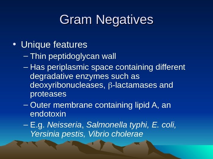
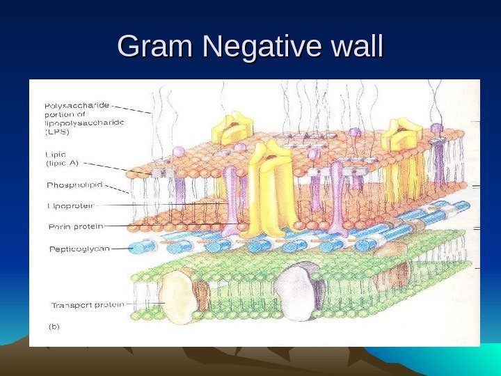
 Gram Negatives • Unique features – Thin peptidoglycan wall – Has periplasmic space containing different degradative enzymes such as deoxyribonucleases, -lactamases and proteases – Outer membrane containing lipid A, an endotoxin – E. g. Neisseria , Salmonella typhi, E. coli, Yersinia pestis, Vibrio cholerae
 Gram Negative wall
 Types of pathogens 2. Parasites (Eukaryotic Pathogen) 1. Fungi e. g. Candida, Aspergillus 2. Protozoa e. g. Plasmodium , Schistosoma 3. Worms e. g. Ascaris, Taenia
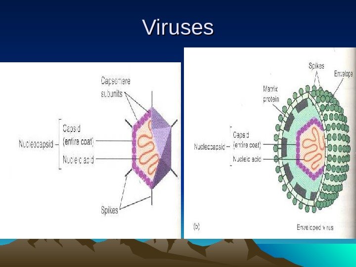
 Types of pathogens 3. Viruses 1. Are pieces of DNA or RNA surrounded by protein coat. The may be 2. Encapsulated e. g. HIV, HBV, measles, mumps, influenza, rabies 3. Non-encapsulated e. g. adenoviruses, HPV, Polio
 Viruses
 Modes of transmission 1. Direct contact e. g. touching, handshaking, or sexual intercourse 2. Indirect contact e. g. food, water or droplets in air; 3. Animal vectors e. g. insect bites in malaria, plague and oncho, dog bite in rabies
 Pathogenesis • Sequence of activities 1. Transmission of causative agent to susceptible host; 2. Adherence of the agent to a target tissue; 3. Colonization and invasion; 4. Damage to host by toxins or other mechanisms; 5. Exit from host; 6. Survival outside host long enough for step 1 to occur.
 Virulent Factors • For all pathogens there is an infective dose and a lethal dose. • Virulent factors that confer pathogenicity include; 1. Pili that facilitate attachment; 2. Capsules that interfere with phagocytosis 3. Exotoxins 4. Endotoxins 5. Proteases that break down antibodies 6. Ability to vary antigens to evade antibodies
 Bacterial Pathogenesis 1. Toxin production. Toxins fall into two categories; exotoxins and endotoxins. 2. Invasiveness, where bacteria grow to large numbers locally and produce enzymes that damage host tissues.
 exotoxins 1. Heat labile (60 -100 degrees for 30 mins) proteins produced and released by both gram positive and gram negative bacteria. 2. Produced by bacteria such as Clostridium (neurotoxins) and Bacillus (enterotoxin) (+) and E. coli and Vibrio (enterotoxin) (-)
 endotoxins 1. Are heat stable (100 degrees for 1 hr) lipopolysaccharide produced only by gram –ve bacteria. They remain attached to cell wall. 2. Cause fever and shock and is of lower toxicity compared to exotoxins. 3. Produced by bacteria such as Salmonella
 cholera 1. Causative Agent: Vibrio cholerae 2. Symptoms: severe diarrhoea up to 20 liters a day of “rice water stool”, vomiting, muscle cramps caused by loss fluid and electrolytes. 3. Pathogenesis: Vibrio adheres to the small intestinal lining, multiply and produce the enterotoxin choleragen which causes the accumulation of c. AMP. An increased secretion of water and electrolyte from the cells results
 Cholera 4. Epidemiology: Feacally contaminated water, crabs and vegetables fertilized with human faeces. Has been eradicated most developed countries but a new strain discovered in 1992 is threatening another pandemic.
 Cholera 5. Incubation period: 12 -48 hours 6. Lab diagnosis: Microscopy, culture of sample from faeces or vomit. 7. Prevention: Purification of water, washing of hands. 8. Treatment: administration of solution of glucose and electrolyte orally or intravenously; tetracycline antibiotic orally
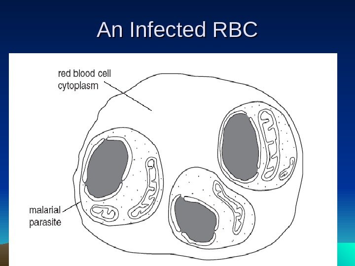
 malaria 1. Causative Agent: Plasmodium ( 4 species) 2. Symptoms (Clinical features): fever, chills, anaemia, headache, nausea, shivering, convulsions (esp. in under 5 yr olds) enlarged spleen. 3. Pathogenesis: site of action of pathogen include: liver, RBC, brain. The vector, female Anopheles mosquito, transfer pathogen during feeding.
 malaria 4. Epidemiology: Endemic in 91 tropical and subtropical countries. Invade the liver 1 st and move to reproduce in RBCs resulting in their rupture and the associated chills. 5. Incubation Period: 1 – 2 weeks. 6. Lab diagnosis: Microscopy.
 An Infected R
 Malaria — Prevention 1. Reduce the number of mosquitoes; destruction of larvae and adult mosquitoes by biological and chemical control methods 2. Avoid being bitten; protective clothing and creams, treated bed nets 3. Use of drugs to prevent infection; chemoprophylaxis
 Malaria -Treatment • Combination therapy: Artesunate Amodiaquine
 Tuberculosis 1. Pathogen: M. tuberculosis (pulmonary TB); M. bovis(GI TB) 2. Transmission: airborne droplets (NB MTB is dessication resistant and survives in dried sputum); unpasteurized milk. 3. Clinical features: prolonged coughing sometimes with bloody sputum, shortness of breath, fever, sweating , weight loss
 Tuberculosis 4. No toxin production. Pathogenicty is by invasiveness that produce characteristic lesions in the lungs. 5. Epidemiology: pathogen triggers acute inflammatory response + forms tubercle – giant cells containing MTB and surrounded by epithelial cells. Tubercles heals by fibrosis and calcification. Can desseminate via bloodstream to other internal organs
