
I ask you to stand for silence minute


")


 is")


. Balanced electrolyte solutions: Ringer‘s lactate, sterofundyne,")
: Haecodez, Refortan, Stabizol, Voluven,")


. The positive effects were exaggerated and did not confirmed in")
 Cryoprecipitate")


")

![Nomogram for calculating the blood volume deficit (Blutvolumendefizit [mL] – right vertical axis) according](https://present5.com/presentacii-2/20171208\14884-traumatic_shock.ppt\14884-traumatic_shock_30.jpg "Nomogram for calculating the blood volume deficit (Blutvolumendefizit [mL] – right vertical axis) according")


")


14884-traumatic_shock.ppt
- Количество слайдов: 45
I ask you to stand for silence minute in memory of Lord Rector of KNMU academician Tsyganenko Anatoly Yakovlevich
The traumatic shock. The prehospital management. The blood replacement in trauma patients. Professor of the department of anesthesiology, traumatology and emergency medicine Fesenko Olga Khamitovna
Specificity of battle trauma The bleeding is the cause of death in 50%. The management of patients on the battle field includes three stages: • The first aid under gunfire according the protocol MARCH (massive bleeding, airway, respirations, circulation, head). • The medical aid after stopping gunfire on the battlefield includes: cardio-pulmonary resuscitation, identification of severity of trauma, immobilization of fractures, wound bandage applying, analgesia, antibiotics. The main goal on this stage – hemodynamic stabilization by stopping the bleeding. Evacuation.
Grading the injured soldiers for evacuation Marking the patients according to the severity of trauma: • black – the pulse and breath are absent– resuscitation on the field; they should not been transported; • red – life-threatening trauma – transportation in the first place; • yellow – serious trauma, but not life-threatening– transportation in the second place; • green – walking patients – transportation last of all.
RTS (Revised trauma score)
Shock Acute hemodynamic instability, which leads to organ dysfunction due to poor perfusion, with poor oxygen delivery and consumption.
Causes of traumatic shock Hypovolemia due to bleeding or dehydration in burned patients; Cardiac failure due to tension pneumotorax or cardiac tamponage; Vasodilatation due to spinal trauma.
Pathogenesis of shock утрата Arterial hypotension Decreased oxygen delivery to organs Decreased oxidative phosphorilation Energy deficit in cells Cell dysfunction and death Disruption of ion channels in cell membrane К Na, H2O ↑ Са ↑ lactate → intracellular acidoses Injuring of organelle (mitochondria, lysosome) Intestinal ischemia ↓barrier function of intestinal epithelium Entering of microbes and toxins into the blood Systemic inflammation Multiple organ dysfunction Human death Other source of infection
Clinical signs of shock Paleness Tachycardia Breathlessness Oliguria → anuria Impairment of consciousness Decreased blood pressure Poor perfusion (after the pressure to the nail the pink color is normally restored in 2 seconds, if the time of restoring is prolonged, this indicates the poor perfusion)
Stages of shock Compensated: the perfusion of vital organs (brain, lungs, heart) is maintained due to peripheral vasoconstriction. Decompensated: the compensatory mechanisms can not maintain adequate perfusion of vital organs. Clinical manifestation of shock. Irreversible multiple organ failure: massive cell death, multiorgan dysfunction.
Hemodynamic parameters
Integral hemodynamic parameters
Receptors of autonomic nervous system
Inotropic and vasopressor drugs
Inotropic and vasopressor drugs
Inotropic and vasopressor drugs
Reserve inotropic drugs
Vasodilatative drugs
Intravenous fluid administration
Cristalloides Normal saline (isotonic 0,9% NaCl). Balanced electrolyte solutions: Ringer‘s lactate, sterofundyne, e.g. Hypertonic NaCl (from 1,6% tо 10%). Dextrose 5-10%, is not recommended in brain edema, increases intracellular volume.
Synthetic colloides Dextrans: Poliglucine, Reopoliglucine Gelatins: Helafusine, e.g. Hydroxyaethylstarchs (HAES): Haecodez, Refortan, Stabizol, Voluven, Tetrastarch, e.g. Combination: HAES + hypertonic NaCl: for “small volume reanimation” – HyperHAES
Polyhydric alcohol solutions: Sorbilact, Reosorbilact, Xylite Crystalloid solutions. Volemic effect is short. Advantages: Energy supply without insulin - useful in patients with intolerance to glucose. Fast volemic effect due to hyperosmolarity. Side-effects: vascular oxalose in brain, liver and kidney → acute renal, hepatic failure. Therefore not used in USA, Europe, Australia. Unfortunately, in Ukraine these solutions are used widely.
Polivinylpirrolidones: peristoy, haemodez The first synthetic colloides. Repeated administration can lead to depression of reticule-endothelial system with severe immune depression. This side-effect is dangerous particularly in neonates and young children. Blastogenic effect. Accumulate in interstitial space leading to edema. Deposit toxins in liver, kidneys, spleen, lungs, bone marrow. Restrained for medical use in USA from 1958, in Ukraine from 1998, in Russia – from 2005.
Perfluorocarbons: Perftoran («blue blood»). The positive effects were exaggerated and did not confirmed in medical practice. Unfortunately, it is still in use in Ukraine.
Blood preparations Whole blood Packed red blood cells Fresh frozen plasma (FFP) Cryoprecipitate Platelets Albumin
Hemorrhagic shock Cause – blood loss. Hemodynamic changes: Decreased volume of blood circulation, central venous pressure (CVP), cardiac index (CI), pulmonary capillary closing pressure (PCCP) Increased peripheral vascular resistance (PVR)
Response to the blood loss Sympathetic activation: centralization of blood circulation, discharge of blood from spleen, liver, lungs, tachycardia, increased oxygen demand in myocardium. Stimulation of renin-angiotensin-aldosterone system: renal vasospasm, increased renal Na reabsorbtion → oliguria. Stimulation of vasopressin synthesis: increased renal water reabsorbtion → oliguria. Stimulation of osmoreceptors in hypothalamus: thirst. Stimulation of glucocorticoid production: increased sensibility of adrenoreceptors, membrane stabilization? Autohemodilution. Stimulation of renal erythropoietin production (in the late stage of shock).
Classification of blood loss severity (American Association of Surgeons)
At the prehospital stage Algover‘s shock index is useful for evaluating the volume of blood loss: Shock index = Pulse rate / Systolic BP, in normal 60 : 120 = 0,5.
Nomogram for calculating the blood volume deficit (Blutvolumendefizit [mL] – right vertical axis) according to the Algover’s index (Schockindex – left vertical axis) and body mass (Korpermasse [kg], inferior horisontal axis)
Shock index: Is not informative in young children and old patients due to the age differences of hemodynamic parameters. Blood loss volume you can evaluate visually in cases of external bleeding. In cases of internal blood loss (intraabdominal, intrathoracic, interstitial bleeding) the shock index is usefull.
Hemorrhagic shock Laboratory dates: Hb ↓. Ht ↓. Lactate ↑. Diuresis ↓. рН ↓.
Prehospital management of hemorrhagic shock Venous access – canulation of 2-3 veins; Crystalloid / colloid infusion; Oxygen inhalation; Analgesia if indicated (prefer non-narcotics); If systolic BP is < 90 mm Hg in spite of rapid infusion – begin vasopressors (norepinephrine or dobutamine; Transportation to the hospital in horizontal Fowler position with raised legs; Stop external bleeding (tourniquet, raised position of damaged extremity, pressing apply to the wound); Transport immobilization; Warming the patient (take off the wet clothes, cover with a blanket).
Infusion rate in continuous bleeding If the bleeding is not stopped the infusion rate must provide the minimally sufficient hemodynamic parameters (systolic BP = 80-90 mm Hg), but not the maximal replacement of blood loss. Such rule of reason decreases blood loss and accelerates the thrombi formation in the affected area. It helps to avoid the dilution syndrome with excessive bleeding.
War – epidemic trauma In the ІІ word war all countries use the blood transfusion. But it doesn’t survive the soldiers in many cases of traumatic shock. The experiments by C. Wiggers explain it: the replacement of blood loss with equivalent volume of blood transfusion in dogs did not survive them. In the late 1940-th in such experiments it was demonstrated that the replacement of blood loss with the twofold normal saline infusion survives dogs without the blood transfusion.
Change of field doctrine On the basis of understanding the pathophisiology of traumatic shock as the fluid sectors’ shift and the significance of plasma volume deficit at the first the field doctrine was changed. In the Korean war for the first stage replacement of blood loss the synthetic colloid Macrodex (the soviet analogue – Poliglucine) was used widely. But it’s maximal dose is restricted and the damages of lung and kidney were found in many cases. In the Vietnam war the rapid Ringer-lactate infusion was provided for the American wounded soldiers in the battlefield. The three vena infusion was continued during transportation on the helicopter to the hospital and to the stopping bleeding in the operating room. Then the blood transfusion began. The excess of fluid was eliminated with diuretics. This “liberal” regimen was provided also in the civil medicine: on the preoperative stage the 3-5-hold crystalloid saline volume to the blood loss was infused).
Change of doctrine The “liberal” regiment of crystalloid infusion gave rise to doubt in early 1990-th. The dilution of blood leads to coagulopathy with increasing blood loss. So there was provided “restrictive” infusion regiment and “permissible hypotension” for the prehospital stage to surgical hemostasis. Acquired immunodeficiency syndrome (AIDS) pandemia and detection of transmissive infections lead to the practice of early fractionating of donor blood. All these result to the fresh whole blood deficit in hospitals. So at the turn of the XX and XXI century the next staging was recommended in the protocols of blood loss replacement in many countries (including Ukraine).
Step-by-step in replacement of blood loss At the first stage: saline cristalloids in restrictive regiment (for detectable pulse on the radial artery. Advantages: replacement of all fluid sectors, enhanced renal perfusion. Side-effects: edema, dangerous in lungs and brain. At the second stage: synthetic colloids (Geleains, hydroxy-ethyl-starch - Tetraspan). Advantages: better volemic effect comparing to crystalloids. Side-effects: coagulopathy, neurotoxicity. At the third stage: transfusion of packed red blood cells (PRBC), that requires some time for detection of blood group and rhesus and compatibility. Indicated in: Hb <70g/l and Ht<21%. At the forth stage: fresh frozen plasma (FFP). In acute massive blood loss the ratio of FFP to CRBC should be 1 : 1. At the fifth stage: platelets if the plates < 50×109/l. Fibrinogen (3-4 g) or cryoprecipitate (50 mg/kg body mass) are recommended in hypofibrinogenemia.
“Damage control” In XX century wars (in Persian Gulf 1990-1991 and in Vietnam 1961-1973) 24% of wounded American soldiers died. In ХХІ century wars (in Iraq and Afghanistan 2003-2009) only 10% of wounded American soldiers died. It was due to the change of medical strategy – american-british innovation called “damage control”.
“Damage control” For surgeons it means the physiological correction, but not anatomical correction. This includes stop bleeding, prevention of infection and further traumatic damage (immobility). For anesthesiologists it means anesthesia for this stage of surgery, “permissible hypotension” before stopping bleeding, “haemostatic resuscitation” and prevention of “lethal triad”: coagulopathy, acidosis and hypothermia.
Complete surgical correction Complete surgical correction may be provided in 1-2 days after the stabilization of the patients physical and metabolic state.
Thank you for your attention! Questions?
THE PLAN of lectures in surgery of extreme conditions and military surgery for the English-speaking medical students of IV course of stomatology faculty (autumn semester of 2012/2013 st. year) Lecture day: MONDAY. Auditorium: The main 9-floor-building of Kharkiv Regional Hospital,
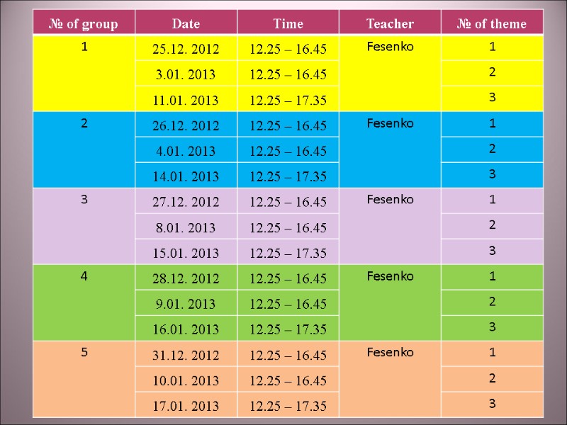
THE PLAN of practical classes in surgery of extreme conditions and military surgery for the English-speaking medical students of IV course stomatology faculty (autumn semester of 2012/2013 st. year) Auditorium: The main 9-floor-building of Kharkiv Regional Hospital, The 7-th floor. (Trinkler str.) Teacher – prof. U.A. Fesenko