НЕРВНАЯ ТКАНЬ2.ppt
- Количество слайдов: 37

Эмбриогенез нервной ткани Эволюция нервной ткани A single neuron on the surface of a microprocessor. A cm 3 of the human brain will contain more than 50 million neurons.

Neurulation D Formation of Neural Crest Cells (makes PNS, endocrine cells, pigment cells, connective tissue). V

Origin of Floor Plate and Neural Crest 4

6 Months • • Nerve cell generation complete Cortex beginning to wrinkle Myelinization Outnumber neurons 10: 1

8 stages of cortical development 1. 2. 3. 4. 5. 6. 7. 8. Neural proliferation Neural migration Neural differentiation Axonal growth Dendritic growth Synaptogenesis Myelination Neuronal death

• Genes 50% of ≈ 20, 000 -25, 000 genes in the human genome are expressed only in Brain

1. Neural proliferation • Begins with neural tube closure New cells born in ventricular layer Зона пролиферации

Neural proliferation • 1 mother cell produces ≈ 10, 000 daughter cells • All neurons (100 billion in total) are produced pre-natally • Rate of proliferation extremely high; thousands/minute

• Neuroblasts give rise to a limited number of daughter cells • Cells have a genetically mediated memory that allows them to remember how many times they have divided

2: Cellular migration • Non-dividing cells migrate from ventricular layer • Creates a radial inside-out pattern of development

• Migrating cells structurally and functionally immature • Importance of radial glial cells

• Genetics and migration • Mutant or “knock-out” mice • Cannot produce a class of proteins called cell adhesion molecules (CAM’s) • Migration is disrupted because cells cannot attach to and migrate along glia

3. Cellular differentiation • Migrating cells structurally and functionally immature • Once new cells reach their destination, particular genes are turned growth of axons, dendrites, and synapses

• • • A schematic representation of early developmental stages of cortical lamination in the telencephalic wall. In the pseudostratified cerebral wall a preplate forms that contains the pioneer neurons. Subsequently, post-mitotic neurons, migrating along radial glial cells, form a cortical plate within the preplate. Abbreviations: V, ventricular zone; PP, preplate; IZ, intermediate zone; SV, subventricular zone; SP, subplate; CP, cortical plate; MZ, marginal zone (Uylings et al. , 1990).

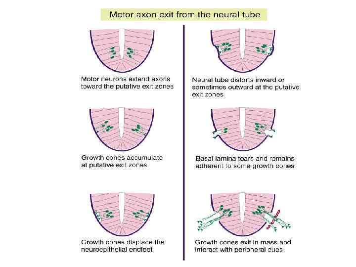
4. Axonal growth • Growth occurs at a growth cone Growth cone

4. Axonal growth • • Growth occurs at a growth cone Axons have specific targets Targets often enormous distances away Some axons extend a distance that is 40, 000 times the width of the cell body it is attached to • Finding targets ? chemical & electrical gradients, multiple branches

5. Dendritic growth • • • Usually begins after migration Slow Occurs at a growth cone Begins prenatally, but continues postnatally Overproduction of branches in development and resultant pruning • Remaining dendrites continue to branch and lengthen

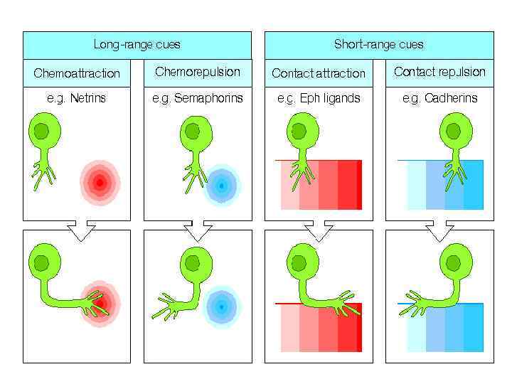
• Growth of dendrites and axons • Undeveloped neuron needs to establish basic “polarity: ” which end is which? • Involves specific proteins • Axons: Affords a sensitivity to chemical signals emitted by targets
 and repellants (orange) located")
Axons locate their target tissues by using chemical attractants (blue) and repellants (orange) located around or on the surface of guide cells. Left: An axon begins to grow toward target tissue. Guide cells 1 and 3 secrete attractants that cause the axon to grow toward them, while guide cell 2 secretes a repellant. Surfaces of guide cells and target tissues also display attractant molecules (blue) and repellant molecules (orange). Right: A day later, the axon has grown around only guide cells 1 and 3.

6. Synaptogenesis • Takes place as dendrites and axons grow • Involves the linking together of the billions of neurons of the brain • 1 neuron makes up to 1000 synapses with other neurons • Neurotransmitters and receptors also required

• The number of synapses reaches a maximum at about 2 years of age • After this, pruning begins • By 16, only half of the original synapses remain

7: Myelinization • The process whereby glial cells wrap themselves around axons • Increases the speed of conduction • Begins before birth in primary motor and sensory areas • Continues into adolescence in certain brain regions (e. g. , frontal lobes)

8: Neuronal death • As many as 50% of neurons created in the first 7 months of life die

• Given the extremely high costs of building and maintaining nervous systems

• Two different types of locomotor systems are found in animals, one muscle-based, the other ciliabased.

• Behaviour evolved before nervous systems. • In single-celled eukaryotes, sensory inputs directly influence the motor behaviour of the сell. • In multicellular organisms, the efficiency of sensory-to-motor transformation (defined as the ratio of sensory cells to total cell number) is low.

• With the advent of neurons, signal amplification and fast, long-range communication between sensory and motor cells became possible. • Тhe first nervous systems consisted of combined sensory-motor neurons, directly translating sensory input into motor output on locomotor ciliated cells and steering muscle cells.
, embedded in")
• Sponge larvae have flask-shaped sensory cells (additional to their photoreceptors), embedded in an epithelial layer of polarized, ciliated cells [36]. These cells express orthologues of bilaterian neurogenic molecules, indicating that they may represent a protoneuron. These cells may also release unknown transmitters to regulate ciliary beating in neighbouring cells in a paracrine fashion. Even if a local release of transmitters is possible in sponges, the effects of such signal transmission can only be local, since sponges have no gap junctions and no axons [
 Primordial myoepithelium with electrically coupled cells.")
• Neuro-muscular hypothesis • • • (1) Primordial myoepithelium with electrically coupled cells. (2) Protomyocytes start to forsake the epithelium, sinking into the interior. (3) Protoneurons evolve, conveying excitation from the exterior to the myocytes. (4) Neurosensory cells and neurons evolve, which make use of action potentials. They are connected to one another and to the myocytes by chemically transmitting, polarized junctions. Electrical coupling persists in many epithelia and muscles. Abbreviations: ap = action potential; ec = electrical coupling; 1 st = primary chemical signal; 2 nd = secondary chemical signal; s = synapse. A,

• Neuroactive substances of invertebrata are classified according to their molecular structure • i) amino acids and their derivatives, which are known as biogenic amines (e. g. , serotonin, histamine) • ii) neuropeptides (e. g. , FMRFamide, allatostatin, tachykinin) • iii) gaseous molecules (e. g. , nitric oxide, carbon monoxide)

Hypothetical stages in the evolution of the endocrine and neuroendocrine systems. Cells releasing endocrine signals are likely to predate the appearance of a nervous system, since they can be found in extant metazoa lacking nerve cells (A). The first nervoussystem is thought to have possessed the structure of a basi-epithelial nerve net, similar to the one still found in present day cnidarians (B). At this stage, neurons and NSCs/endocrine cells most likely had evolved into distinct lineages of sensory cells integrated into the epidermis, the gastrodermis and the nerve net. A central nervous system integrating multimodal sensory input evolved in bilateriananimals (C). Populations of sensory NSCs involved in the regulation of fundamental biological processes, such as feeding and reproduction may have formed specialized complexes in the brain, pharynx, and gut of early bilaterians. During later stages of evolution (shown in (D) for the chordate lineage), NSCs and endocrine cells in general show the tendency of losing their sensory function, delaminating from the surface epithelium (epidermis, pharynx, and intestinal epithelium), and undergoing morphogeneticchanges that produced dedicated endocrine glands, such as the pituitary, thyroid/parathyroid, and pancreatic islets.
 Recording from")
• Electrical responses and nervous systems in the lowest invertebrates. (a) Recording from Paramecium illustrates a graded action potential which uses Ca 2+ ions in the upswing phase and K+ ions during repolarization; three responses of varying size were produced by mechanical taps of increasing intensity to the front of the animal; Vm = membrane potential recorded with a glass microelectrode (b) Hydrozoan showing nerve net underlying the epidermis. (c) Intracellular microelectrode recording from a large neuron of the subumbrellar nerve net of the jellyfish Carybdea; this action potential was generated by a synaptic potential (arrow); RMP approximately − 70 m. V
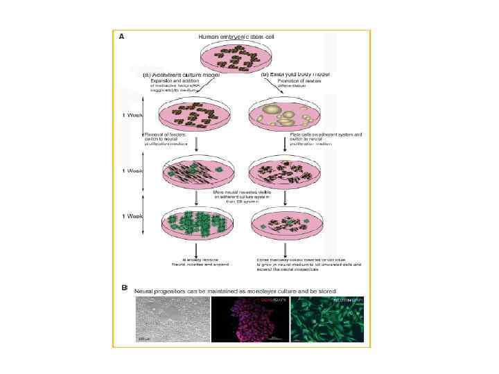
-derived neural progenitors and neurons by phase microscopy")
Characterization human embryonic stem cell (h. ESC)-derived neural progenitors and neurons by phase microscopy and immunocytochemistry.
НЕРВНАЯ ТКАНЬ2.ppt