
Dentistry 07 1 EXCITABLE TISSUES : Nerve And


- Размер: 913 Кб
- Количество слайдов: 39
Описание презентации Dentistry 07 1 EXCITABLE TISSUES : Nerve And по слайдам
Dentistry 07 1 EXCITABLE TISSUES : Nerve And Muscle BY: DR. MAHA HEGAZI, Associate Professor Of Physiology
Dentistry 07 2 Learning objectives: by the end of these lectures the student should know • Morphology of the nerve cell & functional organization of neurons • Excitation & conduction along the nerve (local & propagated action potentials) • Resting membrane potential ( causes & recording) • Action potential (ionic bases & recording) electrical changes that occur on a nerve on stimulation. • Compound action potential • Changes in excitability during electronic potential (local) & action potential • All or non law • Saltatory conduction • Energy sources & metabolism of nerve • Properties of mixed nerve • Nerve types & functions
Dentistry 07 3 Nerve cells: • The neurons are the basic building blocks of the nervous system, their axons may or may not myelinated. • The myelin sheath is produced by the Schwan cells. It envelops the axon except at the ends & the nodes of Ranvier • The impulse is conducted faster in myelinated than unmyelinated nerves.
Dentistry 07 4 Resting Membrane Potential Definition: it is the potential difference recorded across the cell membrane at rest. • Causes: • 80% caused by selective permeability of the cell membrane The K+ diffuses out the cell & Na+ diffuses inside the cell according to concentration gradient. The K+ permeability is 50 -75 folds more than Na+ • 20% is caused by the Na+ K+ pump an active process that needs energy taken from ATP. This is very important to maintain the concentration gradient across the cell membrane
Dentistry 07 5 Resting Membrane Potential (V r )
Dentistry 07 6 Sodium-Potassium Exchange Pump
Dentistry 07 7 • Significance: • PROTEINS have a negative charge & can not leave the cell to the outside • K+ efflux is not accompanied by an equal influx of anions & membrane is maintained in a polarized state with the outside positive relative to the inside making the RMP for a nerve to be — 70 m. V
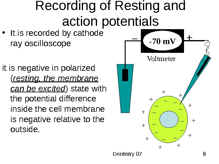
Dentistry 07 8 • It is recorded by cathode ray oscilloscope it is negative in polarized ( resting, the membrane can be excited ) state with the potential difference inside the cell membrane is negative relative to the outside. Recording of Resting and action potentials + + + +++ + – – – –––– –Voltmeter– + 0 m. V-70 m. V +
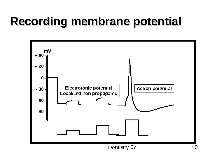
Dentistry 07 9 Excitation & conduction: Nerve cells have low threshold for excitation. The stimulus may be electrical, chemical or mechanical. Two types of potentials may be produced • Local (Non-propagated action potential ) named after its location synaptic, generator or electronic potential • PROPAGATED ACTION POTENTIAL (nerve impulse). Both are due to changes in the conduction of ions across the cell membrane that are produced by alternations in the ion channels
Dentistry 07 10 Recording membrane potential + 60 — + 30 — — 60 — — 90 — m. V Electrotonic potential Localized non propagated Action potential
Dentistry 07 11 • All or non law: • Application of a threshold stimulus either produces a full response or not at all. • Further increase in the intensity of a stimulus produces no increment or other changes in action potential. • The action potential failed to occur if the stimulus is sub-threshold, it produces only local changes with no propagation. • Latent period in a nerve : it is a period corresponding to the time taken from the site of simulation till the recording electrode.
Dentistry 07 12 Stimulation of a nerve produces: • ELECTRICAL CHANGES CALLS ACTION POTENTIAL • EXCITABILITY CHANGES. • THERMAL CHANGES
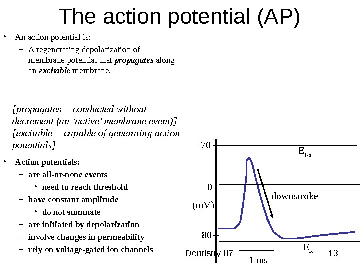
Dentistry 07 13 The action potential (AP) • An action potential is: – A regenerating depolarization of membrane potential that propagates along an excitable membrane. 1 ms+70 0 (m. V) -80 E Na E K downstroke[propagates = conducted without decrement (an ‘active’ membrane event)] [excitable = capable of generating action potentials] • Action potentials: – are all-or-none events • need to reach threshold – have constant amplitude • do not summate – are initiated by depolarization – involve changes in permeability – rely on voltage-gated ion channels
Dentistry 07 14 Threshold and Action Potentials • Threshold – membrane is depolarized by 15 to 20 m. V • Established by the total amount of current flowing through the membrane • Weak (subthreshold) stimuli are not relayed into action potentials • Strong (threshold) stimuli are relayed into action potentials • All-or-none phenomenon – action potentials either happen completely, or not at all
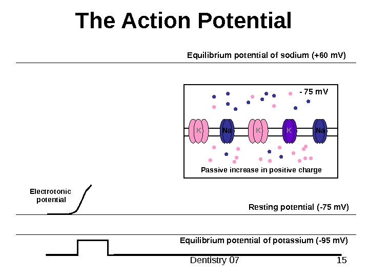
Dentistry 07 15 The Action Potential Resting potential ( -75 m. V ) Equilibrium potential of potassium ( -95 m. V )Equilibrium potential of sodium ( +60 m. V ) Electrotonic potential K Na K K Na Passive increase in positive charge — 75 m. V
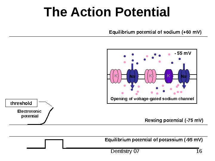
Dentistry 07 16 The Action Potential Resting potential ( -75 m. V ) Equilibrium potential of potassium ( -95 m. V )Equilibrium potential of sodium ( +60 m. V ) Electrotonic potential Opening of voltage — gated sodium channel. K K K — 55 m. V Na Na threshold
Dentistry 07 17 The Action Potential Resting potential ( -75 m. V ) Equilibrium potential of potassium ( -95 m. V )Equilibrium potential of sodium ( +60 m. V ) Electrotonic potential. Depolarisation due to sodium influx Opening of voltage — gated sodium channel. K K K — 40 m. V Na Na
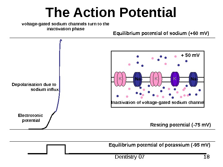
Dentistry 07 18 The Action Potential Resting potential ( -75 m. V ) Equilibrium potential of potassium ( -95 m. V )Equilibrium potential of sodium ( +60 m. V ) Electrotonic potential. Depolarisation due to sodium influx Inactivation of voltage — gated sodium channel. K K K + 50 m. V Na Navoltage — gated sodium channels turn to the inactivation phase
Dentistry 07 19 The Action Potential Resting potential ( -75 m. V ) Equilibrium potential of potassium ( -95 m. V )Equilibrium potential of sodium ( +60 m. V ) Electrotonic potential. Depolarisation due to sodium influx opening of voltage — gated potassium channel. K K + 50 m. V Na Na. K
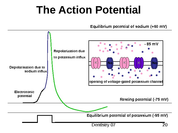
Dentistry 07 20 The Action Potential Resting potential ( -75 m. V ) Equilibrium potential of potassium ( -95 m. V )Equilibrium potential of sodium ( +60 m. V ) Electrotonic potential. Depolarisation due to sodium influx opening of voltage — gated potassium channel. K K — 85 m. V Na Na. KRepolarization due to potassium influx
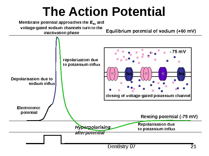
Dentistry 07 21 The Action Potential Resting potential ( -75 m. V )Equilibrium potential of sodium ( +60 m. V ) Electrotonic potential. Depolarisation due to sodium influx K K — 75 m. V Na Na. K closing of voltage — gated potassium channelrepolarization due to potassium influx Repolarisation due to potassium influx. Membrane potential approaches the E Na and voltage — gated sodium channels turn to the inactivation phase Hyperpolarising afterpotential
Dentistry 07 22 The Action Potential Electrotonic potential. Opening of voltage — controlled sodium channel Inactivation of voltage — controlled sodium channel Opening of voltage — controlled potassium channel Resting potential ( -75 m. V )Equilibrium potential of sodium ( +60 m. V ) threshold Hyperpolarization due to more outflux of potassium ions
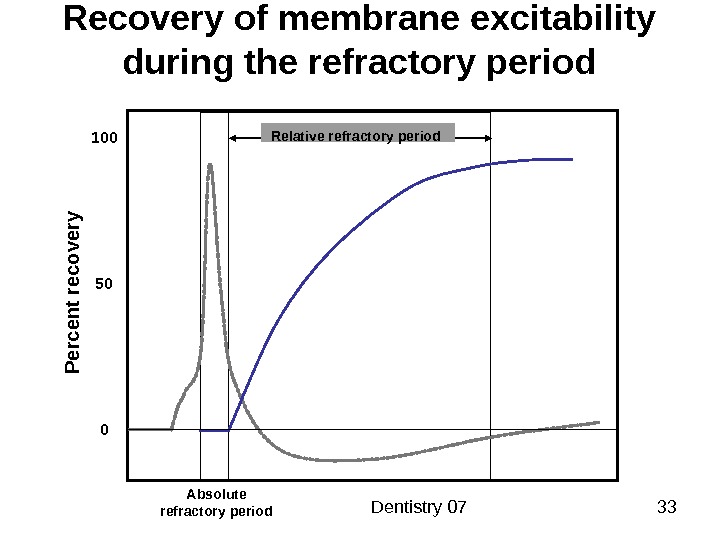
Dentistry 07 23 The Action Potential (excitability changes) Polarized state (resting)Depolarisation ( due to sodium influx ) Resting potential ( -75 m. V )E Na ( +60 m. V ) Hyperpolarising afterpotentialafterdepolarization E K ( -95 m. V )Absolute refractory period Relative refractory period
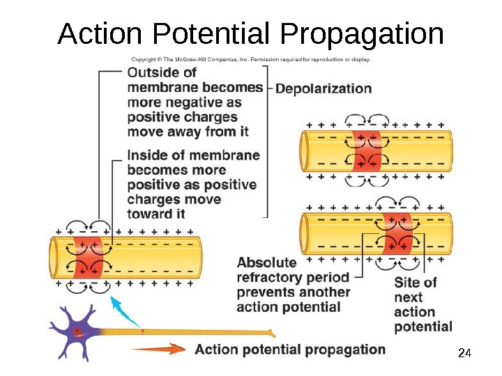
Dentistry 07 24 Action Potential Propagation
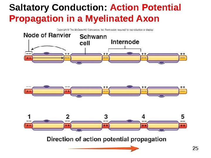
Dentistry 07 25 Saltatory Conduction: Action Potential Propagation in a Myelinated Axon
Dentistry 07 26 Propagation of an Action Potential (Time = 1 ms) • Ions of the extracellular fluid move toward the area of greatest negative charge • A current is created that depolarizes the adjacent membrane in a forward direction • The impulse propagates away from its point of origin
Dentistry 07 27 Properties of action potentials • Action potentials: • are all-or-none events • threshold voltage (usually 15 m. V positive to resting potential) threshold -70+60 m. VS tim ulus 0 • are initiated by depolarization • action potentials can be induced in nerve and muscle by extrinsic (percutaneous) stimulation – • APs do not summate — information is coded by frequency not amplitude.
Dentistry 07 28 Properties of action potentials non-myelinated (squid) 0 800400 • have constant conduction velocity • True for given fibre. Fibres with large diameter conduct faster than small fibres. As general rule: • Impulses are conducted faster in myelinated fibre than non- myelinated fibre Fibre diameter ( m)Velocity (m s-1)0 3 6 9 Myelinated (cat)
Dentistry 07 29 Functions of action potentials • Information delivery to CNS – carriage of all sensory input to CNS. Consider block APs in sensory nerves by local anaesthetics. This usually produces analgesia without paralysis. This is because LAs are more effective against small diameter (large surface area to volume ratio) C fibers than a-motorneurones. • Information encoding – The frequency of APs encodes information (remember amplitude cannot change) — covered in lecture 3. 3.
Dentistry 07 30 Functions of action potentials • Rapid transmission over distance (nerve cell APs) – Note: speed of transmission depends on fiber size and whether it is myelinated. Information of lesser importance carried by slowly conducting unmyelinated fibers. • In non-nervous tissue APs are the initiators of a range of cellular responses – muscle contraction – secretion (eg. Adrenalin from chromaffin cells of medulla)
Dentistry 07 31 Conduction velocity of AP • Compound action potentials can be recorded from nerve truncks • usually done percutaneously from nerves that are close to the surface (eg. Ulnar nerve) • The passage of an action potentials in all the axons in the nerves is seen as a small ( V) voltage signal on body surface
Dentistry 07 32 • as recordings are made further from the site of stimulation the waveform develops into several discrete peaks • Each peak was named: alpha — the first to appear; beta — the next, and so on. • The first signal to arrive at a distant recording site has travelled the fastest! • So each peak represents a set of axons with similar conduction velocity • velocity is calculated from the distance between R 1 and R 3 and the time taken to traverse that distance — distance/time = velocity (ranges from 0. 5 to ~100 ms-1)
Dentistry 07 33 Recovery of membrane excitability during the refractory period Absolute refractory period 100 50 0 Relative refractory period P e rc e n t re c o v e ry
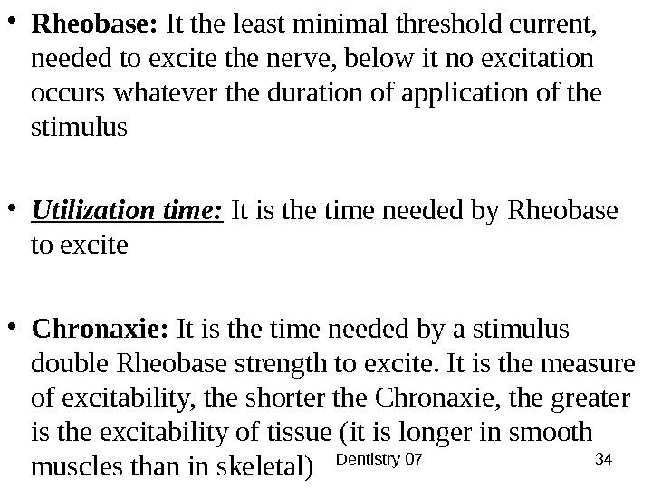
Dentistry 07 34 • Rheobase: It the least minimal threshold current, needed to excite the nerve, below it no excitation occurs whatever the duration of application of the stimulus • Utilization time: It is the time needed by Rheobase to excite • Chronaxie: It is the time needed by a stimulus double Rheobase strength to excite. It is the measure of excitability, the shorter the Chronaxie, the greater is the excitability of tissue (it is longer in smooth muscles than in skeletal)
Dentistry 07 35 Strength — Duration Curve for Action Potential Initiation Duration of stimulus ( msec )5 — 4 — 3 — 2 — 1 — 0 -Intensity of Stimulus ( relative ) Minimal stimulation time Rheobase Chronaxie ( ) Q = I x T m ln Time constant = 1. 44 x chronaxie
Dentistry 07 36 Characteristics of Action Potential • Threshold • All — or — none property
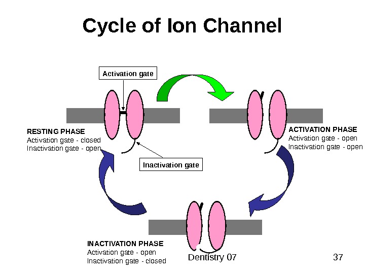
Dentistry 07 37 Cycle of Ion Channel Activation gate Inactivation gate. RESTING PHASE Activation gate — closed Inactivation gate — open INACTIVATION PHASE Activation gate — open Inactivation gate — closed ACTIVATION PHASE Activation gate — open Inactivation gate — open
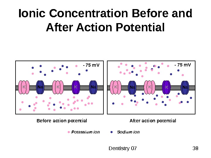
Dentistry 07 38 Ionic Concentration Before and After Action Potential K Na K K — 75 m. V Na Na. K- 75 m. V Before action potential After action potential Potassium ion Sodium ion
Dentistry 07 39 Pump and Maintenance of Membrane Potential K Na K K Potassium ion Sodium ion. K Na KNa Na — K — ATPase pump