
Congenital heart diseases Dr. Owis Khater


- Размер: 3 Mегабайта
- Количество слайдов: 90
Описание презентации Congenital heart diseases Dr. Owis Khater по слайдам
Congenital heart diseases Dr. Owis Khater
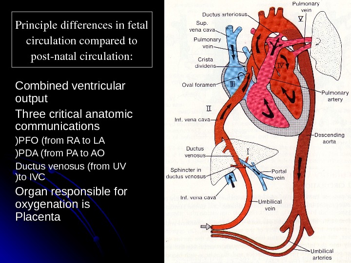
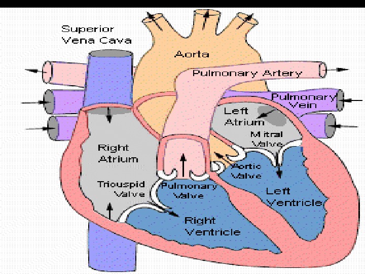
Principledifferencesinfetal circulationcomparedto postnatalcirculation: Combined ventricular output Three critical anatomic communications PFO (from RA to LA (( PDA (from PA to AO (( Ductus venosus (from UV to IVC (( Organ responsible for oxygenation is Placenta
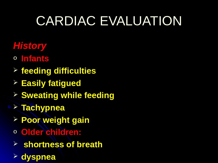
CARDIAC EVALUATION History o Infants feeding difficulties Easily fatigued Sweating while feeding Tachypnea Poor weight gain o Older children: shortness of breath dyspnea
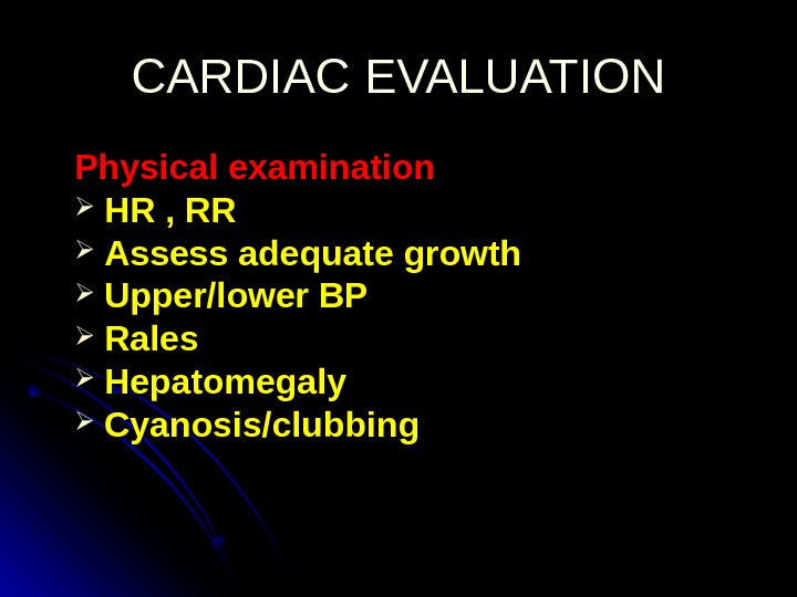
CARDIAC EVALUATION Physical examination HR , RR Assess adequate growth Upper/lower BP Rales Hepatomegaly Cyanosis/clubbing
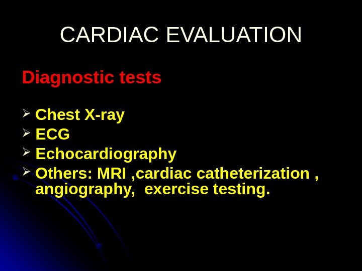
CARDIAC EVALUATION Diagnostic tests Chest X-ray ECGECG Echocardiography Others: MRI , cardiac catheterization , angiography, exercise testing.
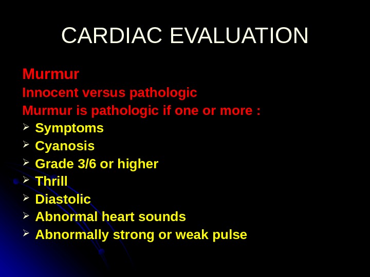
CARDIAC EVALUATION Murmur Innocent versus pathologic Murmur is pathologic if one or more : Symptoms Cyanosis Grade 3/6 or higher Thrill Diastolic Abnormal heart sounds Abnormally strong or weak pulse
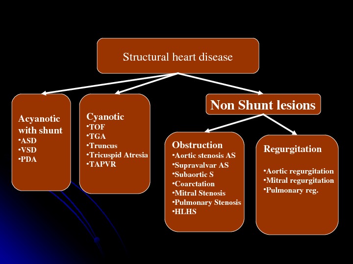
Structuralheartdisease Acyanotic withshunt • ASD • VSD • PDA Cyanotic • TOF • TGA • Truncus • Tricuspid. Atresia • TAPVR Non. Shuntlesions Obstruction • Aorticstenosis. AS • Supravalvar. AS • Subaortic. S • Coarctation • Mitral. Stenosis • Pulmonary. Stenosis • HLHS Regurgitation • Aorticregurgitation • Mitralregurgitation • Pulmonaryreg.
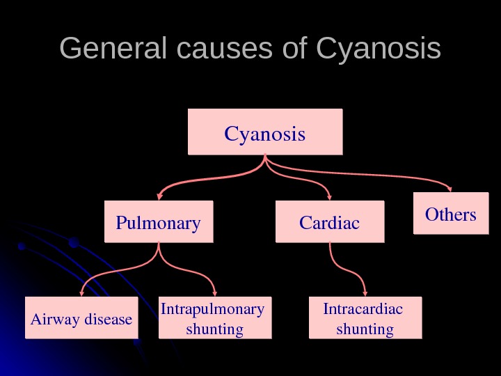
General causes of Cyanosis Pulmonary Cardiac Others Airwaydisease Intrapulmonary shunting Intracardiac shunting. Cyanosis
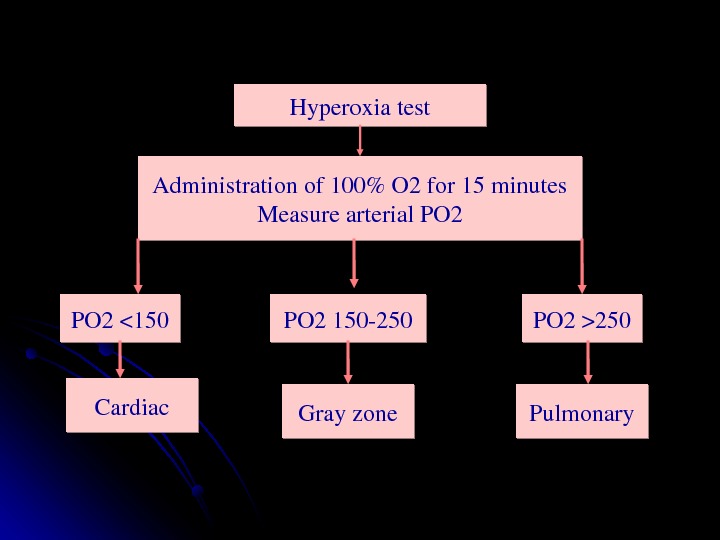
Hyperoxiatest Administrationof 100%O 2 for 15 minutes Measurearterial. PO 2250 PO 2150250 Cardiac Pulmonary Grayzone
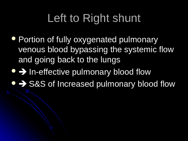
Left to Right shunt Portion of fully oxygenated pulmonary venous blood bypassing the systemic flow and going back to the lungs In-effective pulmonary blood flow S&S of Increased pulmonary blood flow
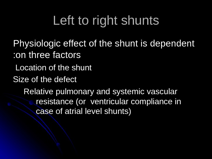
Left to right shunts Physiologic effect of the shunt is dependent on three factors : : Location of the shunt Size of the defect Relative pulmonary and systemic vascular resistance (or ventricular compliance in case of atrial level shunts(
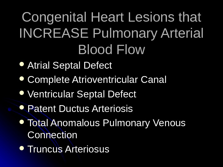
Congenital Heart Lesions that INCREASE Pulmonary Arterial Blood Flow Atrial Septal Defect Complete Atrioventricular Canal Ventricular Septal Defect Patent Ductus Arteriosis Total Anomalous Pulmonary Venous Connection Truncus Arteriosus
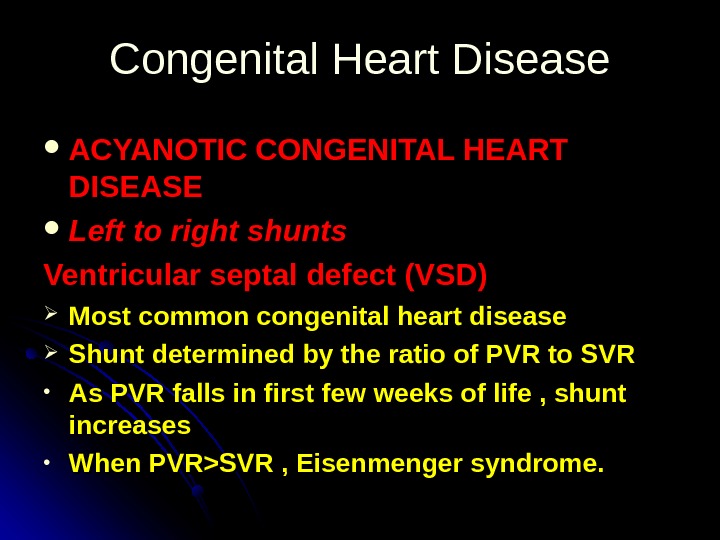
Congenital Heart Disease ACYANOTIC CONGENITAL HEART DISEASE Left to right shunts Ventricular septal defect (VSD) Most common congenital heart disease Shunt determined by the ratio of PVR to SVR • As PVR falls in first few weeks of life , shunt increases • When PVR>SVR , Eisenmenger syndrome.
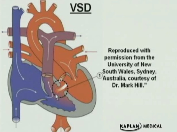
Congenital Heart Disease
Ventricular septal defect (VSD (( Clinical findings Asymptomatic if small defect with normal pulmonary artery pressure (most( Large defect- dyspnea , feeding difficulties , poor growth , sweating , pulmonary infection , heart failure Harsh halosystolic murmur over lower left sternal border +/- thrill Male affected as female Small defect 2 cm
Types of VSD Perimembranous (conoventricular defect( Commonest type of VSD Defect is under the aortic valve There is incidence of aortic valve prolapse and AI Muscular VSD Can be single or multiple Inlet Type VSD (AV canal type( Complete AV canal is common in Trisomy 21 Sub-pulmonary (conal septal hypoplasia VSD( Rare Incidence of aortic valve prolapse
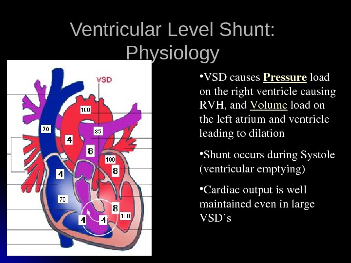
Ventricular Level Shunt: Physiology • VSDcauses Pressure load ontherightventriclecausing RVH, and Volume loadon theleftatriumandventricle leadingtodilation • Shuntoccursduring. Systole (ventricularemptying) • Cardiacoutputiswell maintainedeveninlarge VSD’s
Diagnostic studies ECG: (beyond infancy( Left axis deviation LVHLVH Left atrial dilation Northwest (superior( axis in AV canal defects CXR shows cardiomegally, and increased pulmonary vascular markings in significant VSD’s
Management No restriction from activity No SBE prophylaxis (the newer guidelines( Spontaneous closure is common in small and moderate perimembranous and muscular defects AV canal type VSD’s don’t close spontaneously Surgical treatment is the standard treatment for symptomatic VSD’s
Ventricular septal defect (VSD (( Complications Large defects lead to HF, failure to thrive Endocarditis Pulmonary hypertension
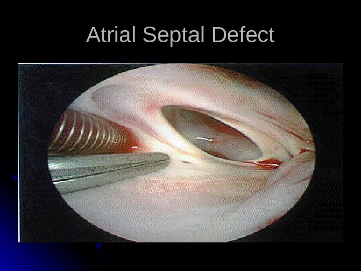
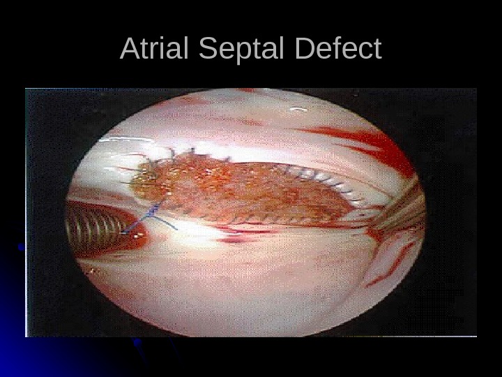
Atrial Septal Defect Acyanotic; asymptomatic, or dyspnea on exertion. Right ventricular lift. Fixed, widely split second heart sound. Female more affected (3 to 1 ( Sporadic or
Types of ASD Ostium secundum ASD: Commonest type Deficiency of septum primum Can be one defect or multiple (fenestrated( Mostly isolated Ostium Primum ASD Also called partial AV canal defect (No VSD component( Frequently associated with cleft mital valve with MR Sinus Venosus ASD SVC type much more common than IVC type Majority associated with partial anomalous pulmonary venous return Coronary sinus ASD Rare Defect is the os of the coronary sinus with partial or complete un-roofing of the sinus
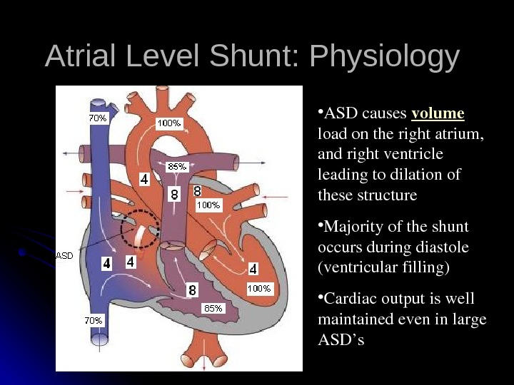
Atrial Level Shunt: Physiology • ASDcauses volume loadontherightatrium, andrightventricle leadingtodilationof thesestructure • Majorityoftheshunt occursduringdiastole (ventricularfilling) • Cardiacoutputiswell maintainedeveninlarge ASD’s
Examination Normal in young infants Prominent RV heave Wide, fixed S 2 Ejection systolic murmur Diastolic rumble
Atrial spetal defect Treatment Most in term infants close spontaneously; symptoms often do not appear until third decade Surgery or transcatheter device closure for all symptomatic patients. Complications Dysrhythmia Low-flow lesions; does not require endocarditic prophylaxis
Management No restriction from activity No SBE prophylaxis No medications Observation for spontaneous closure if secundum type and no significant volume overload on the right ventricle Closure is indicated for significant secundum ASD’s, and all primum and SV ASD’s
Atrial Septal Defect
Atrial Septal Defect
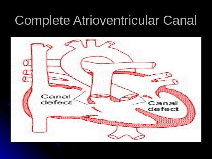
Complete Atrioventricular Canal
Complete Atrioventricular Canal Heart failure common in infancy. Cardiomegaly, blowing pansystolic murmur, other variable murmurs. Deficiencies of both atrial and ventricular septal cushions and abnormalities of both mitral and tricuspid valves.
Complete Atrioventricular Canal Partial and complete AV canal defects frequently accompany Down’s syndrome. Early surgical correction. Reconstruction of the AV valves and closure of the septal defects by a single or double patch technique.
Congenital Heart Disease Patent Ductus Arteriosus (PDA) results when the ductus arteriosus fails to close; this leads to blood flow from the aorta to the pulmonary artery Risk factors More common in girls by 2: 1 Association with maternal rubella infection Common in premature infants
Patent Ductus Arteriosis Murmur usually systolic, sometimes continuous, “machinery” Poor feeding, respiratory distress, and frequent respiratory infections in infants with heart failure. Physical exam and echocardiography.
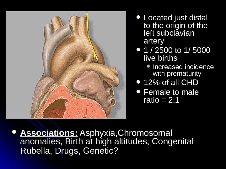
Located just distal to the origin of the left subclavian artery 1 / 2500 to 1/ 5000 live births Increased incidence with prematurity 12% of all CHD Female to male ratio = 2: 1 Associations: Asphyxia, Chromosomal anomalies, Birth at high altitudes, Congenital Rubella, Drugs, Genetic?
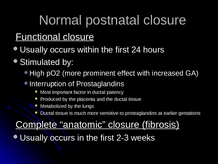
Normal postnatal closure Functional closure Usually occurs within the first 24 hours Stimulated by: High p. O 2 (more prominent effect with increased GA( Interruption of Prostaglandins Most important factor in ductal patency Produced by the placenta and the ductal tissue Metabolized by the lungs Ductal tissue is much more sensitive to prostaglandins at earlier gestations Complete “anatomic” closure (fibrosis( Usually occurs in the first 2 -3 weeks
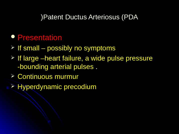
Patent Ductus Arteriosus (PDA (( Presentation If small – possibly no symptoms If large –heart failure, a wide pulse pressure -bounding arterial pulses . . Continuous murmur Hyperdynamic precodium
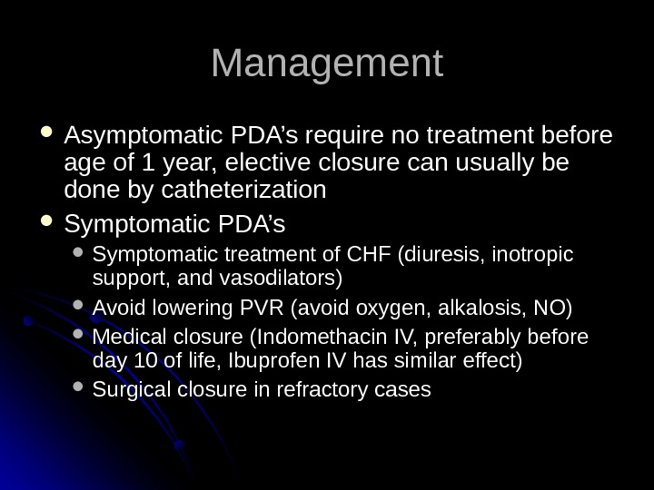
Management Asymptomatic PDA’s require no treatment before age of 1 year, elective closure can usually be done by catheterization Symptomatic PDA’s Symptomatic treatment of CHF (diuresis, inotropic support, and vasodilators( Avoid lowering PVR (avoid oxygen, alkalosis, NO( Medical closure (Indomethacin IV, preferably before day 10 of life, Ibuprofen IV has similar effect( Surgical closure in refractory cases
Cyanotic heart disease (right to left shunt ((
CYANOTIC CONGENITAL HEART DISEASE Common cyanotic heart disease ( 5 Ts & a PP (( TT etralogy of fallot TT ransposition of the great vesseles TT rancus arteriosis TT otal anomalous pulmonary venous return TT ricuspid atresia PP ulmonic stenosis
Congenital Heart Lesions that DECREASE Pulmonary Arterial Blood Flow Tetralogy of Fallot Transposition of the Great Arteries Tricuspid Atresia Ebstein’s Anomaly
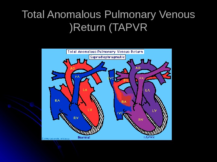
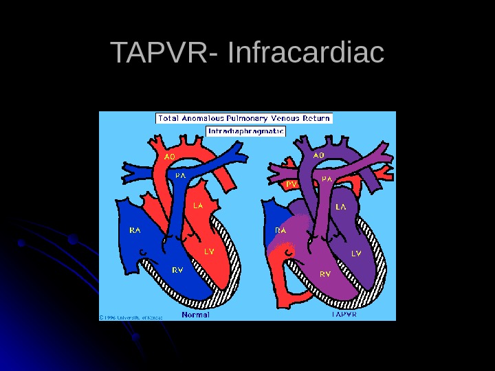
Total Anomalous Pulmonary Venous Connection Pulmonary veins do not make a direct connection with the left atrium. Blood reaches the left atrium only through an atrial septal defect or patent foramen ovale. Pulmonary congestion, tachypnea, cardiac failure, and variable cyanosis.
Total Anomalous Pulmonary Venous Connection Diagnosis by cardiac catherization or echocardiography. Operative repair in all cases.
Total Anomalous Pulmonary Venous Return (TAPVR ((
TAPVR- Infracardiac
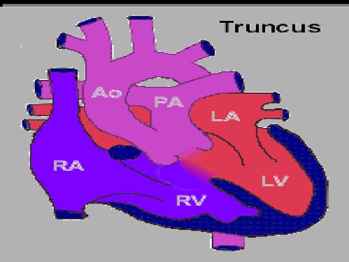
Truncus Arteriosus Single large vessel overrides the ventricular septum and distributes all the blood ejected from the heart. Large VSD is present.
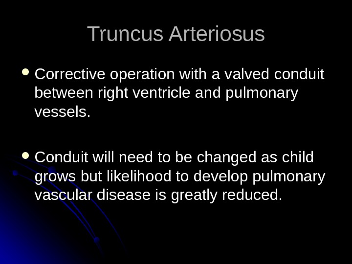
Truncus Arteriosus Corrective operation with a valved conduit between right ventricle and pulmonary vessels. Conduit will need to be changed as child grows but likelihood to develop pulmonary vascular disease is greatly reduced.
Tetralogy of Fallot (TOF (( RVOT obstruction VSDVSD Overriding aorta RV hypertrophy
Tetralogy of Fallot Addition of an atrial septal defect falls in the category of Pentalogy of Fallot. Hypoxic spells and squatting. Cyanosis and clubbing.
Clinical Features Asymptomatic infant with murmur is very common in the usual TOF patients Murmur of RVOT obstruction can be confused with VSD in infancy Cyanosis Typically appears between 6 wks and 6 months in the unrepaired infant Nail beds and mucous membranes May be present at rest or only with agitation/exercise Persistent cyanosis and clubbing if not repaired
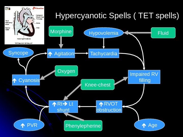
Tachycardia Impaired RV filling RVOT obstruction Rt Lt shunt Agitation Hypovolemia Age PVR Hypercyanotic Spells ( TET spells( Cyanosis. Syncope Fluid. Morphine Oxygen Phenylepherine Knee-chest
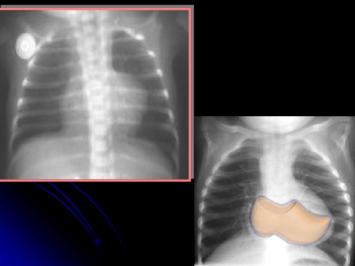
Tetralogy of Fallot Diagnosis Chest X-ray: Boot-shaped heart, dark lung fields ECG: right axis deviation, RVH Echocardiogram (gold standard( Pre-correction complications: cerebral thromboses, brain abscess, bacterial endocarditis , HF. management: surgical repair including closure of VSD and widening of RVOTO
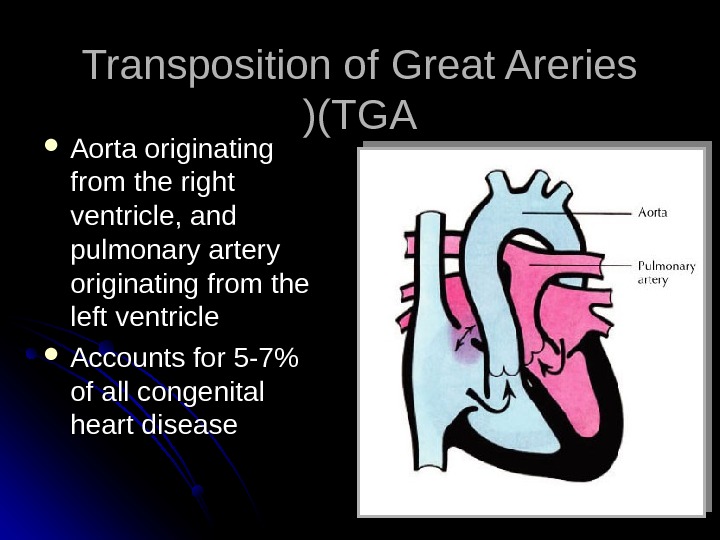
Transposition of Great Areries (TGA (( Aorta originating from the right ventricle, and pulmonary artery originating from the left ventricle Accounts for 5 -7% of all congenital heart disease
TGATGA Survival is dependent on the presence of mixing between the pulmonary and systemic circulation Atrial septal defect is essential for survival 50% of patients have a VSD Usually presents in the first day of life with profound cyanosis More common in boys
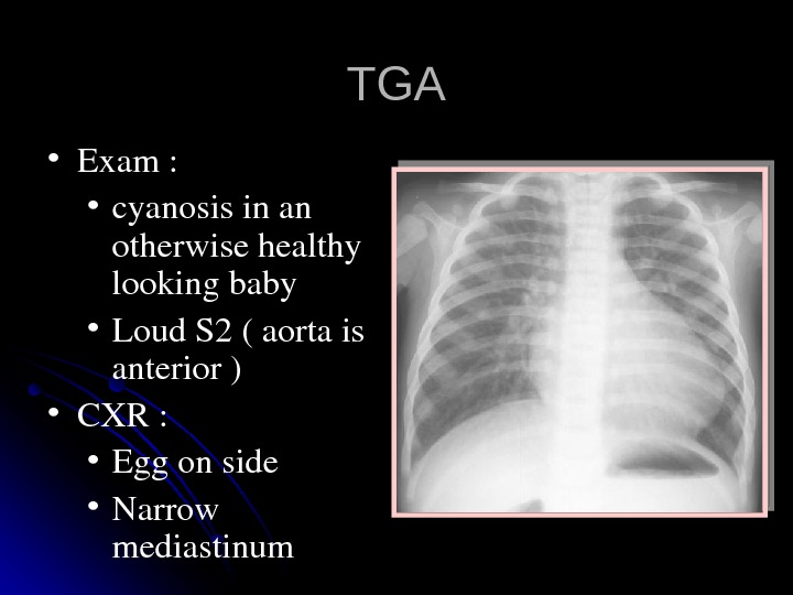
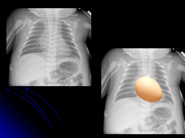
TGATGA • Exam: • cyanosisinan otherwisehealthy lookingbaby • Loud. S 2(aortais anterior) • CXR: • Eggonside • Narrow mediastinum
Transposition of the Great Arteries (TGA (( Management • • prostaglandin E 1 (PGE 1( infusion to keep ductus open until septotomy or surgery • • balloon atrial septostomy with catheter • • surgical correction: arterial switch procedure infants without VSD must be repaired within 2 weeks to avoid weak LV muscle
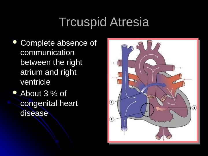
Trcuspid Atresia Complete absence of communication between the right atrium and right ventricle About 3 % of congenital heart disease
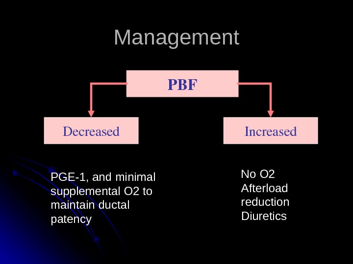
Management PBF Decreased Increased PGE-1, and minimal supplemental O 2 to maintain ductal patency No O 2 Afterload reduction Diuretics
Tricuspid Atresia Repair consists of shunt from right atrium to pulmonary artery or rudimentary right ventricle (Fontan procedure(.
Ebstein’s Anomaly Septal and posterior leaflets of the tricuspid valve are small and deformed, usually displaced toward the right ventricular apex. Most patients have an associated ASD or patent foramen. Cyanosis and arrhythmias in infancy are common.
Ebstein’s Anomaly Right heart failure in half of patients. Operative repair with tricuspid valve replacement.
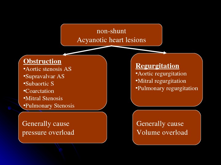
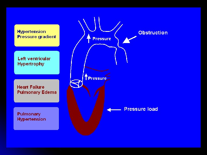
nonshunt Acyanoticheartlesions Obstruction • Aorticstenosis. AS • Supravalvar. AS • Subaortic. S • Coarctation • Mitral. Stenosis • Pulmonary. Stenosis Regurgitation • Aorticregurgitation • Mitralregurgitation • Pulmonaryregurgitation Generallycause pressureoverload Generallycause Volumeoverload
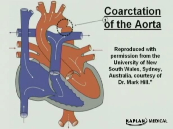
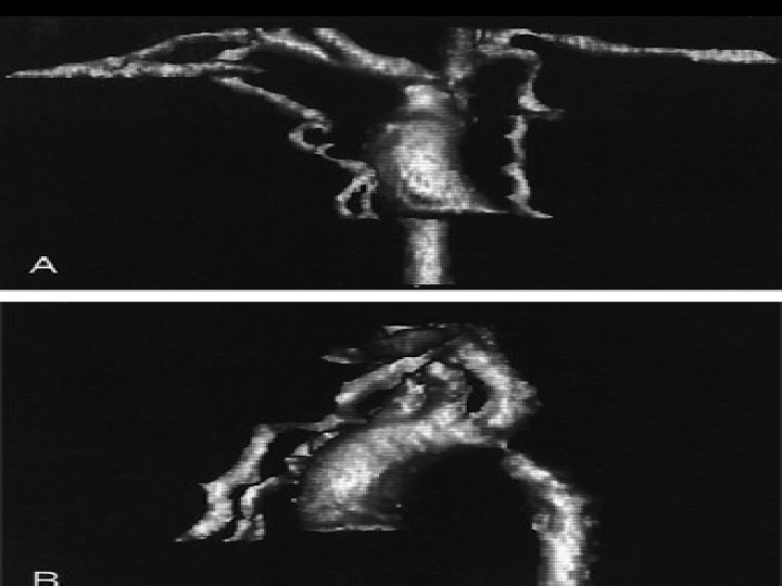
Coarctation of the Aorta Males twice as frequently as females. 98% of all coarctations at segment of aorta adjacent to ductus arteriosus. Produced by both an external narrowing and an intraluminal membrane. Blood flow to the lower body maintained through collateral vessels.
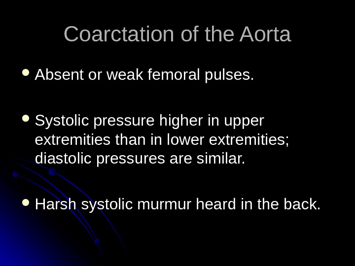
Coarctation of the Aorta Absent or weak femoral pulses. Systolic pressure higher in upper extremities than in lower extremities; diastolic pressures are similar. Harsh systolic murmur heard in the back.
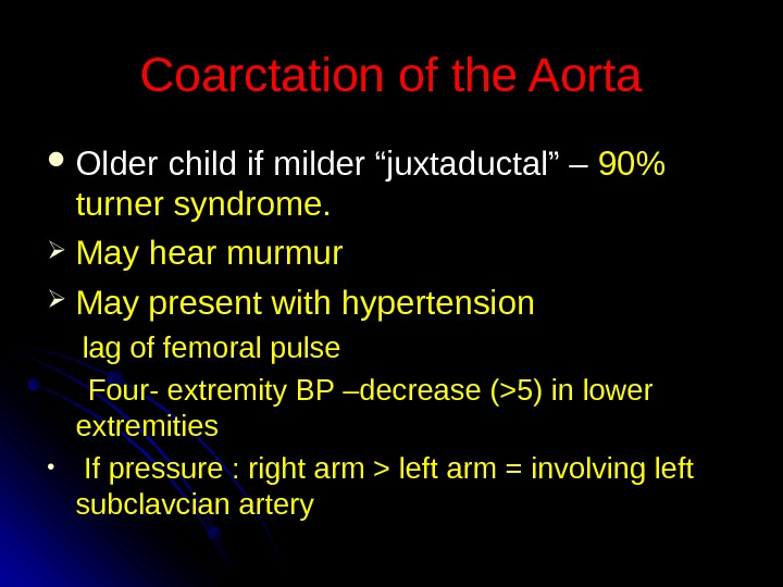
Coarctation of the Aorta Older child if milder “juxtaductal” – 90% turner syndrome. May hear murmur May present with hypertension lag of femoral pulse Four- extremity BP –decrease (>5( in lower extremities • If pressure : right arm > left arm = involving left subclavcian artery
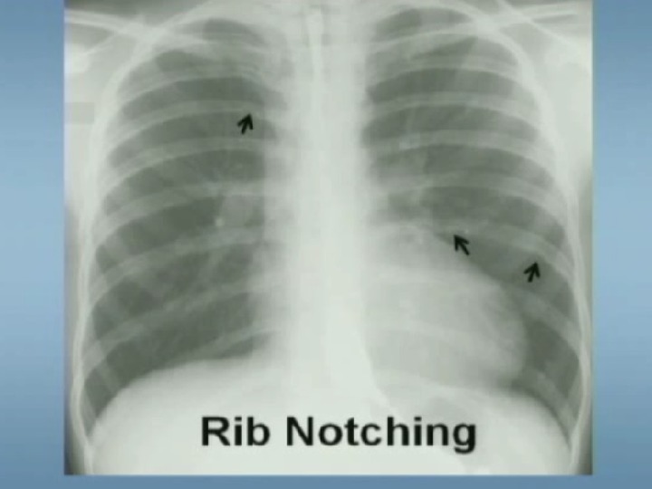
Coarctation of the Aorta Diagnostic tests Chest X-ray Notching of inferior border of ribs Poststenotic dilatation of ascending aorta ECG: RVH early in infancy, LVH later in childhood
Coarctation of the Aorta Management: Neonats : PGE 1 balloon arterioplasty or surgical correction after stabilization Older : treat hypertension then surgery Complications: essential hypertension , aortic aneurysms endocarditis
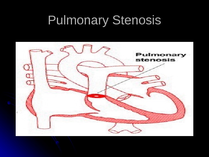
Pulmonary Stenosis
Pulmonary Stenosis No symptoms in mild or moderately severe lesions. Cyanosis and right-sided heart failure in patients with severe lesions. High pitched systolic ejection murmur maximal in second left interspace. Ejection click often present.
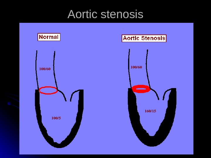
Aortic stenosis 100/60 100/5 100/60 160/
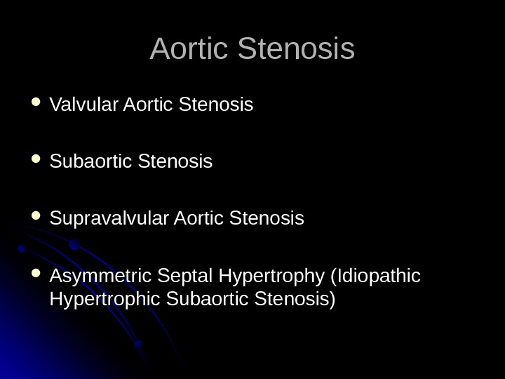
Aortic Stenosis Valvular Aortic Stenosis Subaortic Stenosis Supravalvular Aortic Stenosis Asymmetric Septal Hypertrophy (Idiopathic Hypertrophic Subaortic Stenosis(
Valvular Aortic Stenosis Most common type, usually asymptomatic in children. May cause severe heart failure in infants. Prominent left ventricular impulse, narrow pulse pressure. Harsh systolic murmur and thrill along left sternal border, systolic ejection click.
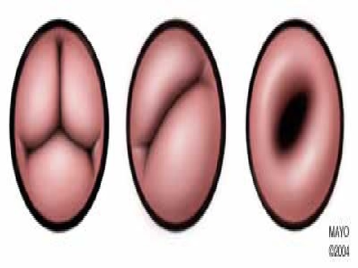
Valvular Aortic Stenosis Predominantly in males Thickened, fibrotic, malformed aortic leaflets. Fused commissures Bicuspid aortic valve.
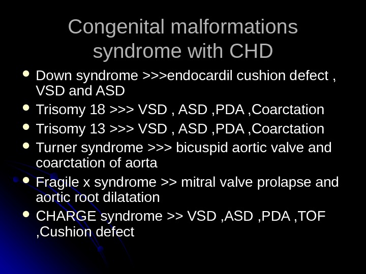
Congenital malformations syndrome with CHD Down syndrome >>>endocardil cushion defect , VSD and ASD Trisomy 18 >>> VSD , ASD , PDA , Coarctation Trisomy 13 >>> VSD , ASD , PDA , Coarctation Turner syndrome >>> bicuspid aortic valve and coarctation of aorta Fragile x syndrome >> mitral valve prolapse and aortic root dilatation CHARGE syndrome >> VSD , ASD , PDA , TOF , Cushion defect
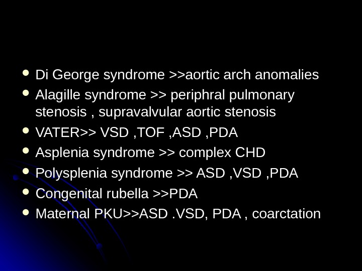
Di George syndrome >>aortic arch anomalies Alagille syndrome >> periphral pulmonary stenosis , supravalvular aortic stenosis VATER>> VSD , TOF , ASD , PDA Asplenia syndrome >> complex CHD Polysplenia syndrome >> ASD , VSD , PDA Congenital rubella >>PDA Maternal PKU>>ASD. VSD, PDA , coarctation
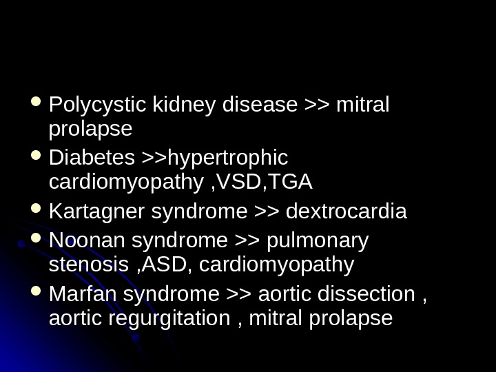
Polycystic kidney disease >> mitral prolapse Diabetes >>hypertrophic cardiomyopathy , VSD, TGA Kartagner syndrome >> dextrocardia Noonan syndrome >> pulmonary stenosis , ASD, cardiomyopathy Marfan syndrome >> aortic dissection , aortic regurgitation , mitral prolapse
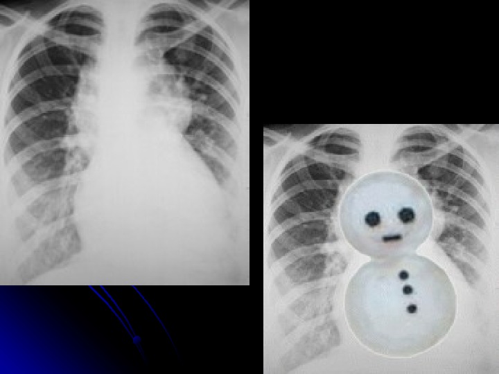
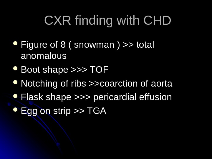
CXR finding with CHD Figure of 8 ( snowman ( >> total anomalous Boot shape >>> TOF Notching of ribs >>coarction of aorta Flask shape >>> pericardial effusion Egg on strip >> TG
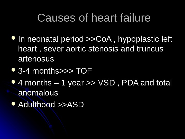
Causes of heart failure In neonatal period >>Co. A , hypoplastic left heart , sever aortic stenosis and truncus arteriosus 3 -4 months>>> TOF 4 months – 1 year >> VSD , PDA and total anomalous Adulthood >>AS
The most common …. …. Murmur >> innocent murmur CHD>>VSD Cyanotic CHD >>in less than 1 month. . TGV, more than 1 month TOF CHD diagnosed at adulthood >>ASD CHD in premature >>> PDA Fatal CHD in neonate >> left hypoplastic heart Causes of plethoric lungs>>> VSD, ASD PDA Dysrhythmia >>> supraventricular tachycardia