
Common Pediatrics Infectious Diseases Dr. Owis khater


common_pediatrics_infectious_diseases.ppt
- Размер: 5.5 Mегабайта
- Количество слайдов: 100
Описание презентации Common Pediatrics Infectious Diseases Dr. Owis khater по слайдам
Common Pediatrics Infectious Diseases Dr. Owis khater
Is a widespread rash , usually viral , and usually occurring in children. It represents either a reaction to a toxin produced by the organism damage to the skin by the organism an immune response. or due to a drug, most commonly antibiotics. Exanthem s
Exanthema 1. 1. Measles. 2. 2. Rubella. 3. 3. Scarlet fever 4. 4. Chicken pox. 5. 5. Erythema infectiosum. 6. 6. Roseola infantum.
Historically, exanthems in children have been numbered in the order they were identified: First disease — Rubeola , Measles (caused by Morbillivirus ) Second disease — rubella, German measles caused rubella virus) Third disease -, scarlet fever, scarlatina, caused by Group A strep- the only bacterial exanthem. Fourth disease- varicella –chicken pox- hepes zoster Fifth disease Erythema infectiosum caused by parvo virus B 19 Sixth disease Roseola infantum. Caused by human herpes virus simplex
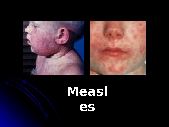
Measl es
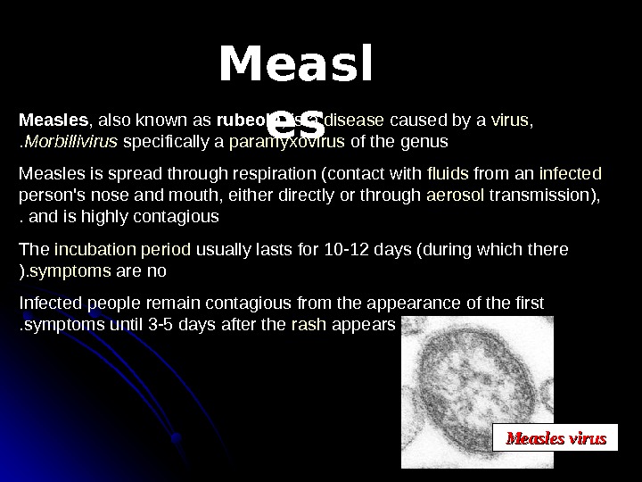
Measl es. Measles , also known as rubeola , is a disease caused by a virus , specifically a paramyxovirus of the genus Morbillivirus. Measles is spread through respiration (contact with fluids from an infected person’s nose and mouth, either directly or through aerosol transmission), and is highly contagious. The incubation period usually lasts for 10 -12 days (during which there are no symptoms). Infected people remain contagious from the appearance of the first symptoms until 3 -5 days after the rash appears. Measles virus
Symptoms fever for at least three days, 40° Celsius. Three C s— cough , coryza (runny nose) conjunctivitis (red eyes) Koplik’s spots seen inside the mouth are pathognomonic transient and may disappear within a day of arising. M aculopapular , erythematous rash that begins several days after the fever starts. It starts on the head before spreading to cover most of the body. The measles rash also classically «stains» by changing colour from red to dark brown before disappearing later. The rash can be itchy.
DIAGNOSIS A detailed history. vaccination history, contact history, and travel history. Clinical diagnosis of measles requires a history of fever of at least three days together with at least one of the three C s. Observation of Koplik’s spots is also diagnostic of measles. Laboratory diagnosis of measles can be done with confirmation of positive measles Ig. M antibodies or isolation of measles virus RNA from respiratory specimens. Positive contact with other patients known to have measles adds strong epidemiological evidence to the diagnosis.
Measles
TREATMENT No specific therapy for measles. Adequate hydration and antipyretics. Avoid strong light : photophobia. IV ribavirin in severe cases. High dose vit A supplementation for 6 M to 2 years old need hospitilization , HIV-infected infants, and infant in endemic areas in devoloping countries.
Complications Otitis media. the most common Interstitial pneumonia. giant cell pneumonia. Activate latent T. B. Myocarditis , and mesentric lymphadenopathy. Encephalomyelitis. Subacute scelorosing panencephlitis. .
Prognosis Death due to bronchopneumonia or encephlitis, and subacute scelorosing panencephlitis. Mortality rate is 15%with 20% to 30% of survivor having serious neurological sequele. Prevented by vaccine.
Prevention & vaccination MMR vaccine first dose 12 to 15 months of age. second dose at school entry People who have measles should limit their contact with others. Exposure within 3 days —— vaccine within 6 days —— immunoglobin
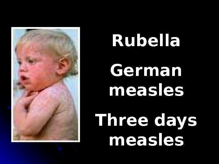
Rubella German measles Three days measles
Rubella ( German Measles ) ) Rubella is also called as 3 day Measles or German Measles. . Family – Togaviridae Genus — Rubivirus In general belong to Togavirus group
Rubella Virus Rubella virus are ss – RNA virus Diameter 50 – 70 nm Enveloped Spherical Virus carry hemagglutinin Virus multiply in the cytoplasam of infected cell. .
Clinical findings Malaise Low grade fever Morbilliform rash Rash starts on Face Extremities Rarely lasts more than 5 days No features of the rash give clues to definitive diagnosis of Rubella. .
Systemic events of Rubella Infection
Adults and children: swollen glands or lymph nodes (may persist for up to a week) fever (rarely rises above 38 degrees Celsius rash (Appears on the face and then spreads to the trunk and limbs. It appears as pink dots under the skin. It appears on the first or third day of the illness but it disappears after a few days with no staining or peeling of the skin) Forchheimer’s sign occurs in 20% of cases, and is characterized by small, red papules on the area of the soft palate cojunctivities
Other manifestations and complications May produce transient Arthritis, in women in particular. . Serious complications areare Thrombocytopenia Purpura Encephalits
Classical Triad of Rubella Classical Triad Cataract Cardiac abnormalities Deafness Other manifestations Growth retardation Rash Hepatosplenomegaly Jaundice Meingoencephalitis CNS defects lead to moderate to profound mental retardation
Diagnosis of Congenital Rubella Syndrome Demonstration of Rubella antibodies of Ig. M in a new born is diagnostic value. As Ig. M group donot cross the placenta and they are produce in the infected fetus , ,
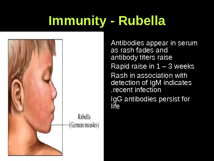
Immunity — Rubella Antibodies appear in serum as rash fades and antibody titers raise Rapid raise in 1 – 3 weeks Rash in association with detection of Ig. M indicates recent infection. . Ig. G antibodies persist for life
Immunity — Protects One attack of Rubella infection, protects for life Immune mothers transfer antibodies to off springs who are in turn are protected for 4 – 6 months. .
Treatment, Prevention, Control No specific treatment is available CRS can be prevented by effective immunization of the young children and teenage girls, remain the best option to prevent Congenital Rubella Syndrome. . The component of Rubella in MMR vaccine protects the vaccinated
MMR Vaccine The MMR vaccine is a mixture of three live attenuated viruses , administered via injection for immunization against measles , , mumps and rubella. It is generally administered to children around the age of one year, with a second dose before starting school (i. e. age 4/5). The second dose is not a booster ; it is a dose to produce immunity in the small number of persons (2 -5%) who fail to develop measles immunity after the first dose In the United States, the vaccine was licensed in 1963 and the second dose was introduced in the mid 1990 s. It is widely used in all National, Universal Immunization programmes
Chickenpox-varicella
Etiology Chickenpox and zoster are caused by varicella -zoster virus. (VZV) , an enveloped , double stranded DNA virus that is a member of the herpes virus family. Human are the only natural host.
Etiology cont …… After resolution of chickenpox, the virus persists in latent phase in the dorsal root ganglia cell. Its highly communicable with secondary attack rate of more than 90%.
The Stages of Chickenpox Incubation Period Usually (14 -17 days) Prodrome (1 – 3 days) Vesicles Pustules Scabs Recovery typically 7 days after rash appears (ranges 5 — 35 days)
Clinical Features Mild prodrome (fever, malaise) for 1 -2 days Successive crops (2 -4 days) of pruritic vesicles Generally appear first on head; most concentrated on trunk Can spread over the entire body causing between 250 to 500 itchy blisters Generally mild in healthy children
NEONATAL CHICKENPOX. . Birth within 1 wk before or after the onset of maternal varicella frequently results in the newborn developing varicella, which may be severe. The initial infection is intrauterine, although the newborn often develops clinical chickenpox postpartum. The risk to the newborn is dependent on the amount of maternal anti-VZV antibody that the fetus acquired transplacentally before birth.
1. Damage to Sensory Nerves. Cicatricial skin lesions 2. Hypopigmentation. Damage to Optic Stalk and Lens Vesicle. Microphthalmia. Cataracts. Chorioretiniti s. optic atrophy. Damage to Brain/Encephalitis, Microcephaly. Hydrocephaly 3. Calcifications. Aplasia of brain 4. Damage to Cervical or Lumbosacral Cord 5. Hypoplasia of an extremity. Motor and sensory deficits 6. Absent deep tendon reflexes. Anisocoria. Horner syndrome, Anal/urinary sphincter dysfunction Stigmata of Varicella-Zoster Virus Fetopathy : :
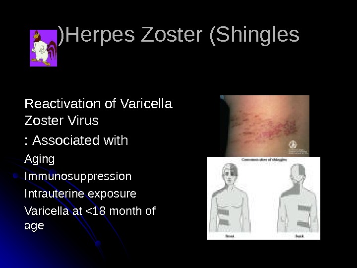
Herpes Zoster (Shingles )) Reactivation of Varicella Zoster Virus Associated with : : Aging Immunosuppression Intrauterine exposure Varicella at <18 month of ageage
Groups at Increased Risk of Complications Normal Adults Immunocompromised persons Newborns with maternal rash onset within 5 days before to 48 hours after delivery
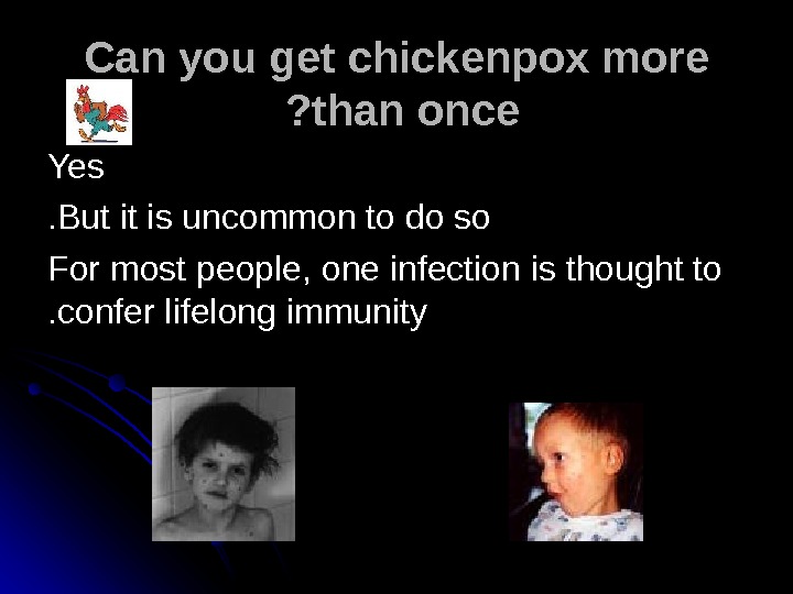
Can you get chickenpox more than once ? ? Yes But it is uncommon to do so. . For most people, one infection is thought to confer lifelong immunity. .
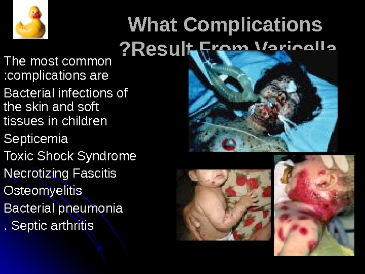
What Complications Result From Varicella ? ? The most common complications are : : Bacterial infections of the skin and soft tissues in children Septicemia Toxic Shock Syndrome Necrotizing Fascitis Osteomyelitis Bacterial pneumonia Septic arthritis. .
What home treatments are available for chickenpox ? ? Fingernails trimmed short Calamine lotion and Aveeno (oatmeal) baths may help relieve some of the itching Aspirin or aspirin-containing products to relieve your child’s fever are not recommended. . The use of aspirin has been associated with development of Reye syndrome (a severe disease affecting all organs — most seriously affecting the liver and brain, that may cause death ). ). The use of non aspirin medications such as acetaminophen is recommended. .
Varicella Vaccine Recommendations Routine vaccination at 12 to 18 months of ageage Recommended for all susceptible children by the 13 thth birthday Persons >> 13 years of age without history of Varicella Two doses separated by 4 – 8 weeks
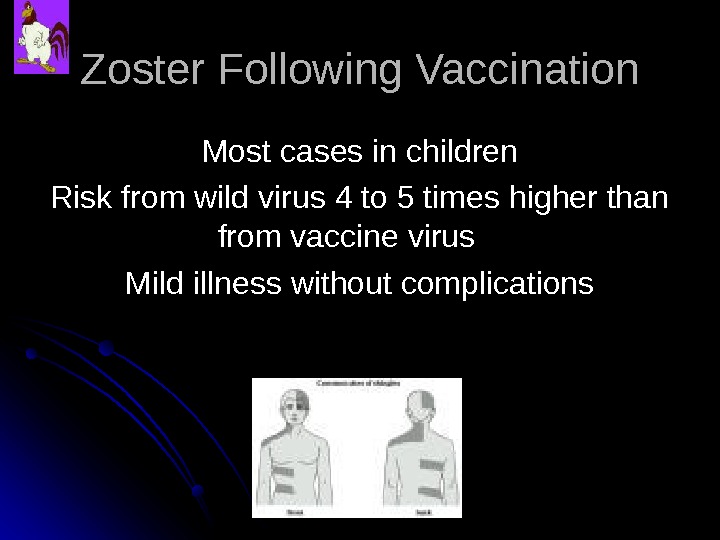
Zoster Following Vaccination Most cases in children Risk from wild virus 4 to 5 times higher than from vaccine virus Mild illness without complications
Varicella Zoster Immune Globulin (VZIG )) May modify or prevent disease if given <96 hours after exposure Indications : : Immunocompromised persons Newborn of mothers with onset 5 days before to 2 days after birth Premature infants with postnatal exposure Susceptible adults and pregnant women
Erythema infectiosum fifth disease
Erythema infectiosum fifth disease : : Caused by human parvovirus B 19. . In children between three and 12 years of age, although it can present as a rheumatic syndrome in adults. . The prodrome : fever, anorexia, sore throat and abdominal pain. . Once the fever resolves, the classic bright-red facial rash (“ slapped cheek ”) appears. . Exanthem progresses to a diffuse, lacy, reticular rash that may wax and wane for six to eight weeks . .
Erythema infectiosum
The incubation period is usually 7 -10 days but can be 4 -21 days. The mechanism producing the dermatologic and rheumatologic features is unknown but thought to represent antigen-antibody (Ag-Ab) complexes in the skin and joints. Arthropathy is observed most commonly in adult women and occurs in fewer than 10% of children. It is a symmetric polyarthritis, usually involving finger joints. The onset of joint symptoms occurs 2 -3 weeks after exposure
Roseola infantum
Background Roseola infantum is the sixth of the traditional exanthems of childhood. The condition is an acute benign disease of childhood characterized by a history of a prodromal febrile illness lasting approximately 3 days, followed by the appearance of a faint pink maculopapular rash.
Causes : : HHV-6 was identified as the etiologic agent in 1988. This large, double-stranded (DNA) virus is a member of the Herpesviridae family. The incubation period is approximately 9 days (range, 5 -15 d).
Mortality/Morbidity: Roseola is usually a self-limited illness with no sequelae. The major morbidity associated with roseola is seizures (6 -15%) during the febrile phase of the illness. Encephalitis, fulminant hepatitis, and disseminated infection with HHV-6 are extremely rare manifestations.
History: Most cases present within the first 2 years of life, with peak occurrence in infants aged 9 m-2 y Roseola is typically characterized by a history of high fever followed by characteristic rash. Fever (often up to 40°C) Rash (fades within a few hours to 2 d) Maculopapular or erythematous Typically beginning on the trunk and may spread to involve the neck and extremities Nonpruritic Blanches on pressure
Medication : To date, no controlled antiviral trials exist against HHV-6. . Prevention : Because of the ubiquity of the virus, isolation of patients with HHV-6 infection is probably unnecessary
Scarlet Fever
Scarlet fever Is an exotoxin-mediated disease caused by Group A streptococcal infection that occurs most often in association with a sore throat and rarely with impetigo or other streptococcal infections. Scarlet fever is not rheumatic fever. Rheumatic fever is the autoimmune disease that occurs after infection with Group A strep that causes damage to the heart valves. The disease was once greatly feared and killed many thousands of people. Today, however, it is fairly easy to treat with modern antibiotics.
Clinical manifestation Scarlet fever generally has a 1 — to 4 -days incubation period. Emergence of the illness tends to be abrupt, usually manifested by sudden onset of fever associated with sore throat, headache, nausea, vomiting, abdominal pain, myalgias, and malaise. The characteristic rash appears 12 -48 hours after onset of fever. In the untreated patient, fever peaks by the second day (as high as 103 -104°F) and gradually returns to normal in 5 -7 days. Fever abates within 12 -24 hours after initiation of antibiotic therapy.
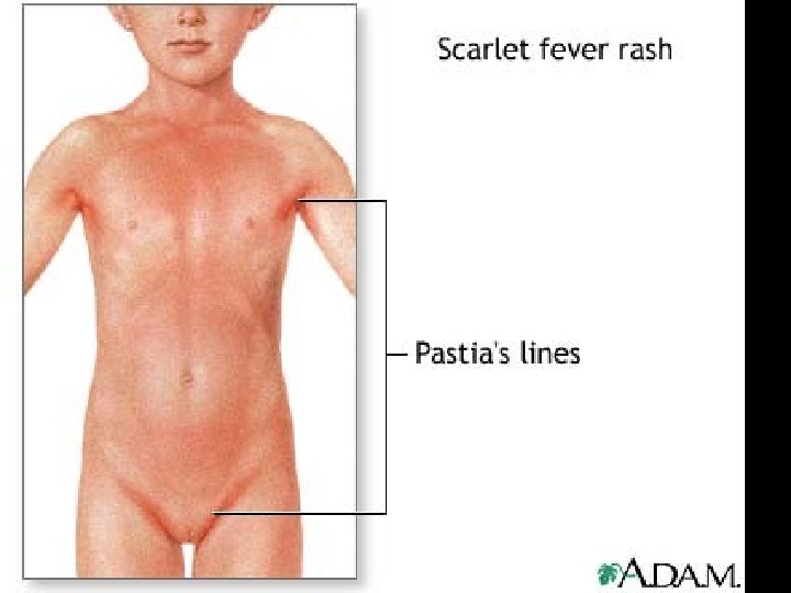
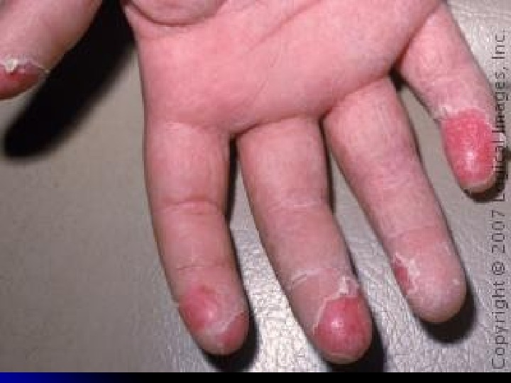
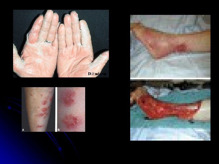
Skin rash scarlatina generally starts on the chest, axilla , and behind the ears worse in the skin folds Pastia lines (small linear petechiae) appear and persist after the rash is gone Scarlet fever also produces a bright red tongue with a » strawberry » appearance. The area around the mouth is usually pale (circumoral pallor) After about a week, the skin often desquamates or peels, usually in the groin, axilla, and on tips of fingers and toes
Complications arise from suppurative complications such as; *peritonsillar abscess, *sinusitis, *bronchopneumonia, *and meningitis, or problems associated with immune system as rheumatic fever or glomerulonephritis
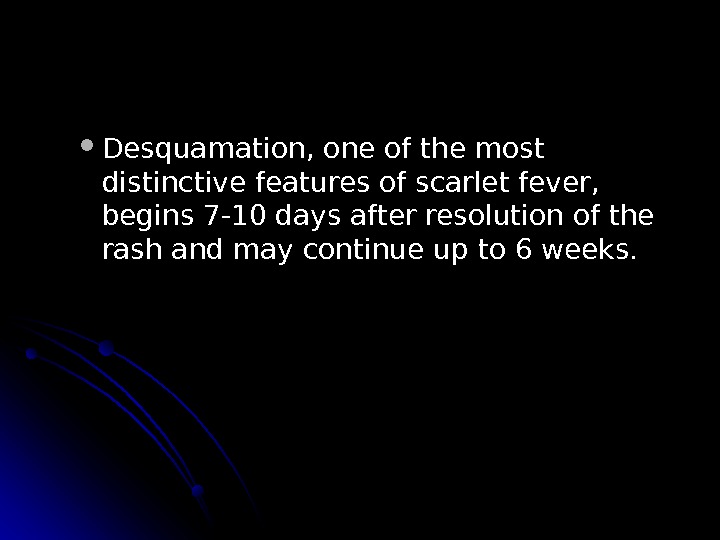
Desquamation, one of the most distinctive features of scarlet fever, begins 7 -10 days after resolution of the rash and may continue up to 6 weeks.
Lab Studies: 1. Throat culture remains the “gold standard” for confirmation of group A streptococcal upper respiratory infection. ASOT antistreptolysine o titere
treatment penicillin Pediatric Dose 12 year: Administer as in adults Adult Dose 250 mg PO tid/qid for 10 d Contraindications Documented hypersensitivity
TOXIC SHOCK SYNDROME AND SCALDED SKIN SYNDROME Staphylococcus aureus exotoxins responsible for classic toxic shock syndrome and scalded skin syndrome. . Presention : hypotension, erythema, fever and multisystem dysfunction. . The rash : diffuse and can present as bullous impetigo, scarlatiniform lesions or diffuse erythema. . The mucous membranes : spared
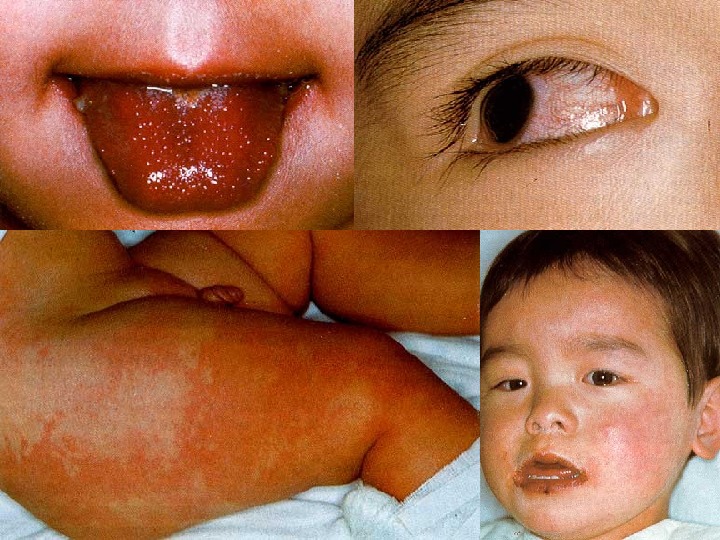
Coxsackie viruses and other enteroviruses Hand-foot-and-mouth disease: the children develop fever and rash. The rash includes blisters to the mouth and tongue, to the hands and the feet. . Herpangina causes a fever, sore throat, and painful blisters or ulcers to the back of the mouth. .
Hand-Foot-Mouth Disease Enteroviruses coxsackieviruses A and B echoviruses Vesicular lesions, may be petechial Associated with aseptic meningitis, myocarditis
Infectious Mononucleosis Acute, self limited illness Epstein-Barr virus Oral transmission – incubation 30 -50 days Fever, fatigue, pharyngitis, LA, splenomegaly, atypical lymphocytosis Exanthem is seen in 10 -15% Erythematous, maculopapular, morbilliform, scarlatiniform, urticarial, hemorrhagic, or even nodular
Impetigo Superficial infection of the dermis Two types : : Impetigo contagiosa Bullous impetigo Etiology Group A ß hemolytic streptococcus Coagulase positive S. aureus Treatment : Keflex, erythromycin, Bactroban
Rocky Mountain Spotted Fever Most common rickettsial infection in US Abrupt fever, headache, and myalgia Rash from extremities towards trunk Macules petechiae Treatment Tetracycline Doxycycline Chloramphenicol
Periorbital- Orbital Cellulitis S. aureus, S. pneumoniae , and HIBHIB CBC, blood culture, CT LPLP ? ? IV antibiotics Admit
Kawasaki Syndrome Unknown etiology Peak incidence 18 -24 months Clinical findings : : Fever for at least five days Conjunctivitis Polymorphous rash Oral cavity changes Cervical adenopathy
Erythema Toxicum Neonatorum Impressive title — harmless skin condition Erythematous macule with a central tiny papule, seen anywhere — except the palms and soles. . The lesions are packed with eosinophils, and there may be accompanying eosinophilia in the blood count. . The cause is unknown, and no treatment is required as the rash disappears after 1 -2 weeks. .
Miliaria Prickly heat, sweat rash Many red macules with central papules, vesicles or pustules are present. . These may be on the trunk, diaper area, head or neck. .
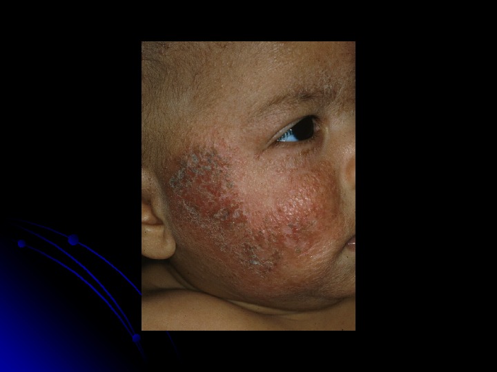
Infantile Atopic Dermatitis Cause is unknown Red, itchy papules and plaques that ooze and crust Sites of Predilection Face in the young Extensor surfaces of the arms and legs 8 -10 momo. . Antecubital and popliteal fossa , neck, face in older
Eczema- Treatment Avoidance or elimination of predisposing factors Hydration and lubrication of dry skin Anti-pruritic agents Topical steroids
Seborrheic Dermatitis Common, generally self-limiting Its cause remains ill-understood There is a genetic predisposition Most frequent between the ages of 1 to 6 momo. . Greasy, salmon-colored scaling eruption Hair-bearing and intertriginous areas The rash causes no discomfort or itching
Seborrheic Dermatitis-Treatment Anti-seborrheic shampoo Topical steroids
Cytomegalovirus (CMV )) Most common congenital viral infection ~~ 40, 000 infants per year in the U. S. . Mild, self limiting illness Transmission can occur with primary infection or reactivation of virus 40%40% risk of transmission in primary infxn Studies suggest increased risk of transmission later in pregnancy However, more severe sequalae associated with earlier acquisition
Clinical Manifestations 90%90% are asymptomatic at birth !! Up to 15% develop symptoms later, notably sensorineural hearing loss Symptomatic infection SGA, HSM, petechiae, jaundice, chorioretinitis, periventricular calcifications , , neurological deficits >> 80%80% develop long term complications Hearing loss, vision impairment, developmental delay
Ventriculomegaly and calcifications of congenital CMV
Diagnosis Maternal Ig. G shows only past infection Infection common – this is useless Viral isolation from urine or saliva in 1 stst 3 weeks of life Afterwards may represent post-natal infection Viral load and DNA copies can be assessed by PCRPCR Less useful for diagnosis, but helps in following viral activity in patient Serologies not helpful given high antibody in population
Treatment Ganciclovir x 6 wks in symptomatic infants Studies show improvement or no progression of hearing loss at 6 mos No other outcomes evaluated (development, etc. ). ) Neutropenia often leads to cessation of therapy Treatment currently not recommended in asymptomatic infants due to side effects Area of active research to include use of valgancyclovir, treating asx patients, etc. .