

кольпоскопия норма и патология VESNA KESIC.ppt
- Количество слайдов: 106
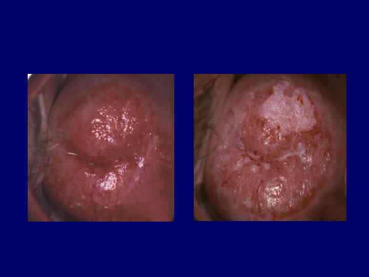
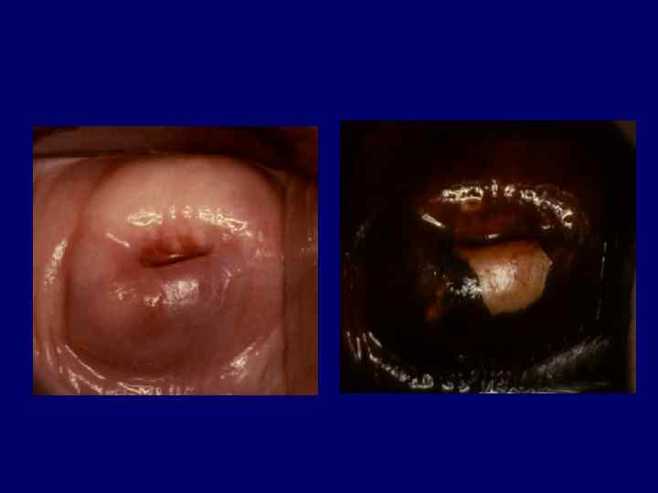
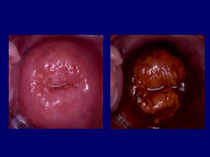
 Schiller test (Lugol’s solution)") Colposcopy Acetic acid test (3 -5% acetic acid) Schiller test (Lugol’s solution)
Colposcopy Acetic acid test (3 -5% acetic acid) Schiller test (Lugol’s solution)
 • Squamous epithelium • Columnar epithelium • Squamo-columnar junction • Metaplasia • Transformation Zone
 Collumnar epithelium Squamous epithelium
 Squamo-collumnar junction- SCJ
 Metaplasia a physiological and benign process whereby the columnar epithelium is gradually replaced by squamous epithelium Transformation zone the area where metaplasia takes place
 The result of normal metaplasia is a normal Transformation zone
 Immature metaplastic cells are susceptible to the development of atypical cellular changes
 The process of transformation from normal cells to atypical cells occurs under the influence of Human papillomavirus (HPV) and cofactors
 If atypical metaplasia takes place an abnormal Transformation zone develops
 N O R M A L M E T A P L A S I A A T Y P I C A L
 In colposcopy, it is essential to asses whether Transformation zone is normal or abnormal
 Colposcopic Classification I Normal colposcopic findings") International Federation for Cervical Pathology and Colposcopy (IFCPC) Colposcopic Classification I Normal colposcopic findings II Abnormal colposcopic findings III Colposcopic findings suggestive of invasive cancer IN Unsatisfactory colposcopy V Miscellaneous findings
International Federation for Cervical Pathology and Colposcopy (IFCPC) Colposcopic Classification I Normal colposcopic findings II Abnormal colposcopic findings III Colposcopic findings suggestive of invasive cancer IN Unsatisfactory colposcopy V Miscellaneous findings
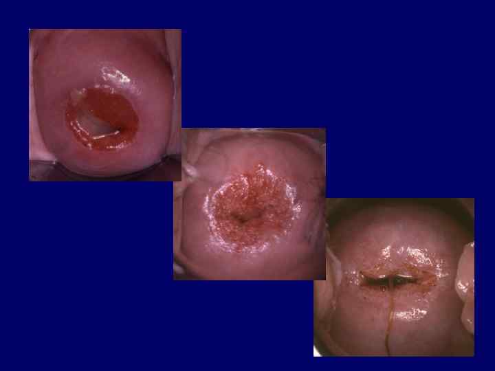
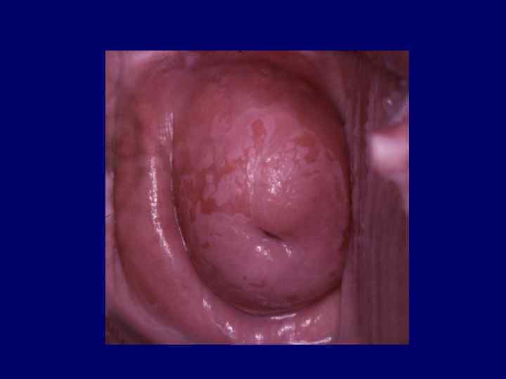
 Components of a normal Transformation zone • Islands of columnar epithelium • Cleft openings • Nabothian cysts
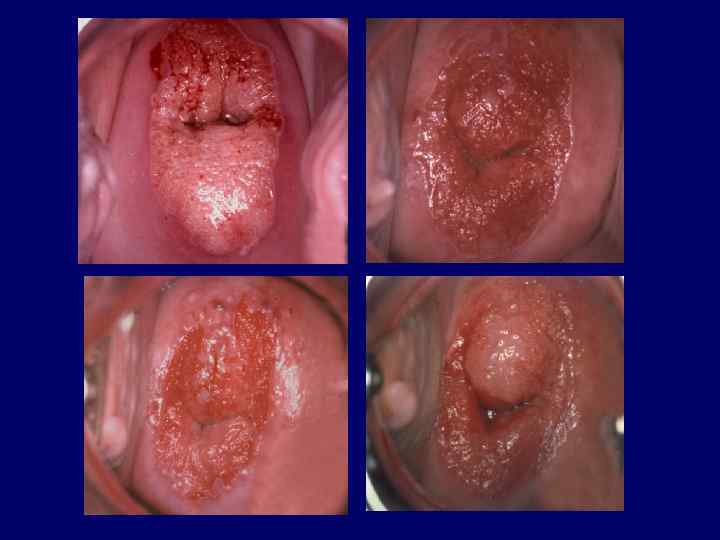
 The abnormal Transformation zone is manifested as a wide spectrum of epithelial and vascular findings
 colposcopic findings") Abnormal transformation zone is presented by abnormal (atypical) colposcopic findings
Abnormal transformation zone is presented by abnormal (atypical) colposcopic findings
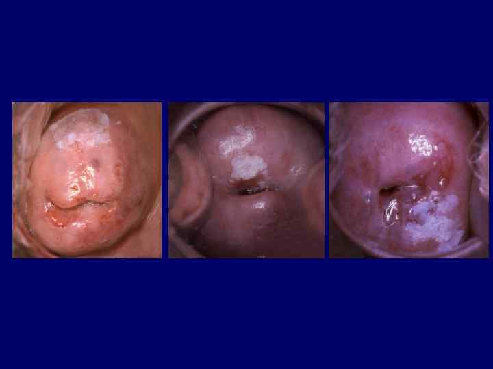
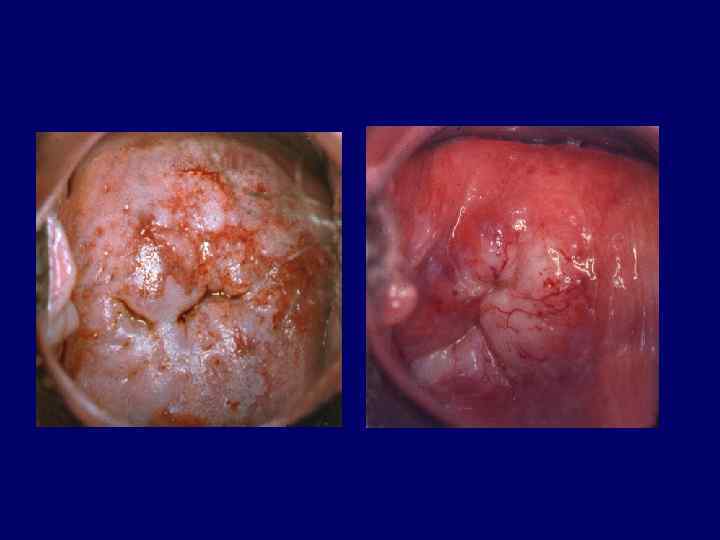
 Abnormal colposcopic findings • Leukoplakia • Acetowhite epithelium • Punctation • Mosaic • Iodine negativity • Atypical vessels
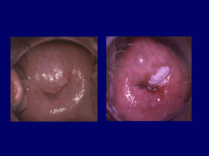
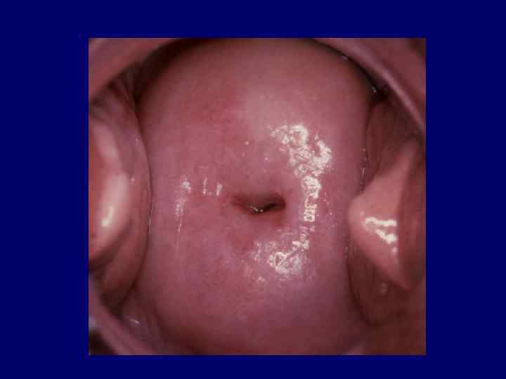
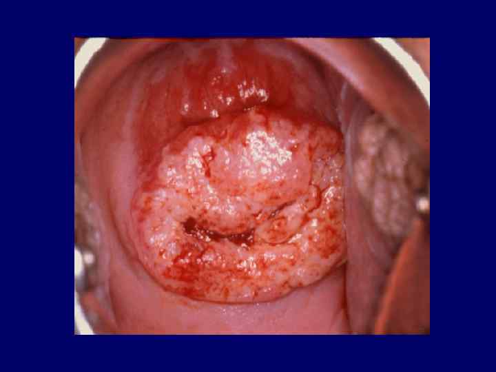
 Leukoplakia or white plaque is visible grossly as a white often raised area that is not necessarily confined to TZ
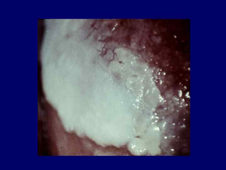
 Leukoplakia • HPV infection • Keratinizing CIN • Keratinizing cancer • Chronic trauma • Radiotherapy • Immature metaplasia
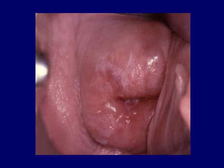
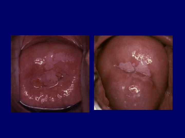
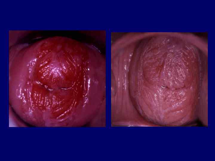
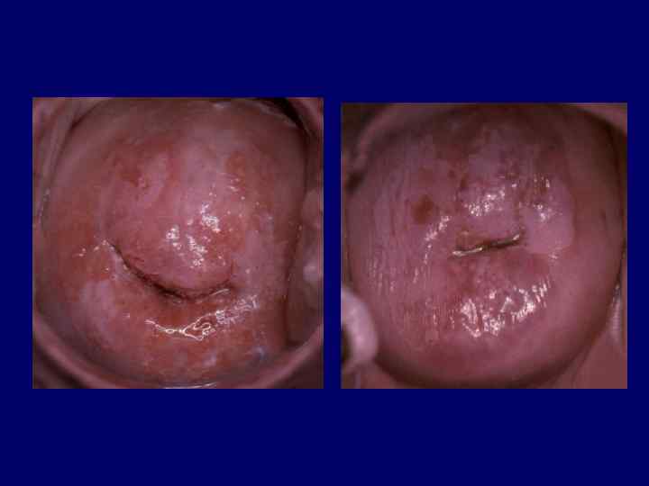
 Acetowhite epitehlium Appears grossly normal but turns white after application of 3% to 5% acetic acid
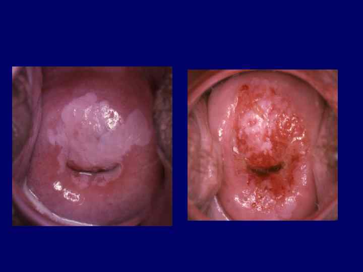
 Acetowhite epithelium • HPV infection • Immature squamous metaplasia • Regenerative or reparative changes • Congenital Transformation zone • Inflammation • CIN • Adenocarcinoma • Invasive squamous carcinoma
 Any cells with an enlarged nucleus such as metaplatic cells or cells traumatized by infection or friction, may exibit varying degrees of acetowhiteness
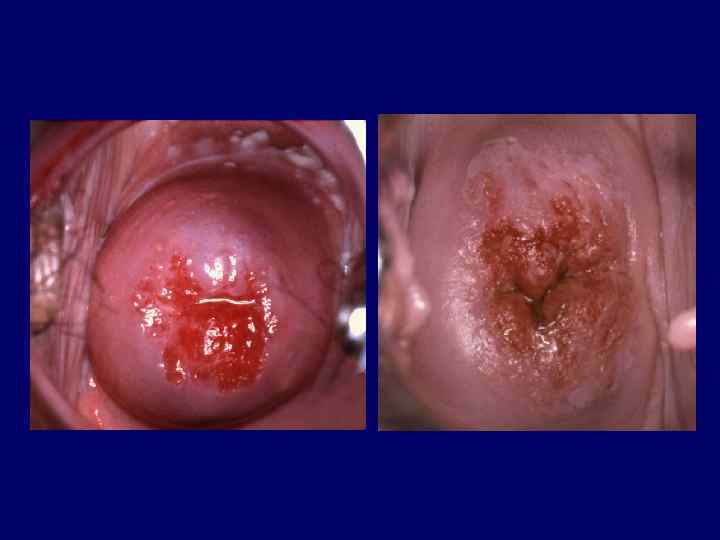
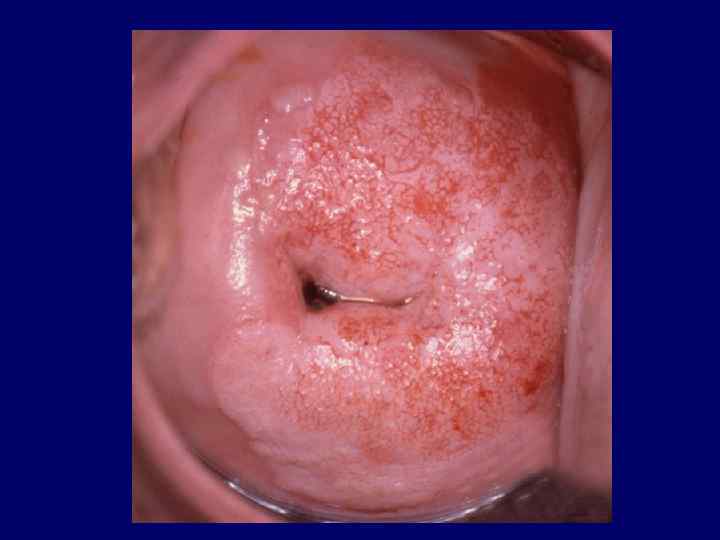
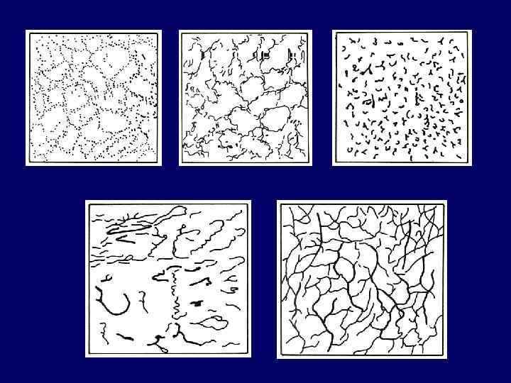
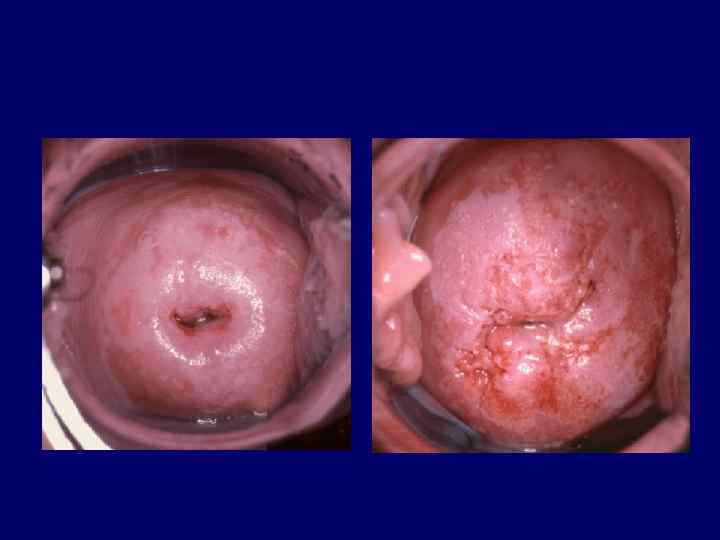
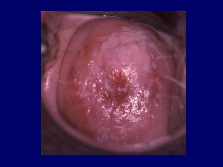
 Punctation a focal colposcopic pattern in which cappilaries appear in stippled pattern. Mozaik a focal colposcopic appearance in which the new vessel formation appears as a rectangular pattern like mosaic
 Punctation colposcopic finding reflecting the capillaries in the stromal papillae that are seen end-on and penetrate the epithelium
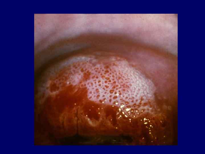
 Mosaic colposcopic finding reflecting the islands of squamous epithelium, encircled by blood vessels in a basket-like arrangement
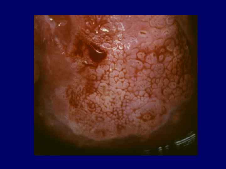
 Punctation and Mosaic • Inflammation • Rapidly growing metaplastic epithelium • CIN • Invasive squamous cancer • Recurrence of cervical cancer
 If the punctation or mosaic is not located in a field of acetowhite epithelium, it is unlikely to be associated with CIN
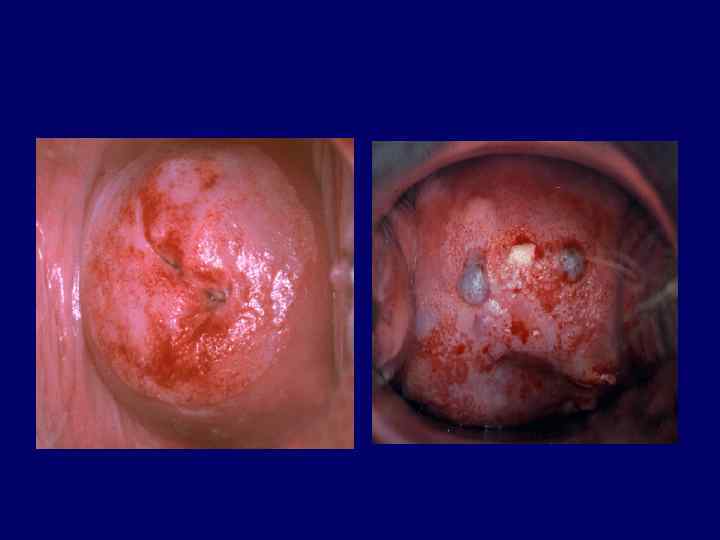
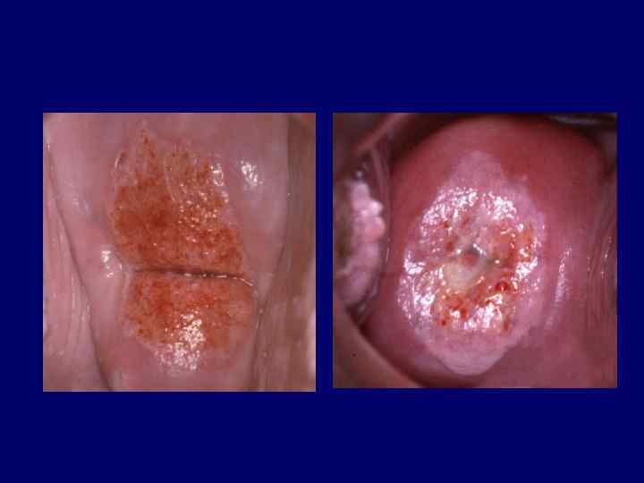
") Iodine negativity • Immature metaplasia • Cervical intraepithelial neoplasia • Low estrogen status (atrophy)
Iodine negativity • Immature metaplasia • Cervical intraepithelial neoplasia • Low estrogen status (atrophy)
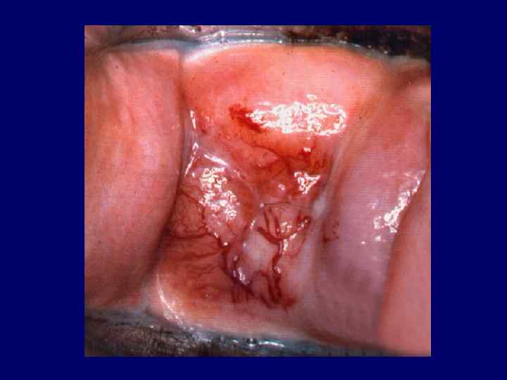
 Atypical vessels • Irregular vessels with an abrupt and interrupted course • Appearing as commas, corkscrw capillaries or spaghetti-like forms
 Atypical vessels are the hallmark of invasion, but can be associated with other conditions such as • Inflammation • Postirradiation effect • Rapidly growing metaplastic epitheluim • Normal epithelium • Systemic diseases
 Development of abnormal colposcopic features may be the result of: • Immature physiologic metaplasia • Papilloma virus infection • Developing CIN
 a grading system used to evaluate the severity of the colposocpic") Colposcopic index (score) a grading system used to evaluate the severity of the colposocpic findings
Colposcopic index (score) a grading system used to evaluate the severity of the colposocpic findings
 A number of scoring systems have been introduced: • Coppleson & Pixley • Burghardt • Rubin & Barbo • Reid
 Grading of colposcopical findings • Vascular pattern • Intercapillary distance • Color tone and opacity • Surface pattern • Borders with normal tissue
 Colour • Severe abnormalities become whiter than minor lesions • They tend to become white more quickly • Retain their whiteness longer than the mild lesions
 Borders A clear zone of demarcation exists between the native squamous epithelium and high grade CIN lesion. Mild changes usually have a less distinct outline
 Surface pattern More uneven and elevated contours are, the higher grade the lesion is.
 Intercapillary distance • Increases as the lesion becomes more severe. • The larger vessels and further apart they lie, the more severe is the lesion
 Ideally, colposcopic scoring should allow categorizing the colposcopic pattern as: • Normal • Insignificant • Clinically significant
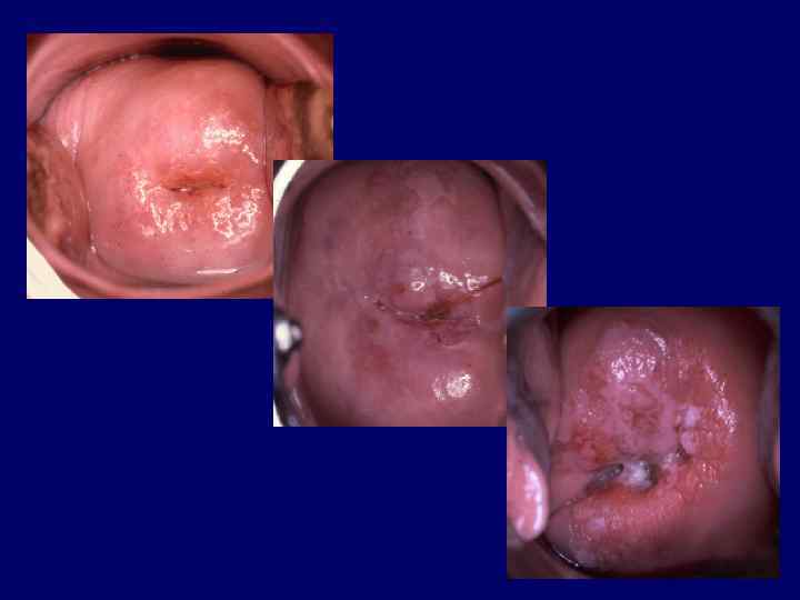
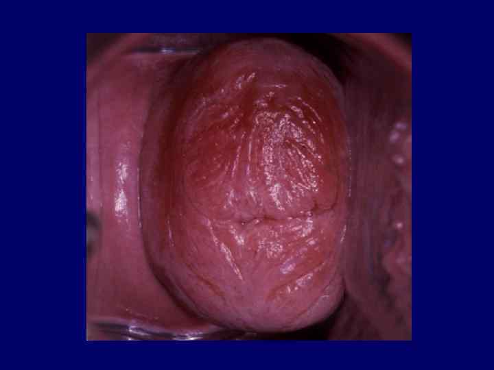
 Colposcopic features suggestive of metaplastic changes • A smooth surface with fine, uniform-caliber vessels • Mild acetowhite change • Negative or partial positivity with Lugol’s iodine
 As the metaplastic cells transform into mature squamous cells, the coloration is indistinquisable from the mature ectocervix
 • A smooth surface with") Colposcopic features suggestive of low grade disease (minor changes) • A smooth surface with an irregular outer border • Slight acetowhite change slow to appear and quick to dissapear • Mild, often speckled iodine partial posivitity • Fine punctation and fine regular mosaic
Colposcopic features suggestive of low grade disease (minor changes) • A smooth surface with an irregular outer border • Slight acetowhite change slow to appear and quick to dissapear • Mild, often speckled iodine partial posivitity • Fine punctation and fine regular mosaic
 The subtle differences between the features of squamous metaplasia and those of low-grade intraepithelial lesions make both the colposcopic and histologic diagnosis of these conditions difficult
 It is easier to determine that a cervix is either normal or very abnormal, than it is to distinguish between minor degrees of change
 Misinterpretation of trivial changes as atypical findings can lead to mismanagement and overtreatment of the patient
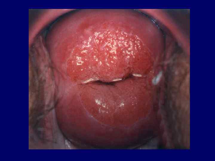
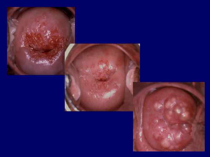
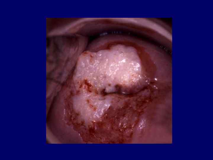
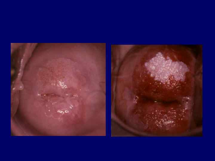
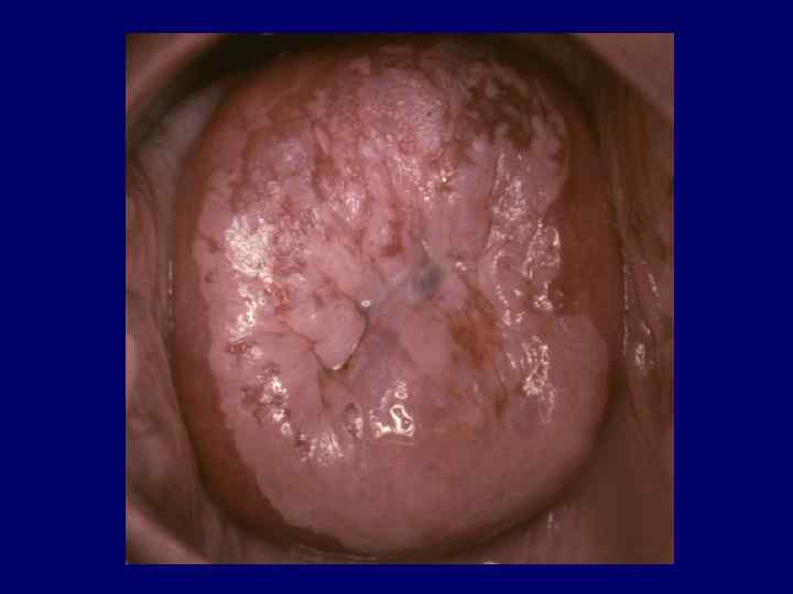
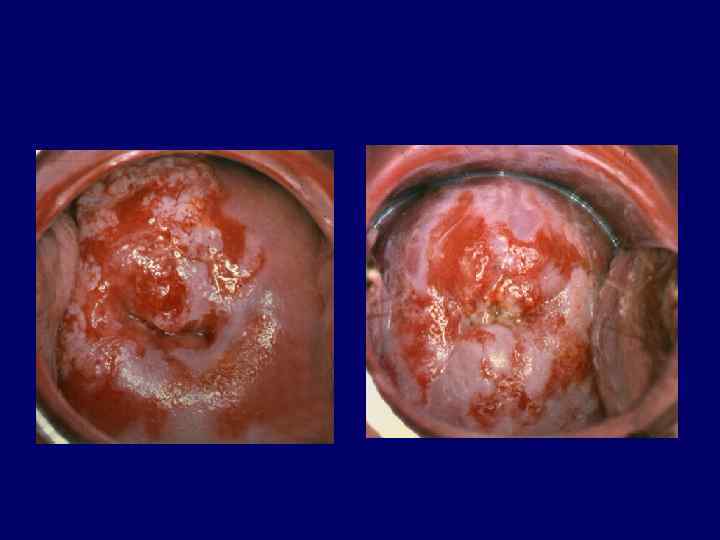
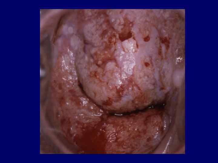
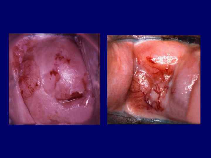
 • A generally smooth surface") Colposcopic features suggestive of high- grade disease (major changes) • A generally smooth surface with sharp outer border • Dense acetowhite change, may be oyster white appears early slow to resolve • Iodine negativity • Coarse punctation and wide irregular mosaic of different size
Colposcopic features suggestive of high- grade disease (major changes) • A generally smooth surface with sharp outer border • Dense acetowhite change, may be oyster white appears early slow to resolve • Iodine negativity • Coarse punctation and wide irregular mosaic of different size
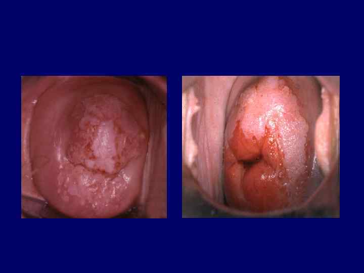
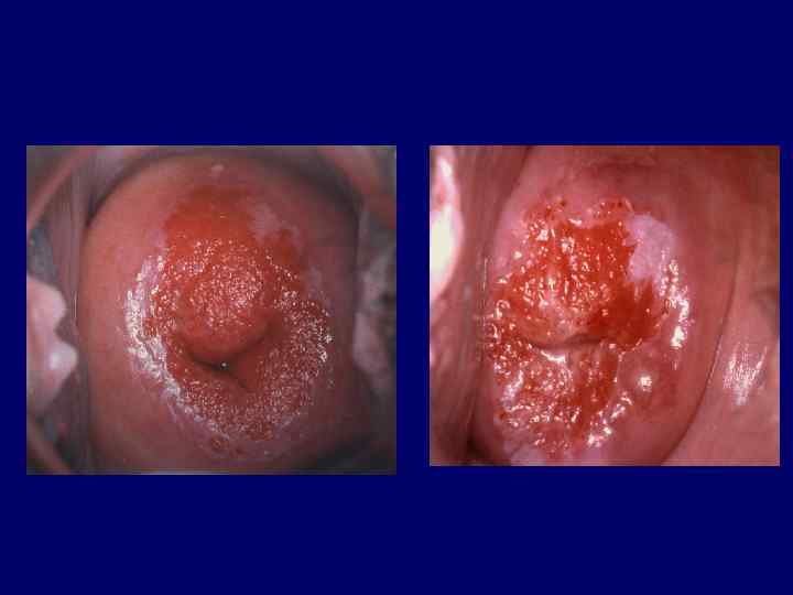
 Signs of microinvasion • Yellow discoloration • Ulceration • Thickened areas • Nodularity • Abnormal vascularity • Rapid increase in size
 There is a direct relationship between the size of a lesion and the likelihood of invasion
 Early stromal invasion is more common when there are different types of epithelia (complex colposcopic changes)
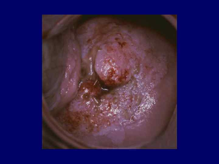
 Microinvasion should be suspected when relatively flat lesions display focal collections of atypical vessels
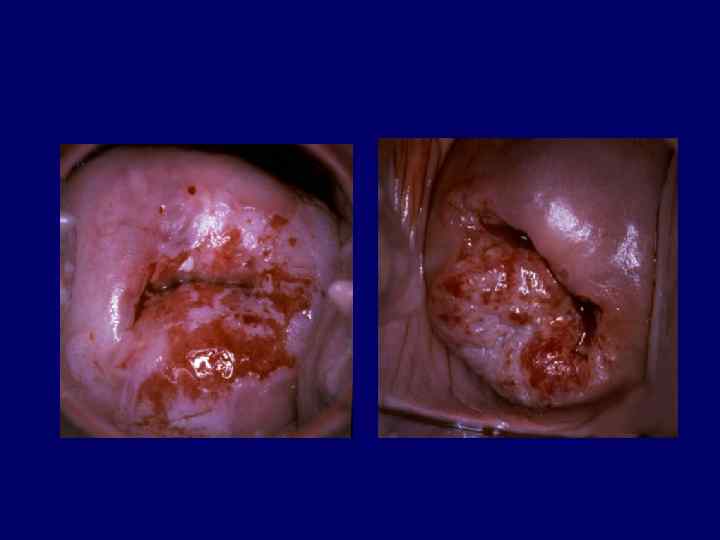
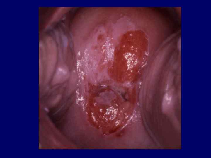
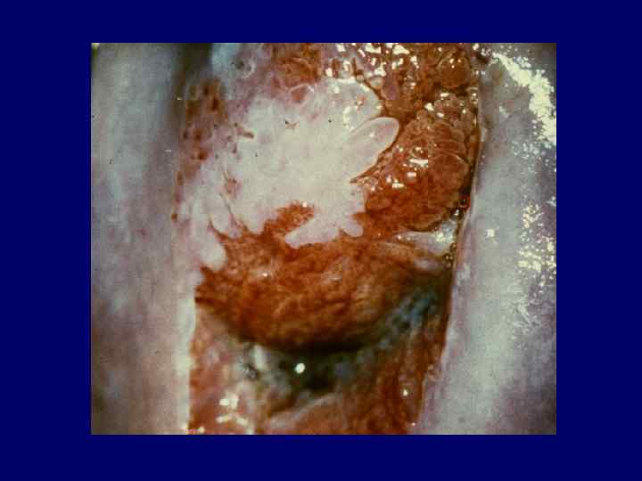
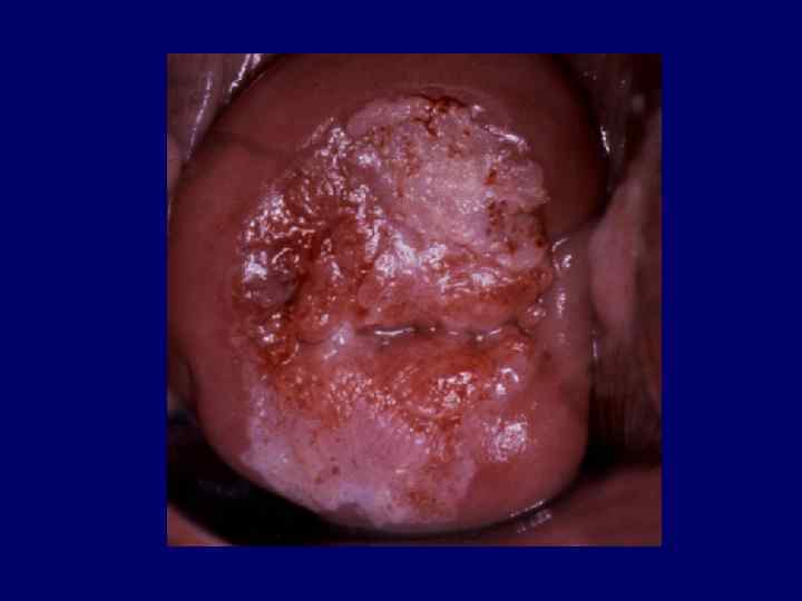
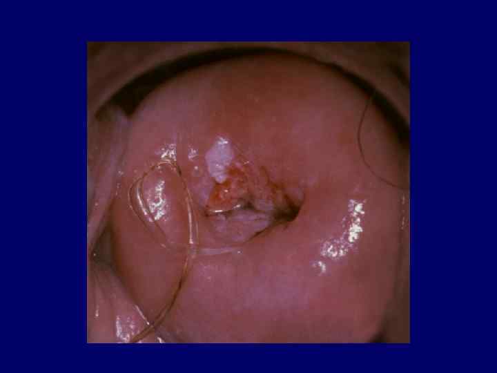
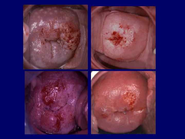
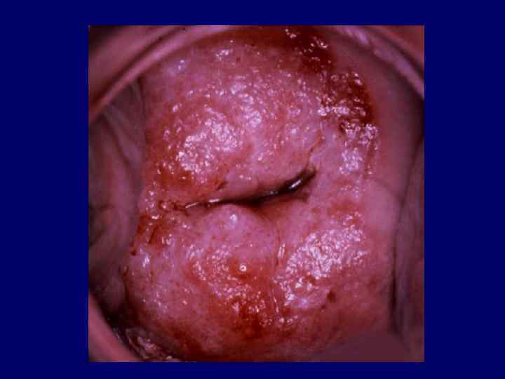
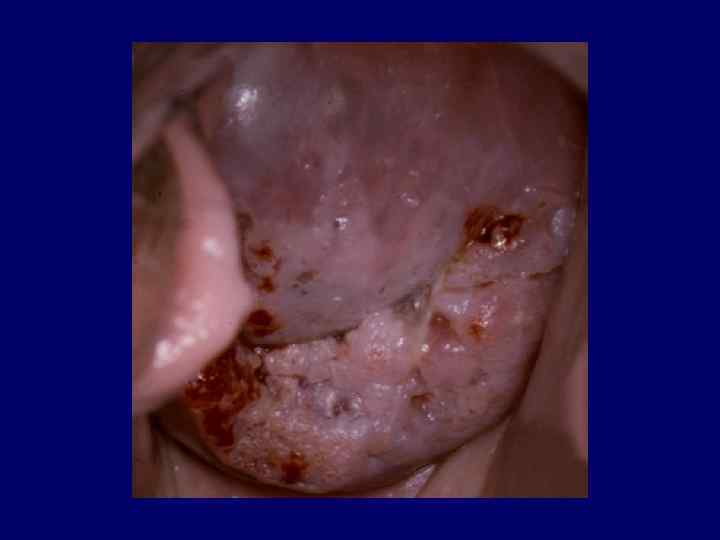
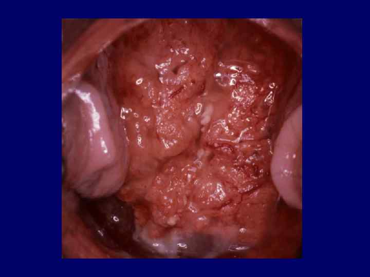
 Colposcopic features suggestive of invasive cancer • Irregular surface, erosion or ulceration • Dense acetowhite change • Wide irregular punctation and mosaic • Atypical vessels
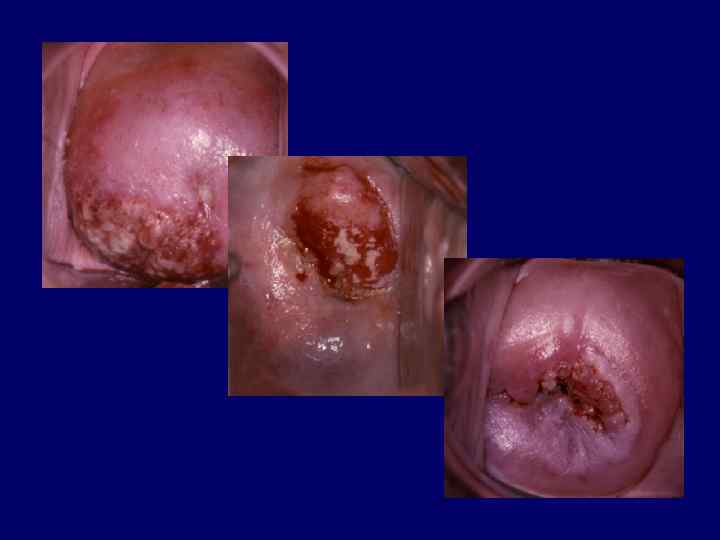
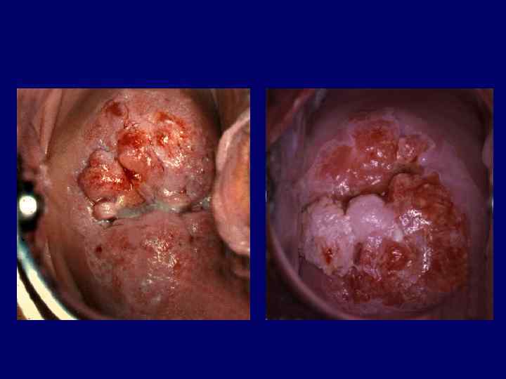
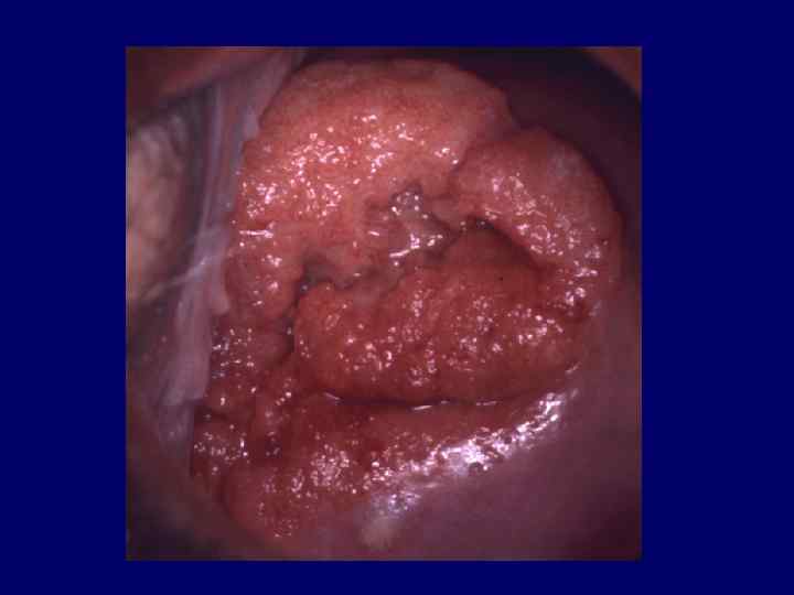
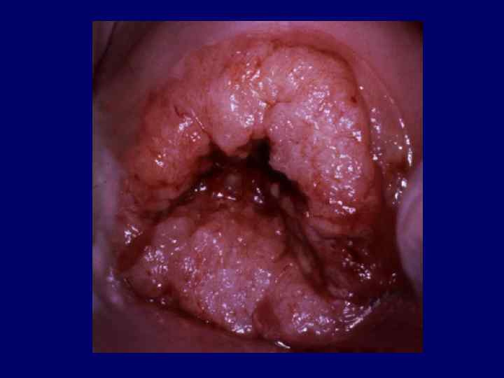
 In most cases biopsy is mandatory to establish the correct diagnosis
 The primary goal of the colposcopist is to ensure that invasive disease is not missed