

Chronic lymphocytic leukemia touro.pptx
- Количество слайдов: 38
 Chronic lymphocytic leukemia
 Chronic Lymphocytic Leukemia, Small Lymphocytic Lymphoma and Monoclonal Bcell Lymphocytosis: Concept • Disorders of “mature” CD 5+ B lymphocytes • SLL and CLL = counterparts (lymph nodes and blood) of the same tumor • MBL: Clinical situation not fulfilling CLL criteria that may or (more frequently) may not evolve to CLL
 Monoclonal B lymphocytosis Sm. Ig weak, CD 5+, CD 19+, CD 23+, CD 20 weak <5000/micro. L >5000/micro. L Spleen/liver Enlarged lymph nodes NO→MBL YES→SLL CLL
 CLL Most frequent leukemia in adults – 30% of all adult leukemias Incidence in western world: 4 -5 new cases/ 100000/year, 10 times lower in Asia - around 0. 48/100000/year Median age at presentation 72; 9% diagnosed between ages 45 – 54, 20% - 55 -64 years old, 27% - 65 -74 years old, 29% - 75 -84 years old, 13% - above 85 years old Median age of CLL patients in clinical trials is 60!! Male : Female 1. 3 -1. 5: 1
 Aetiology The cause of CLL is unknown There is increased incidence in farmers, rubber manufacturing workers, asbestos workers, and tire repair workers Genetic factors have been postulated to play a role in high incidence of CLL in some families
 B-cell development CLL MCL stem cell memory B-cell mature naive B-cell germinal center B-cell lymphoid precursor progenitor-B LBL, ALL pre-B immature B-cell MZL CLL MM DLBCL, FL, BL, HL plasma cell
 viral (mononucleosis) Malignant causes B-cell T-cell leukemic phase") Differential diagnosis Infectious causes bacterial (tuberculosis) viral (mononucleosis) Malignant causes B-cell T-cell leukemic phase of non-Hodgkin lymphomas Hairy-cell leukemia Waldenstrom macroglobulinemia Large granular lymphocytic leukemia
Differential diagnosis Infectious causes bacterial (tuberculosis) viral (mononucleosis) Malignant causes B-cell T-cell leukemic phase of non-Hodgkin lymphomas Hairy-cell leukemia Waldenstrom macroglobulinemia Large granular lymphocytic leukemia
 Approximately 40% of CLL patients are asymptomatic at diagnosis In symptomatic") Clinical findings (1) Approximately 40% of CLL patients are asymptomatic at diagnosis In symptomatic cases the most common complaint is fatigue Less often the initial complaint are enlarged nodes, the development of an infection (bacterial) or bleeding diathesis (thrombocytopenia)
Clinical findings (1) Approximately 40% of CLL patients are asymptomatic at diagnosis In symptomatic cases the most common complaint is fatigue Less often the initial complaint are enlarged nodes, the development of an infection (bacterial) or bleeding diathesis (thrombocytopenia)
 Most symptomatic patients have enlarged lymph nodes (more commonly cervical and") Clinical findings (2) Most symptomatic patients have enlarged lymph nodes (more commonly cervical and supraclavicular) and splenomegaly, hepatomegaly may occur The lymph nodes are usually discrete, freely movable, and non tender Less common manifestation are infiltration of tonsils, mesenteric or retroperitoneal lymphadenopathy, and skin infiltration Patients may present with features of anaemia, and bruising or bleeding
Clinical findings (2) Most symptomatic patients have enlarged lymph nodes (more commonly cervical and supraclavicular) and splenomegaly, hepatomegaly may occur The lymph nodes are usually discrete, freely movable, and non tender Less common manifestation are infiltration of tonsils, mesenteric or retroperitoneal lymphadenopathy, and skin infiltration Patients may present with features of anaemia, and bruising or bleeding
 Investigations Pre-treatment studies of patients with CLL should include examination of: complete blood count peripheral blood smear reticulocyte count Coomb’s test renal and liver function tests - LDH serum protein electrophoresis immunoglobulin levels plasma 2 micro globulin level If available immunophenotyping should be carried out to confirm the diagnosis Bone marrow biopsy and cytogenetic analysis is not routinely performed at diagnosis of CLL BM or blood cytogenetics (FISH)
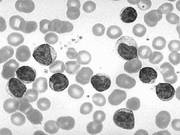
 The blood lymphocyte count above 5, 0 G/L In most patients") Laboratory findings (1) The blood lymphocyte count above 5, 0 G/L In most patients the leukemic cells have the morphologic appearance of normal small lymphocytes In the blood smears are commonly seen ruptured lymphocytes (“basket” or “smudge” cells) Careful examination of the blood smear can usually differentiate CLL, and the diagnosis can be confirmed by immunophenotyping
Laboratory findings (1) The blood lymphocyte count above 5, 0 G/L In most patients the leukemic cells have the morphologic appearance of normal small lymphocytes In the blood smears are commonly seen ruptured lymphocytes (“basket” or “smudge” cells) Careful examination of the blood smear can usually differentiate CLL, and the diagnosis can be confirmed by immunophenotyping
 Clonal expansion of B Lymphocytes In B-cell CLL clonality is confirmed") Laboratory findings (2) Clonal expansion of B Lymphocytes In B-cell CLL clonality is confirmed by the expression of either or light chains on the cell surface membrane the presence of unique idiotypic specificities on the immunoglobulin produced by CLL cells by immunoglobulin gene rearrangements typical B-cell CLL are unique in being CD 19+ and CD 5+ Hypogammaglobulinemia or agammaglobulinemia are often observed 10 - 25% of patients with CLL develop autoimmune haemolytic anaemia, with a positive direct Coombs’ test or immune thrombocytopenia The marrow aspirates shows greater than 30% of the nucleated cells as being lymphoid
Laboratory findings (2) Clonal expansion of B Lymphocytes In B-cell CLL clonality is confirmed by the expression of either or light chains on the cell surface membrane the presence of unique idiotypic specificities on the immunoglobulin produced by CLL cells by immunoglobulin gene rearrangements typical B-cell CLL are unique in being CD 19+ and CD 5+ Hypogammaglobulinemia or agammaglobulinemia are often observed 10 - 25% of patients with CLL develop autoimmune haemolytic anaemia, with a positive direct Coombs’ test or immune thrombocytopenia The marrow aspirates shows greater than 30% of the nucleated cells as being lymphoid
 Immunophenotyping Marker CLL CD 19 PLL ++ HCL-V SLVL FL MCL ++ +++ ++ CD 20 Dim +++ +++ ++ s. Ig Dim +++ +++ ++ CD 5 ++ -/+ - - -/+ ++ CD 10 - -/+ -/+ - CD 22 +/-dim ++ +++ ++ CD 23 ++ -/+ - - CD 25 -/+ +/- +++ - +/- - - CD 103 - - +++ +/- - -
 Staging Rai 0 – lymphocytosis. I – lymphocytosis + lymph nodes. II – lymphocytosis + spleen or liver ± LN. III – lymphocytosis + Hb<10, ± LN, spleen, liver. IV – lymphocytosis + PLT<100000, ± LN, spleen, liver Binet Stage A – lymph node areas ≤ 2 ; Hb>10; PLT≥ 100000. Stage B – lymph node areas ≥ 3; Hb>10; PLT>100000. Stage C – Hb<10; PLT<100000. LN areas – cervical, axillary, inguinofemoral, spleen, liver
 stage median survival (years) 0 >10 I") Prognosis according to stage Rai classification (1975) stage median survival (years) 0 >10 I > 8 II 6 III 2 IV < 2 Binet classification (1981) stage median survival (years) A > 10 B 7 C 2
Prognosis according to stage Rai classification (1975) stage median survival (years) 0 >10 I > 8 II 6 III 2 IV < 2 Binet classification (1981) stage median survival (years) A > 10 B 7 C 2
 Genomic aberrations Have pathogenetic and clinical relevance. Identifiable by FISH in 80% of CLL cases. Provide insights into the pathogenesis, they point to loci of candidate genes (17 p 13: P 53; 11 q 22 -q 23: ATM). Identify subgroups with distinct clinical features – marked lymphadenopathy (11 q-), resistance to treatment (17 p-). Define specific subgroups that differ in the rate of disease progression (time from diagnosis to treatment) and overall survival.
 Genomic aberrations by FISH Study 13 q- 13 qsingle 11 q- +12 q 17 p- 6 q- VH VH unmutated Single center 55% 36% 18% 16% 7% 7% 56% 44% CLL 1 59% 40% 13% 4% 2% 41% 59% CLL 4 53% 34% 21% 11% 3% 9% 69% 31% CLL 3 52% 27% 22% 12% 3% 6% 68% 32% CLL 2 H 48% 14% 32% 18% 27% 9% 81% 19%
 Markers of poor prognosis in CLL Advanced Rai or Binet stage Functional capacity, age , gender Peripheral lymphocyte doubling time <6 months Diffuse marrow histology Increased number of prolymphocytes or cleaved cells Poor response to chemotherapy High 2 - microglobulin level Abnormal karyotyping Molecular – Ig. VH mutation, ZAP-70, CD 38 New markers under investigation
 Risk Stratification Diagnosis TP 53 analysis Age, gender, function, stage, comorbidities Ig. VH mutation TP 53 intact TP 53 defective FISH Molecular Very High Risk
 Risk factors – multivariate analysis VH unmutated & VH 3 -21 usage 17 p deletion 11 q deletion Age Lymphocyte count LDH When the model included cytogenetics and Ig. VH mutation status, the clinical stage lost it’s significance.
 Surrogate markers for Ig. VH mutation status CD 38 expression (Damle et al, Blood, 1999), correlation with unmutated Ig. VH and adverse prognosis. ZAP-70 – a tyrosine kinase expressed in B-CLL cells, correlates with unmutated Ig. VH and adverse prognosis (Crespo et al, NEJM, 2003). BUT – subsequent studies yielded controversial results. (1) Differences between laboratories; (2) the expression levels may change over time (CD 38); (3) careful separation of T-cells is necessary (ZAP-70); (4)different cut-off values for “+” and “-” (CD 38 and ZAP-70); (5)10 -30% discordance with mutation status (both).
") Genomic aberrations and Ig. VH mutation status Aberration Mutated unmutated P-value homology <98% n=132(44%) homology ≥ 98% n=168(56%) Clonal aberrations 80% 84% . 37 13 q deletion 65% 48% . 004 13 q del single 50% 26% <. 001 Trisomy 12 15% 19% . 44 11 q deletion 4% 27% <. 001 17 p deletion 3% 10% . 03 17 p or 11 q del 7% 35% <. 001
Genomic aberrations and Ig. VH mutation status Aberration Mutated unmutated P-value homology <98% n=132(44%) homology ≥ 98% n=168(56%) Clonal aberrations 80% 84% . 37 13 q deletion 65% 48% . 004 13 q del single 50% 26% <. 001 Trisomy 12 15% 19% . 44 11 q deletion 4% 27% <. 001 17 p deletion 3% 10% . 03 17 p or 11 q del 7% 35% <. 001
 Risk for progression in early stage CLL Risk factors: Doubling time <12 months; Diffuse BM infiltration pattern; High tyrosine kinase (>7 U/l); High β 2µG (>3. 5 mg/l) Those patients have high incidence of “bad” cytogenetics (17 p-; 11 q-) and unmutated Ig. VH.
 Genomic aberrations – prognostic relevance
 Prognostic factors Mutation status of Ig. VH gene – 50% mutated. Unmutated Ig. VH gene – pregerminal center B-lymphocytes, unfavorable. Mutated Ig. VH gene – post germinal center B-lymphocytes, favorable. Independent risk factor for all stages at diagnosis.
 Targeting of BCR signaling as a therapeutic strategy in CLL. Red symbols and letters indicate new therapeutics as discussed in the text. Hallek M Hematology 2013; 2013: 138 -150 © 2013 by American Society of Hematology
 Inhibitory signaling axes in CLL. Up-regulation of CD 200, CD 274, and CD 276 induces impaired actin polymerization and immunological synapse formation in CLL T cells. Gribben J G , and Riches J C Hematology 2013; 2013: 151157 © 2013 by American Society of Hematology
 Nucleoside analogs (cladribine, fludarabine) Biological response modifiers, immunomodulators Monoclonal") Treatment Alkylating agents (chlorambucil, cyclophosphamide) Nucleoside analogs (cladribine, fludarabine) Biological response modifiers, immunomodulators Monoclonal antibodies – anti. CD 20, anti. CD 52, anti. CD 23, anti. CD 37 etc. Chemoimmunotherapy (CIT) Bone marrow transplantation Systemic complications requiring therapy antibiotics immunoglobulin steroids blood products
Treatment Alkylating agents (chlorambucil, cyclophosphamide) Nucleoside analogs (cladribine, fludarabine) Biological response modifiers, immunomodulators Monoclonal antibodies – anti. CD 20, anti. CD 52, anti. CD 23, anti. CD 37 etc. Chemoimmunotherapy (CIT) Bone marrow transplantation Systemic complications requiring therapy antibiotics immunoglobulin steroids blood products
 CLL -Treatment Rai st. 0 -2 or Binet st. A-B observe every 3 -6 months, treat if disease progress, short doubling time, symptomatic, recurrent infections, ITP, AIHA Advanced stage, symptomatic patient needs treatment at diagnosis (5 -10% of the patients) High and very high risk early asymptomatic patients should not be treated outside of a clinical trial Low and intermediate-low risk symptomatic patients – B symptoms (weight loss, fever, night swetts), progressive lymphadenopathy, fatigue – need treatment
 Categories of patients CLL treatment “Go-Go” – fit, functionally independent with no or mild comorbidities and normal life expectancy should receive the most effective treatment – CIT: FCR or investigational alternative BR, FR with aim to prolong PFS and possibly OS “Slow Go” – medically less-fit patients – should be recruited into clinical trials. Can receive clorambucil±Rituximab, Bendamustine, clorambucil+ofatumomab or GA 101, dose-reduced FCR, Pentostatin+Rituximab±CTX (PR or PCR) “No Go” – unfit, with >3 comorbidities, dependent with short life expectancy – palliative treatment only
 Cll treatment No known defect in TP 53, “Go Go” – FCR or clinical trial Defective TP 53 – no standard of care. CIT provide low RR, rare durable responses. Therapies with TP 53 -independent action: high dose steroids, Alemtuzumab, combinations FLU-CAM, HD steroids+monoclonal Ab’s, provide short term responses, severe immune suppression. Novel agents – BCR pathway inhibitors Early Allogeneic transplantation for fit younger patients with a suitable donor
") Relapsed/refractory disease If response duration > 1 year retreatment with CIT (FCR, etc. ) Bendamustine + Rituximab Ofatumomab Investigational combinations Novel agents Allogeneic SCT – Reduced Intensity Conditioning
Relapsed/refractory disease If response duration > 1 year retreatment with CIT (FCR, etc. ) Bendamustine + Rituximab Ofatumomab Investigational combinations Novel agents Allogeneic SCT – Reduced Intensity Conditioning
 Novel drugs for CLL Ibrutinib – BTK inhibitor Idelalisib – PI 3 K inhibitor Lenalidomide – immune modulator (IMID) Alvocidib (flavopiridol) – CDK inhibitor Ofatumomab – human anti-CD 20 monoclonal Ab Veltuzumab – humanized anti-CD 20 monoclonal Ab HCD-122 – human anti-CD 40 monoclonal Ab TRU-016 – anti-CD 37 Ig. G fusion protein Obatoclax – BCL-2 inhibitor
 Novel drugs for CLL Fostamatinib – SYK inhibitor Everolimus – m. TOR inhibitor Ai. X – AKT inhibitor PGG β-glucan – Complement receptor 3 agonist 17 -DMAG – HSP 90 inhibitor Dasatinib – tyrosine kinase inhibitor Plerixafor – CXCL 12 inhibitor ABT-263/ABT-737 – BCL 2 and BCLXL inhibitors CAL-101 – PI 3 K inhibitor
 Richter’s Syndrome In 3 -5% the disease undergoes a transformation into aggressive lymphoma - diffuse large cell or immunoblastic, rare Hodgkin lymphoma or T-cell lymphoma Severe B-symptoms, increased LDH, progressive lymphadenopathy The prognosis is poor, median survival <6 months
 of second malignancy Most frequent") Second Malignancies Incidence of 8. 9% (28% increased risk) of second malignancy Most frequent cancers associated with CLL are - skin, lung, gastrointestinal tumors (carcinoma of colon) There is no relationship between the course of CLL, it’s treatment and the incidence of second cancers
Second Malignancies Incidence of 8. 9% (28% increased risk) of second malignancy Most frequent cancers associated with CLL are - skin, lung, gastrointestinal tumors (carcinoma of colon) There is no relationship between the course of CLL, it’s treatment and the incidence of second cancers
 תודה רבה