
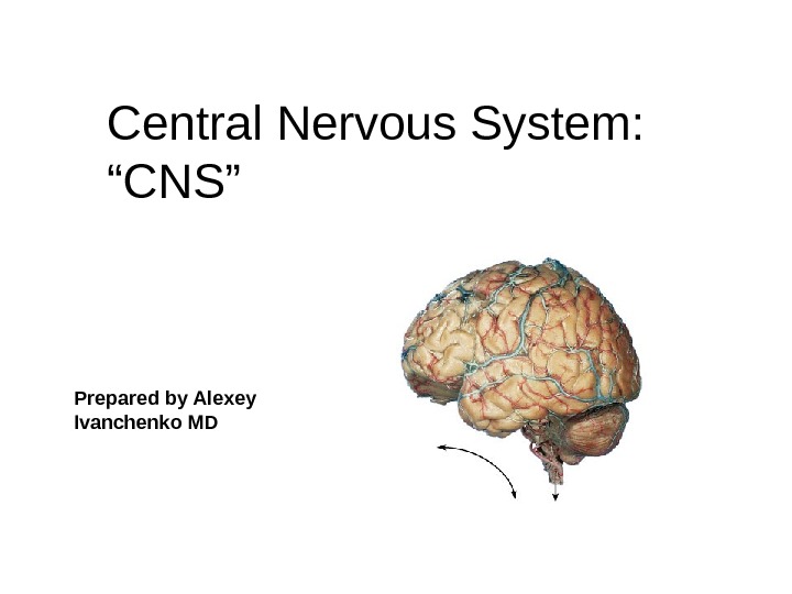
Central Nervous System: “CNS” Prepared b y Alexey


- Размер: 5.9 Mегабайта
- Количество слайдов: 102
Описание презентации Central Nervous System: “CNS” Prepared b y Alexey по слайдам
Central Nervous System: “CNS” Prepared b y Alexey Ivanchenko M
The Spinal Cord Foramen magnum to L 1 or L 2 Runs through the vertebral canal of the vertebral column Functions 1. Sensory and motor innervation of entire body inferior to the head through the spinal nerves 2. Two-way conduction pathway between the body and the brain 3. Major center for reflexes
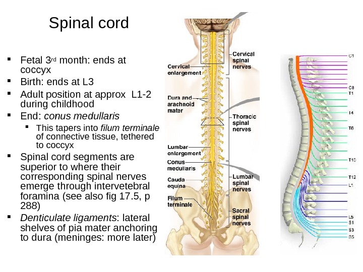
Fetal 3 rd month: ends at coccyx Birth: ends at L 3 Adult position at approx L 1 -2 during childhood End: conus medullaris This tapers into filum terminale of connective tissue, tethered to coccyx Spinal cord segments are superior to where their corresponding spinal nerves emerge through intervetebral foramina (see also fig 17. 5, p 288) Denticulate ligaments : lateral shelves of pia mater anchoring to dura (meninges: more later) Spinal cord
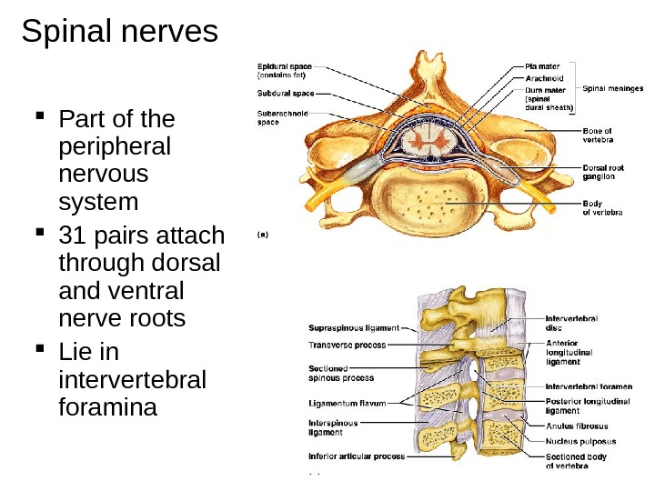
Spinal nerves Part of the peripheral nervous system 31 pairs attach through dorsal and ventral nerve roots Lie in intervertebral foramina
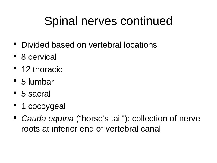
Spinal nerves continued Divided based on vertebral locations 8 cervical 12 thoracic 5 lumbar 5 sacral 1 coccygeal Cauda equina (“horse’s tail”): collection of nerve roots at inferior end of vertebral canal
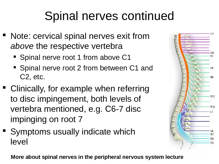
Spinal nerves continued Note: cervical spinal nerves exit from above the respective vertebra Spinal nerve root 1 from above C 1 Spinal nerve root 2 from between C 1 and C 2, etc. Clinically, for example when referring to disc impingement, both levels of vertebra mentioned, e. g. C 6 -7 disc impinging on root 7 Symptoms usually indicate which level More about spinal nerves in the peripheral nervous system lecture
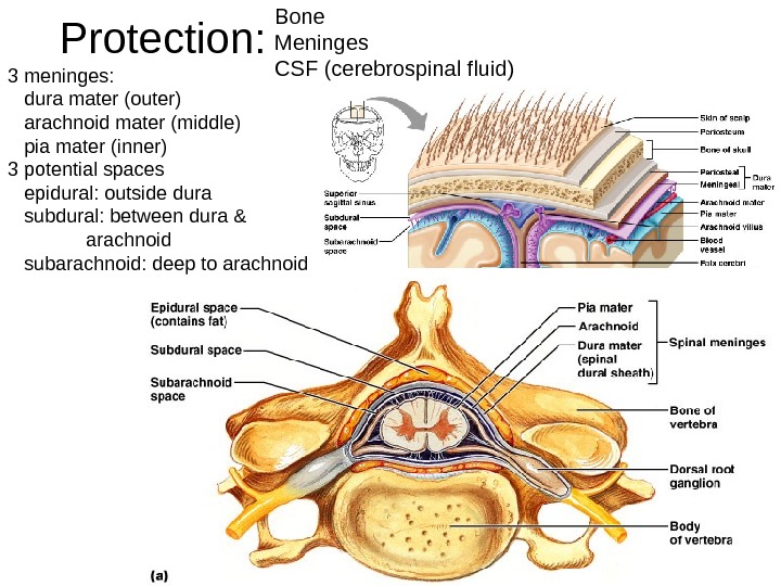
Protection: Bone Meninges CSF (cerebrospinal fluid) 3 meninges: dura mater (outer) arachnoid mater (middle) pia mater (inner) 3 potential spaces epidural: outside dura subdural: between dura & arachnoid subarachnoid: deep to arachnoid
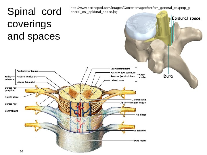
Dura mater Arachnoid mater Pia mater Spinal cord coverings and spaces http: //www. eorthopod. com/images/Content. Images/pm/pm_general_esi/pmp_g eneral_esi_epidural_space. jpg
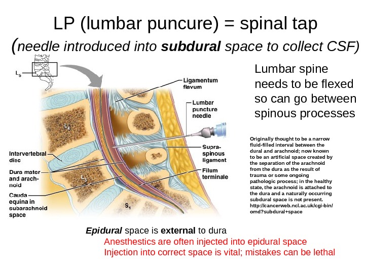
LP (lumbar puncure) = spinal tap ( needle introduced into subdural space to collect CSF) Lumbar spine needs to be flexed so can go between spinous processes Epidural space is external to dura Anesthestics are often injected into epidural space Injection into correct space is vital; mistakes can be lethal Originally thought to be a narrow fluid-filled interval between the dural and arachnoid; now known to be an artificial space created by the separation of the arachnoid from the dura as the result of trauma or some ongoing pathologic process; in the healthy state, the arachnoid is attached to the dura and a naturally occurring subdural space is not present. http: //cancerweb. ncl. ac. uk/cgi-bin/ omd? subdural+space
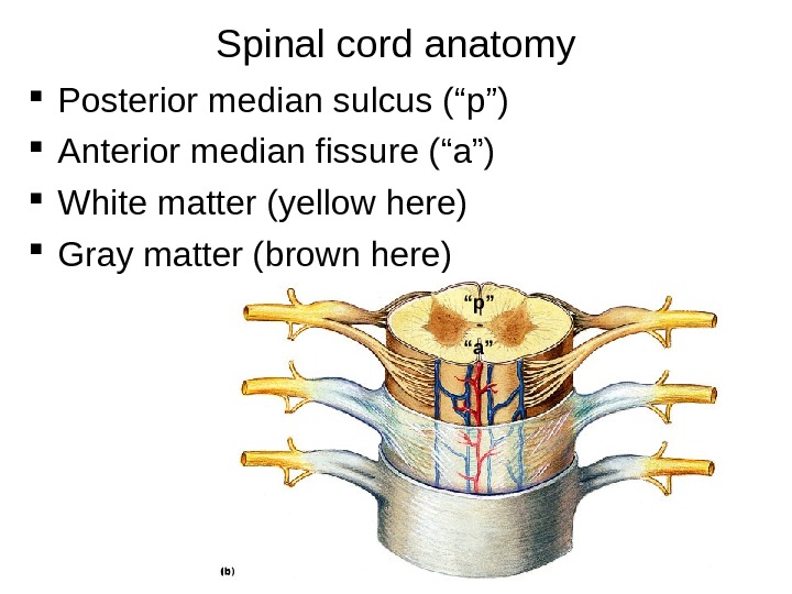
Spinal cord anatomy Posterior median sulcus (“p”) Anterior median fissure (“a”) White matter (yellow here) Gray matter (brown here) “ p” “ a”
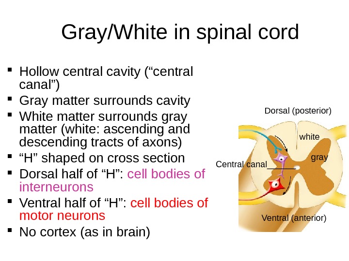
Gray/White in spinal cord Hollow central cavity (“central canal”) Gray matter surrounds cavity White matter surrounds gray matter (white: ascending and descending tracts of axons) “ H” shaped on cross section Dorsal half of “H”: cell bodies of interneurons Ventral half of “H”: cell bodies of motor neurons No cortex (as in brain) Dorsal (posterior) white gray Ventral (anterior)Central canal______
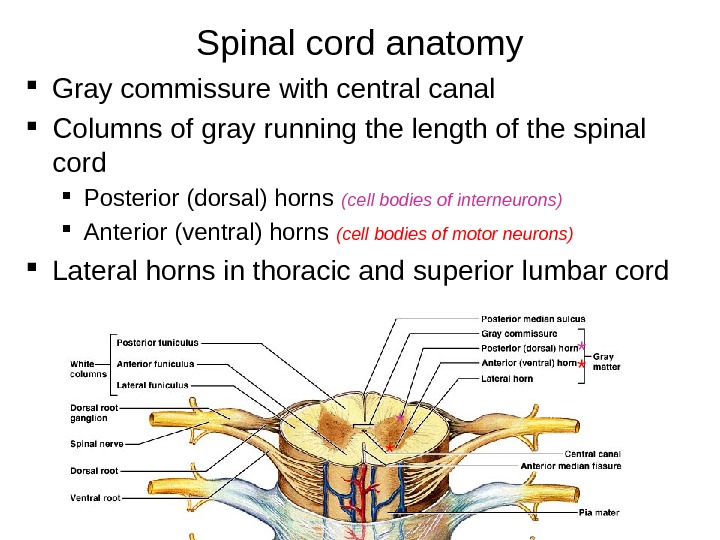
Spinal cord anatomy Gray commissure with central canal Columns of gray running the length of the spinal cord Posterior (dorsal) horns (cell bodies of interneurons) Anterior (ventral) horns (cell bodies of motor neurons) Lateral horns in thoracic and superior lumbar cord ** * *
White matter of the spinal cord (myelinated and unmyelinated axons) Ascending fibers: sensory information from sensory neurons of body up to brain Descending fibers: motor instructions from brain to spinal cord Stimulates contraction of body’s muscles Stimumulates secretion from body’s glands Commissural fibers: white-matter fibers crossing from one side of cord to the other Most pathways cross (or decussate ) at some point Most synapse two or three times along the way, e. g. in brain stem, thalamus or other
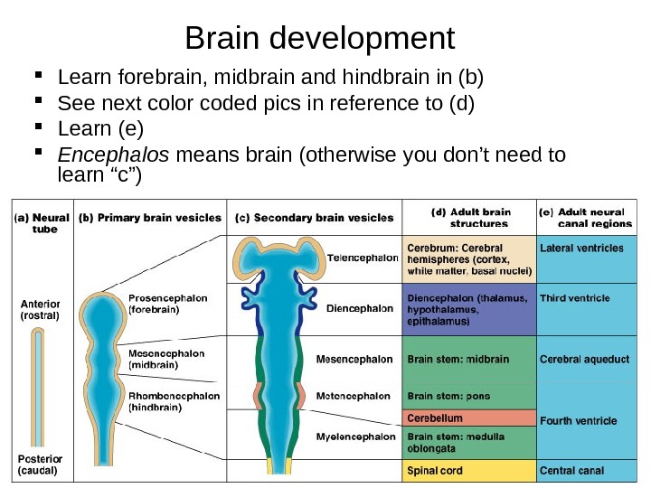
The Brain : embryonic development Develops from neural tube Brain subdivides into Forebrain Midbrain Hindbrain These further divide, each with a fluid filled region: ventricle, aqueduct or canal Spinal cord also has a canal Two major bends, or flexures, occur (midbrain and cervical)
Brain development Learn forebrain, midbrain and hindbrain in (b) See next color coded pics in reference to (d) Learn (e) Encephalos means brain (otherwise you don’t need to learn “c”)
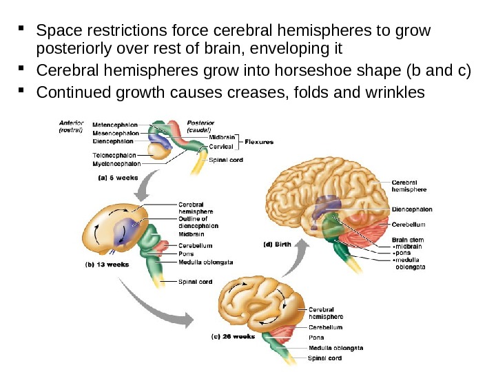
Space restrictions force cerebral hemispheres to grow posteriorly over rest of brain, enveloping it Cerebral hemispheres grow into horseshoe shape (b and c) Continued growth causes creases, folds and wrinkles
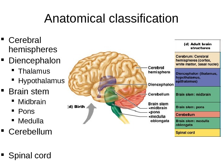
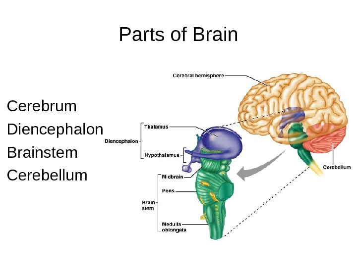
Anatomical classification Cerebral hemispheres Diencephalon Thalamus Hypothalamus Brain stem Midbrain Pons Medulla Cerebellum Spinal cord
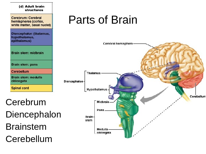
Parts of Brain Cerebrum Diencephalon Brainstem Cerebellum
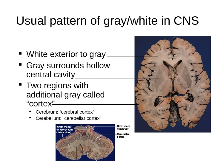
Usual pattern of gray/white in CNS White exterior to gray Gray surrounds hollow central cavity Two regions with additional gray called “cortex” Cerebrum: “cerebral cortex” Cerebellum: “cerebellar cortex” _______________________
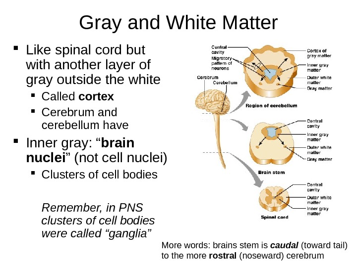
Gray and White Matter Like spinal cord but with another layer of gray outside the white Called cortex Cerebrum and cerebellum have Inner gray: “ brain nuclei ” (not cell nuclei) Clusters of cell bodies Remember, in PNS clusters of cell bodies were called “ganglia” More words: brains stem is caudal (toward tail) to the more rostral (noseward) cerebrum
Ventricles Central cavities expanded Filled with CSF (cerebrospinal fluid) Lined by ependymal cells (these cells lining the choroid plexus make the CSF: see later slides) Continuous with each other and central canal of spinal cord In the following slides, the ventricles are the parts colored blue
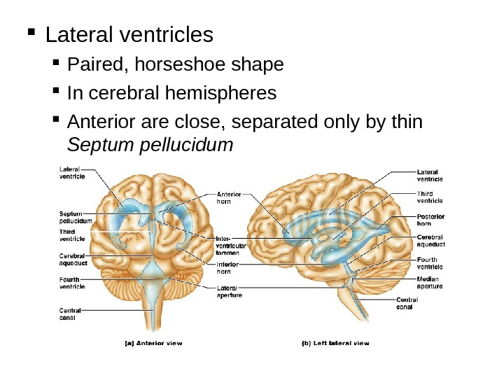
Lateral ventricles Paired, horseshoe shape In cerebral hemispheres Anterior are close, separated only by thin Septum pellucidum
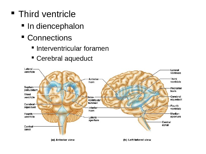
Third ventricle In diencephalon Connections Interventricular foramen Cerebral aqueduct
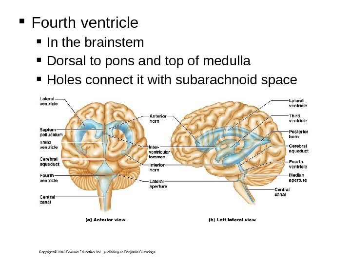
Fourth ventricle In the brainstem Dorsal to pons and top of medulla Holes connect it with subarachnoid space
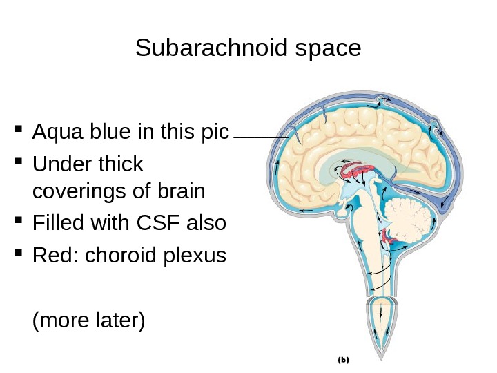
Subarachnoid space Aqua blue in this pic Under thick coverings of brain Filled with CSF also Red: choroid plexus (more later) ____
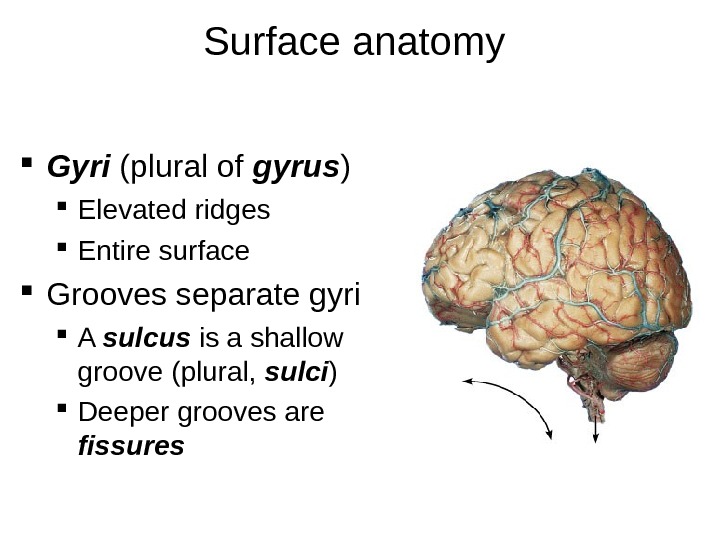
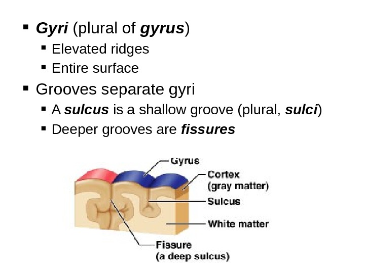
Surface anatomy Gyri (plural of gyrus ) Elevated ridges Entire surface Grooves separate gyri A sulcus is a shallow groove (plural, sulci ) Deeper grooves are fissures
Gyri (plural of gyrus ) Elevated ridges Entire surface Grooves separate gyri A sulcus is a shallow groove (plural, sulci ) Deeper grooves are fissures
Parts of Brain Cerebrum Diencephalon Brainstem Cerebellum
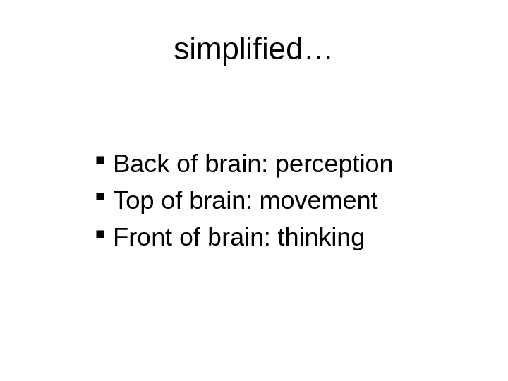
simplified… Back of brain: perception Top of brain: movement Front of brain: thinking
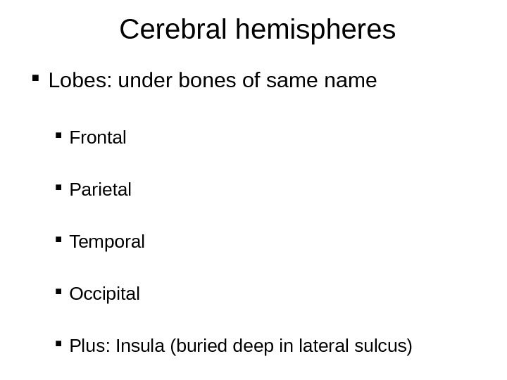
Cerebral hemispheres Lobes: under bones of same name Frontal Parietal Temporal Occipital Plus: Insula (buried deep in lateral sulcus)
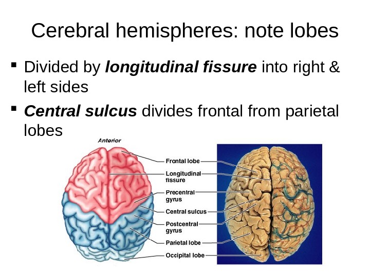
Cerebral hemispheres: note lobes Divided by longitudinal fissure into right & left sides Central sulcus divides frontal from parietal lobes
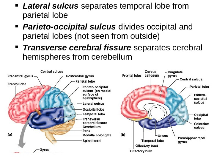
Lateral sulcus separates temporal lobe from parietal lobe Parieto-occipital sulcus divides occipital and parietal lobes (not seen from outside) Transverse cerebral fissure separates cerebral hemispheres from cerebellum
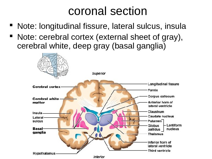
coronal section Note: longitudinal fissure, lateral sulcus, insula Note: cerebral cortex (external sheet of gray), cerebral white, deep gray (basal ganglia)
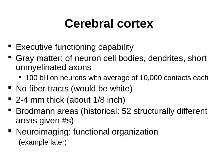
Cerebral cortex Executive functioning capability Gray matter: of neuron cell bodies, dendrites, short unmyelinated axons 100 billion neurons with average of 10, 000 contacts each No fiber tracts (would be white) 2 -4 mm thick (about 1/8 inch) Brodmann areas (historical: 52 structurally different areas given #s) Neuroimaging: functional organization (example later)
Prenatal life: genes are responsible for creating the architecture of the brain Cortex is the last to develop and very immature at birth Birth: excess of neurons but not inter-connected 1 st month of life: a million synapses/sec are made; this is genetic 1 st 3 years of life: synaptic overgrowth (connections) After this the density remains constant though some grow, some die Preadolescence: another increase in synaptic formation Adolescence until 25: brain becomes a reconstruction site Connections important for self-regulation (in prefrontal cortex) are being remodeled: important for a sense of wholeness Causes personal turbulence Susceptible to stress and toxins (like alcohol and drugs) during these years; affects the rest of one’s life The mind changes the brain (throughout life) Where brain activation occurs, synapses happen When pay attention & focus mind, neural firing occurs and brain structure changes (synapses are formed) Human connections impact neural connections (ongoing experiences and learning include the interpersonal ones) adapted from Dr. Daniel Siegel, UCL
Cerebral cortex All the neurons are interneurons By definition confined to the CNS They have to synapse somewhere before the info passes to the peripheral nerves Three kinds of functional areas Motor areas: movement Sensory areas: perception Association areas: integrate diverse information to enable purposeful action
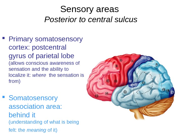
Sensory areas Posterior to central sulcus Primary somatosensory cortex: postcentral gyrus of parietal lobe (allows conscious awareness of sensation and the ability to localize it: where the sensation is from) Somatosensory association area: behind it (understanding of what is being felt: the meaning of it)
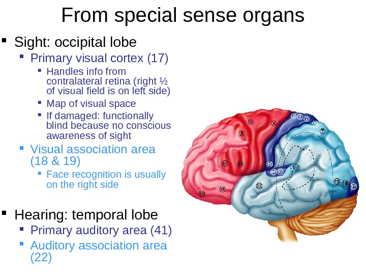
From special sense organs Sight: occipital lobe Primary visual cortex (17) Handles info from contralateral retina (right ½ of visual field is on left side) Map of visual space If damaged: functionally blind because no conscious awareness of sight Visual association area (18 & 19) Face recognition is usually on the right side Hearing: temporal lobe Primary auditory area (41) Auditory association area (22)
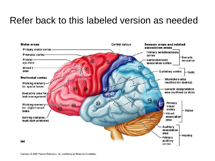
Refer back to this labeled version as needed
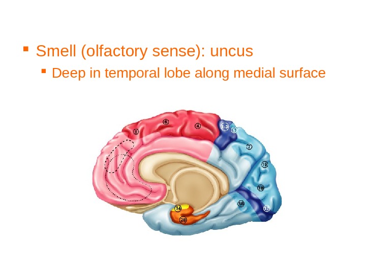
Smell (olfactory sense): uncus Deep in temporal lobe along medial surface
f. MRI: functional magnetic resonance imaging Cerebral cortex of person speaking & hearing Activity (blood flow) in posterior frontal and superior temporal lobes respectively
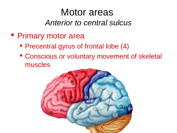
Motor areas Anterior to central sulcus Primary motor area Precentral gyrus of frontal lobe (4) Conscious or voluntary movement of skeletal muscles
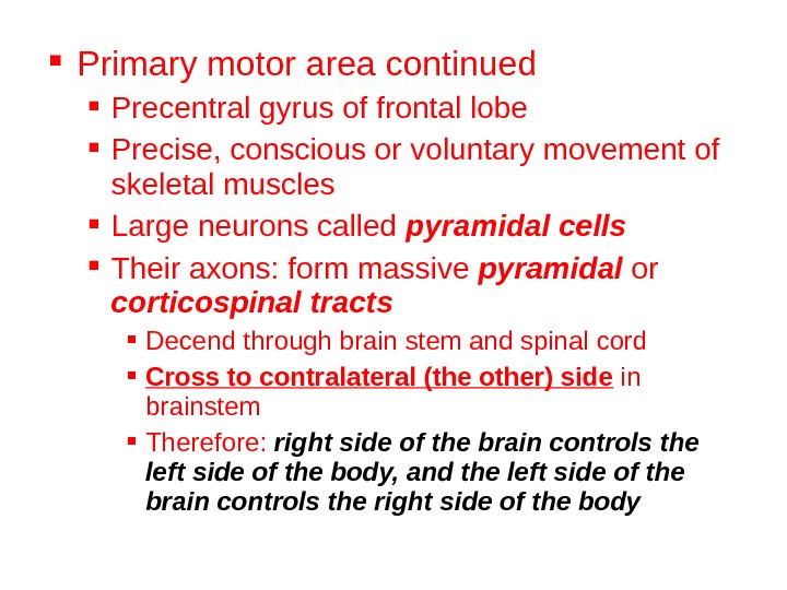
Primary motor area continued Precentral gyrus of frontal lobe Precise, conscious or voluntary movement of skeletal muscles Large neurons called pyramidal cells Their axons: form massive pyramidal or corticospinal tracts Decend through brain stem and spinal cord Cross to contralateral (the other) side in brainstem Therefore: right side of the brain controls the left side of the body, and the left side of the brain controls the right side of the body
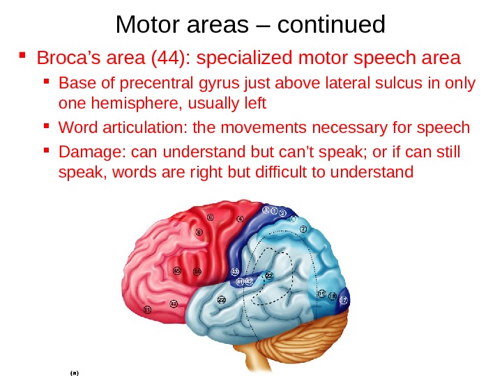
Motor areas – continued Broca’s area (44): specialized motor speech area Base of precentral gyrus just above lateral sulcus in only one hemisphere, usually left Word articulation: the movements necessary for speech Damage: can understand but can’t speak; or if can still speak, words are right but difficult to understand
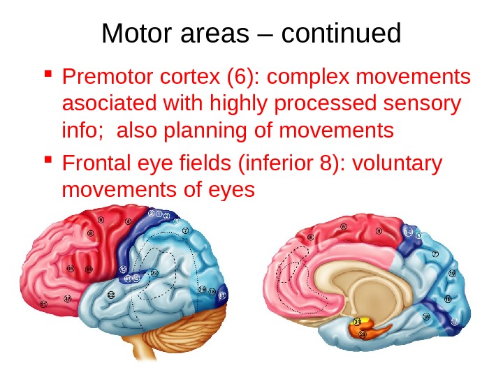
Motor areas – continued Premotor cortex (6): complex movements asociated with highly processed sensory info; also planning of movements Frontal eye fields (inferior 8): voluntary movements of eyes
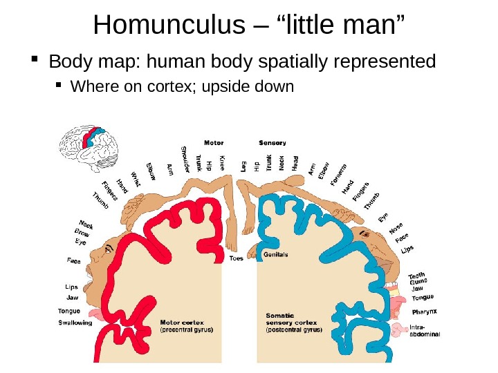
Homunculus – “little man” Body map: human body spatially represented Where on cortex; upside down
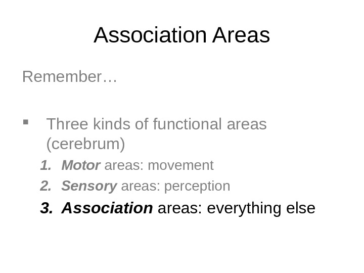
Association Areas Remember… Three kinds of functional areas (cerebrum) 1. Motor areas: movement 2. Sensory areas: perception 3. Association areas: everything else
Association Areas Tie together different kinds of sensory input Associate new input with memories Is to be renamed “higher-order processing“ areas
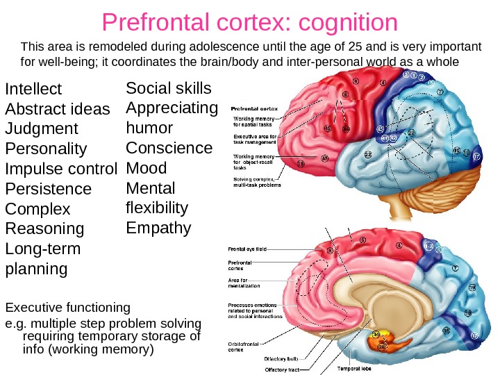
Prefrontal cortex: cognition Executive functioning e. g. multiple step problem solving requiring temporary storage of info (working memory)This area is remodeled during adolescence until the age of 25 and is very important for well-being; it coordinates the brain/body and inter-personal world as a whole Social skills Appreciating humor Conscience Mood Mental flexibility Empathy. Intellect Abstract ideas Judgment Personality Impulse control Persistence Complex Reasoning Long-term planning
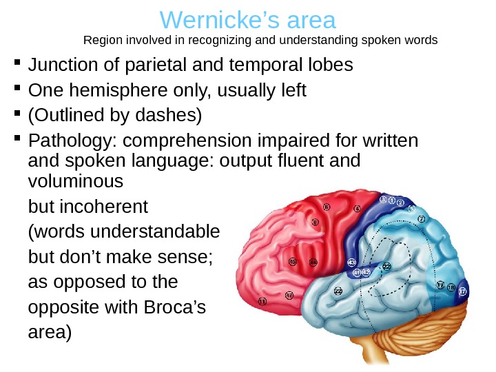
Wernicke’s area Junction of parietal and temporal lobes One hemisphere only, usually left (Outlined by dashes) Pathology: comprehension impaired for written and spoken language: output fluent and voluminous but incoherent (words understandable but don’t make sense; as opposed to the opposite with Broca’s area) Region involved in recognizing and understanding spoken words
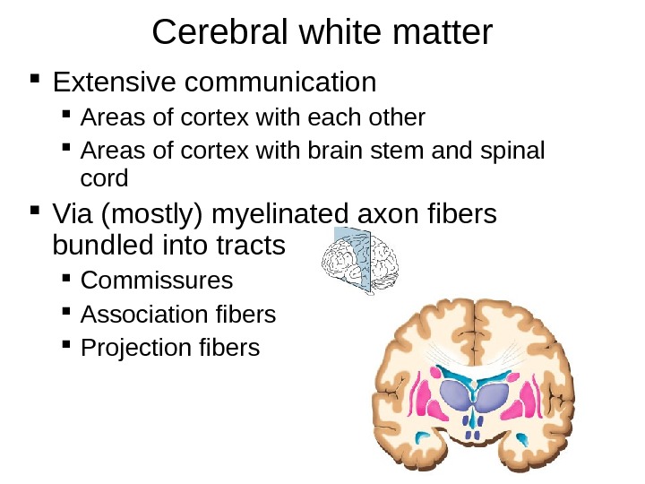
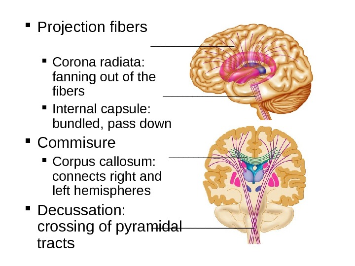
Cerebral white matter Extensive communication Areas of cortex with each other Areas of cortex with brain stem and spinal cord Via (mostly) myelinated axon fibers bundled into tracts Commissures Association fibers Projection fibers
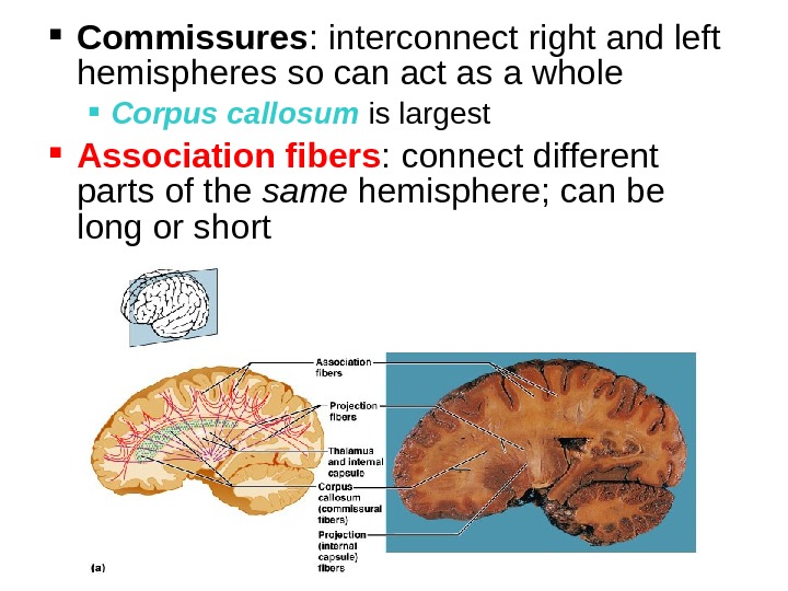
Commissures : interconnect right and left hemispheres so can act as a whole Corpus callosum is largest Association fibers : connect different parts of the same hemisphere; can be long or short
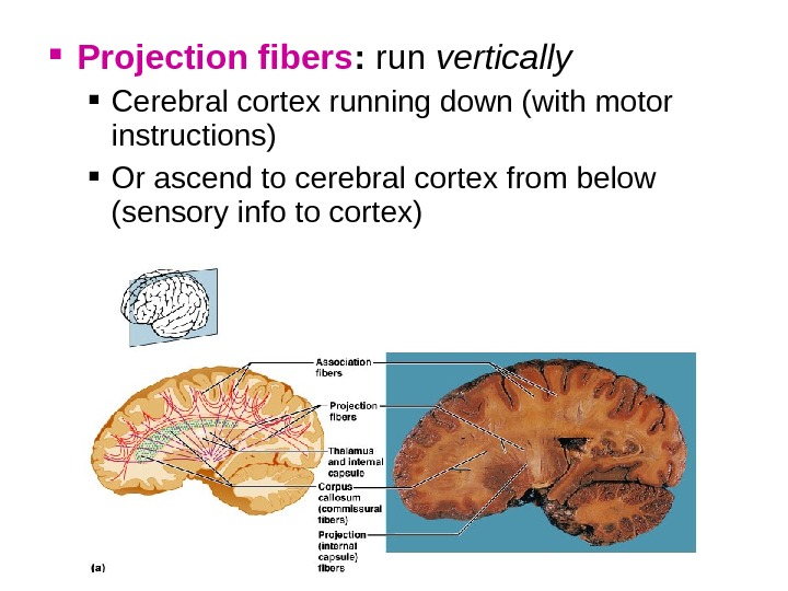
Projection fibers : run vertically Cerebral cortex running down (with motor instructions) Or ascend to cerebral cortex from below (sensory info to cortex)
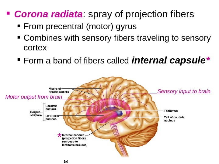
Corona radiata : spray of projection fibers From precentral (motor) gyrus Combines with sensory fibers traveling to sensory cortex Form a band of fibers called internal capsule * ______Sensory input to brain Motor output from brain_____ *
Projection fibers Corona radiata: fanning out of the fibers Internal capsule: bundled, pass down Commisure Corpus callosum: connects right and left hemispheres Decussation: crossing of pyramidal tracts _____________________
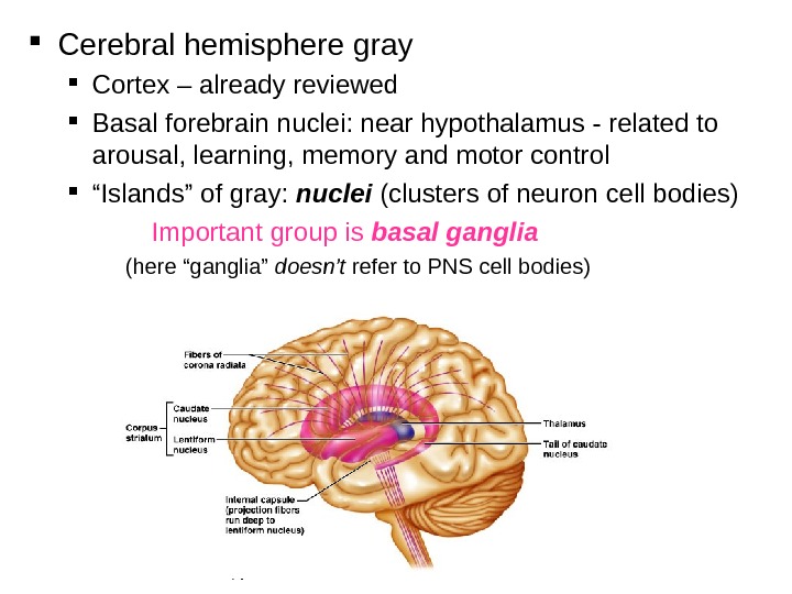
Cerebral hemisphere gray Cortex – already reviewed Basal forebrain nuclei: near hypothalamus — related to arousal, learning, memory and motor control “ Islands” of gray: nuclei (clusters of neuron cell bodies) Important group is basal ganglia (here “ganglia” doesn’t refer to PNS cell bodies)
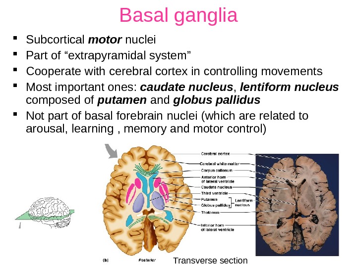
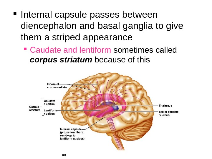
Basal ganglia Subcortical motor nuclei Part of “extrapyramidal system” Cooperate with cerebral cortex in controlling movements Most important ones: caudate nucleus , lentiform nucleus composed of putamen and globus pallidus Not part of basal forebrain nuclei (which are related to arousal, learning , memory and motor control) Transverse section
Internal capsule passes between diencephalon and basal ganglia to give them a striped appearance Caudate and lentiform sometimes called corpus striatum because of this
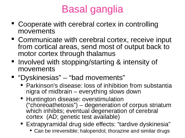
Basal ganglia Cooperate with cerebral cortex in controlling movements Communicate with cerebral cortex, receive input from cortical areas, send most of output back to motor cortex through thalamus Involved with stopping/starting & intensity of movements “ Dyskinesias” – “bad movements” Parkinson’s disease: loss of inhibition from substantia nigra of midbrain – everything slows down Huntington disease: overstimulation (“choreoathetosis”) – degeneration of corpus striatum which inhibits; eventual degeneration of cerebral cortex (AD; genetic test available) Extrapyramidal drug side effects: “tardive dyskinesia” Can be irreversible; haloperidol, thorazine and similar drugs
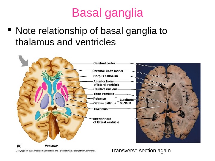
Basal ganglia Note relationship of basal ganglia to thalamus and ventricles Transverse section again
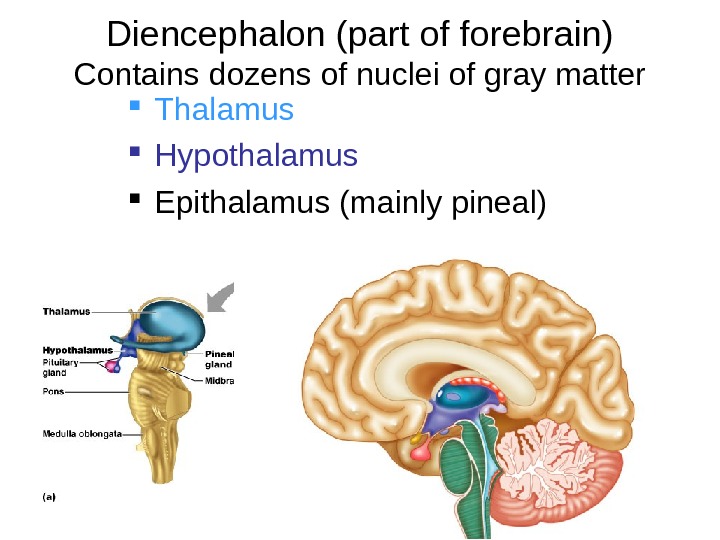
Diencephalon (part of forebrain) Contains dozens of nuclei of gray matter Thalamus Hypothalamus Epithalamus (mainly pineal)
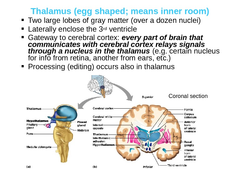
Thalamus (egg shaped; means inner room) Two large lobes of gray matter (over a dozen nuclei) Laterally enclose the 3 rd ventricle Gateway to cerebral cortex: every part of brain that communicates with cerebral cortex relays signals through a nucleus in the thalamus (e. g. certain nucleus for info from retina, another from ears, etc. ) Processing (editing) occurs also in thalamus Coronal section
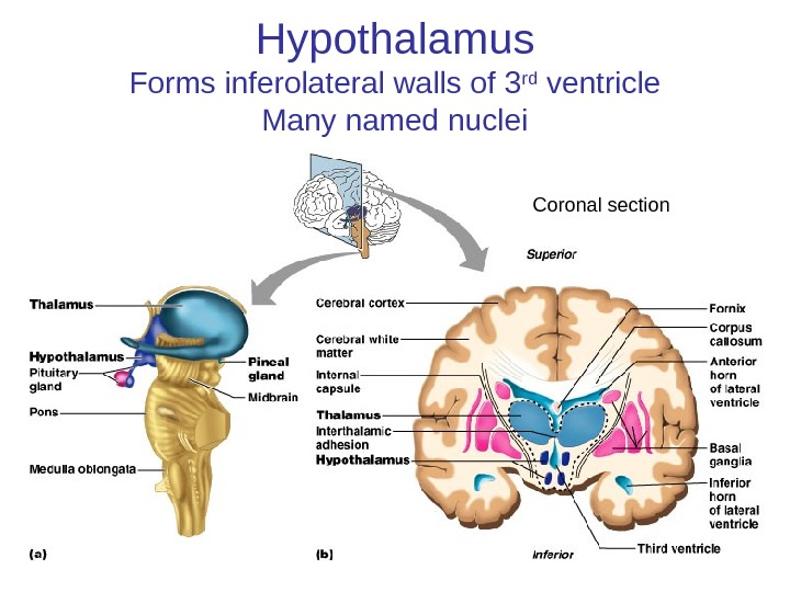
Hypothalamus Forms inferolateral walls of 3 rd ventricle Many named nuclei Coronal section
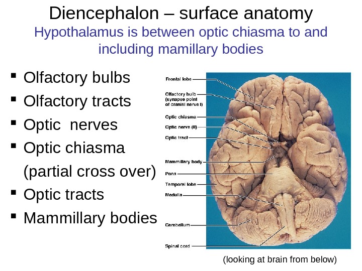
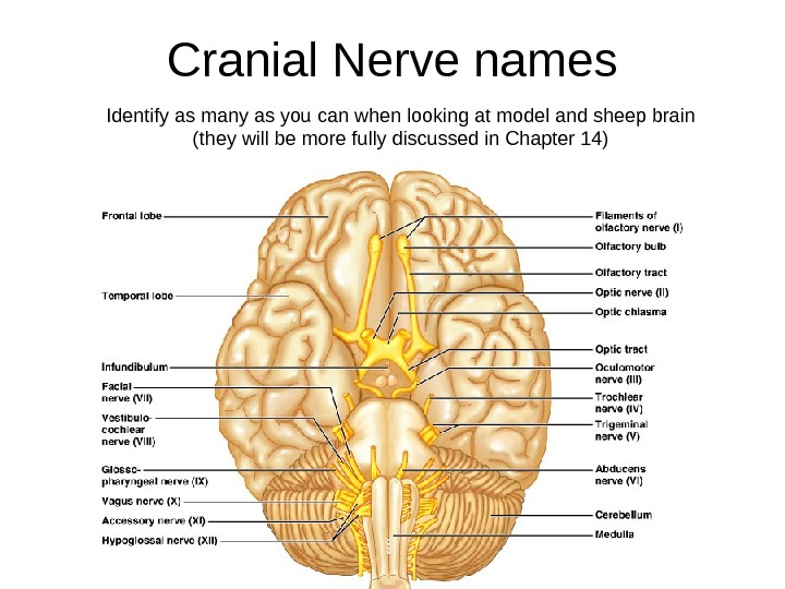
Diencephalon – surface anatomy Hypothalamus is between optic chiasma to and including mamillary bodies Olfactory bulbs Olfactory tracts Optic nerves Optic chiasma (partial cross over) Optic tracts Mammillary bodies (looking at brain from below)
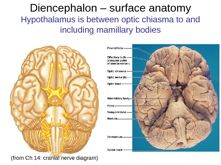
Diencephalon – surface anatomy Hypothalamus is between optic chiasma to and including mamillary bodies (from Ch 14: cranial nerve diagram)
Cranial Nerve names Identify as many as you can when looking at model and sheep brain (they will be more fully discussed in Chapter 14)
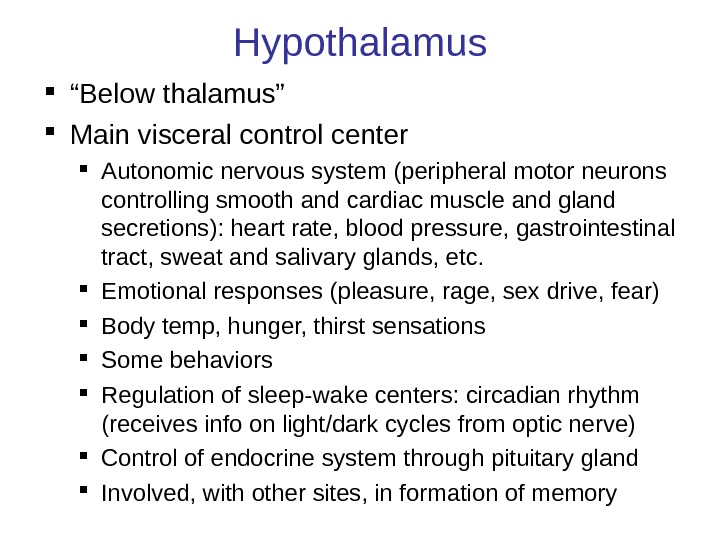
Hypothalamus “ Below thalamus” Main visceral control center Autonomic nervous system (peripheral motor neurons controlling smooth and cardiac muscle and gland secretions): heart rate, blood pressure, gastrointestinal tract, sweat and salivary glands, etc. Emotional responses (pleasure, rage, sex drive, fear) Body temp, hunger, thirst sensations Some behaviors Regulation of sleep-wake centers: circadian rhythm (receives info on light/dark cycles from optic nerve) Control of endocrine system through pituitary gland Involved, with other sites, in formation of memory
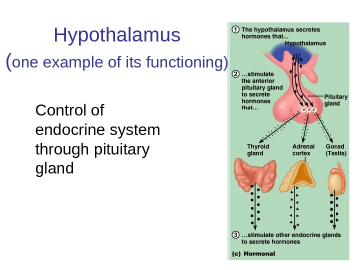
Hypothalamus ( one example of its functioning) Control of endocrine system through pituitary gland
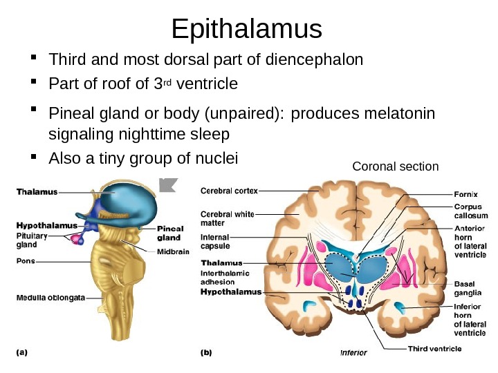
Epithalamus Third and most dorsal part of diencephalon Part of roof of 3 rd ventricle Pineal gland or body (unpaired): produces melatonin signaling nighttime sleep Also a tiny group of nuclei Coronal section
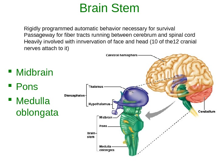
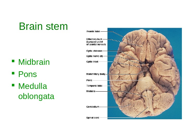
Brain Stem Midbrain Pons Medulla oblongata Rigidly programmed automatic behavior necessary for survival Passageway for fiber tracts running between cerebrum and spinal cord Heavily involved with innvervation of face and head (10 of the 12 cranial nerves attach to it)
Brain stem Midbrain Pons Medulla oblongata
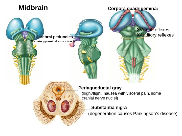
__ Cerebral peduncles ____ Contain pyramidal motor tracts Corpora quadrigemina: X Visual reflexes X Auditory reflexes. Midbrain ______ Substantia nigra (degeneration causes Parkingson’s disease)_______ Periaqueductal gray (flight/flight; nausea with visceral pain; some cranial nerve nuclei)
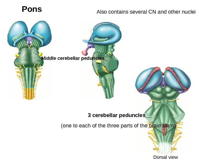
__Middle cerebellar peduncles _Pons 3 cerebellar peduncles__ Also contains several CN and other nuclei (one to each of the three parts of the brain stem) Dorsal view
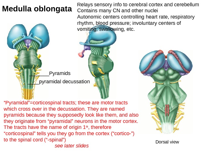
Medulla oblongata Relays sensory info to cerebral cortex and cerebellum Contains many CN and other nuclei Autonomic centers controlling heart rate, respiratory rhythm, blood pressure; involuntary centers of vomiting, swallowing, etc. Dorsal view_______Pyramids ____pyramidal decussation “ Pyramidal”=corticospinal tracts; these are motor tracts which cross over in the decussation. They are named pyramids because they supposedly look like them, and also they originate from “pyramidal” neurons in the motor cortex. The tracts have the name of origin 1 st , therefore “corticospinal” tells you they go from the cortex (“cortico-”) to the spinal cord (“-spinal”) see later slides
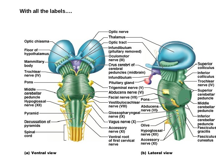
With all the labels….
Brain Stem in mid-sagittal plane Note cerebral aqueduct and fourth ventricle * * *
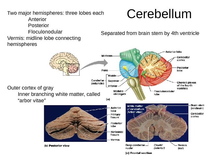
Cerebellum Two major hemispheres: three lobes each Anterior Posterior Floculonodular Vermis: midline lobe connecting hemispheres Outer cortex of gray Inner branching white matter, called “arbor vitae” Separated from brain stem by 4 th ventricle
Functions of cerebellum Smooths, coordinates & fine tunes bodily movements Helps maintain body posture Helps maintain equilibrium How? Gets info from cerebrum re: movements being planned Gets info from inner ear re: equilibrium Gets info from proprioceptors (sensory receptors informing where the parts of the body actually are) Using feedback, adjustments are made Also some role in cognition Damage: ataxia, incoordination, wide-based gait, overshooting, proprioception problems
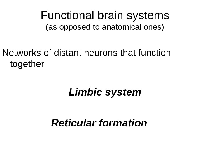
Functional brain systems (as opposed to anatomical ones) Networks of distant neurons that function together Limbic system Reticular formation
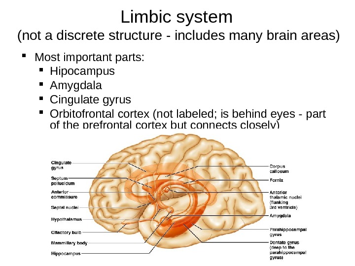
Limbic system (not a discrete structure — includes many brain areas) Most important parts: Hipocampus Amygdala Cingulate gyrus Orbitofrontal cortex (not labeled; is behind eyes — part of the prefrontal cortex but connects closely)
Limbic system continued Called the “emotional” brain Is essential for flexible, stable, adaptive functioning Links different areas so integration can occur Integration: separate things are brought together as a whole Processes emotions and allocates attentional resources Necessary for emotional balance, adaptation to environmental demands (including fearful situations, etc. ), for creating meaningful connections with others (e. g. ability to interpret facial expressions and respond appropriately), and more…
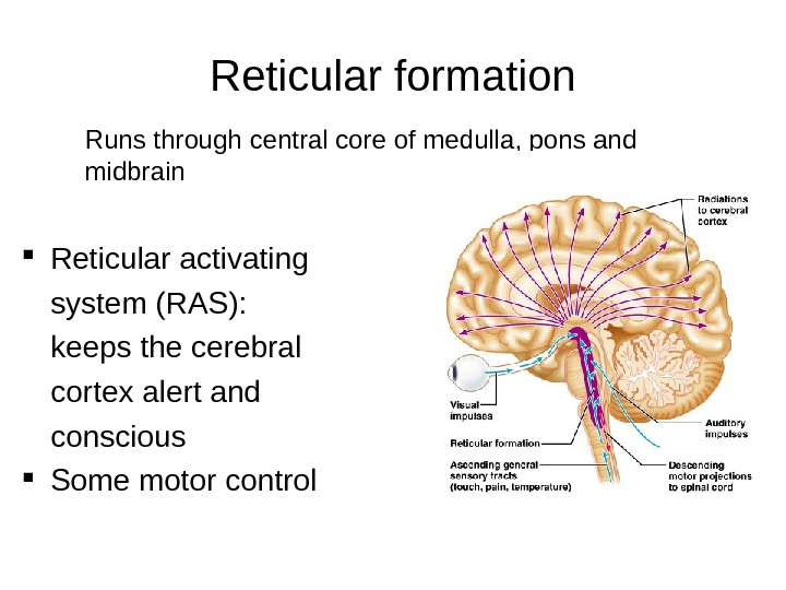
Reticular formation Runs through central core of medulla, pons and midbrain Reticular activating system (RAS): keeps the cerebral cortex alert and conscious Some motor control
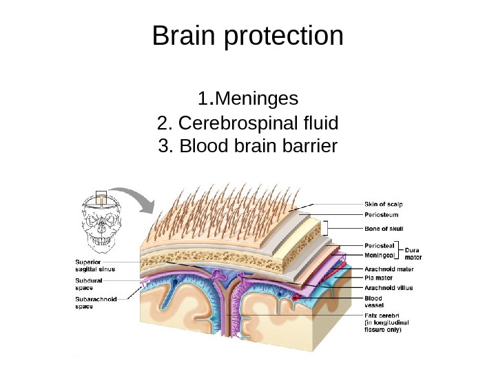
Brain protection 1. Meninges 2. Cerebrospinal fluid 3. Blood brain barrier
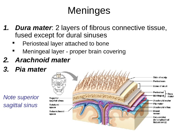
Meninges 1. Dura mater : 2 layers of fibrous connective tissue, fused except for dural sinuses Periosteal layer attached to bone Meningeal layer — proper brain covering 2. Arachnoid mater 3. Pia mater Note superior sagittal sinus
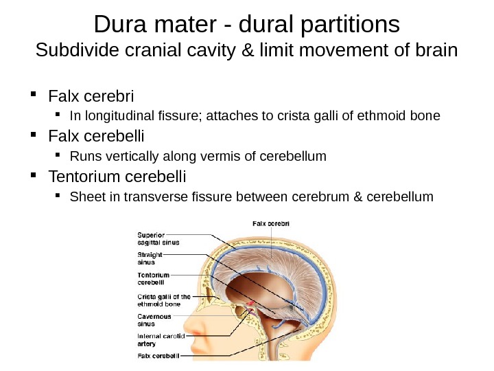
Dura mater — dural partitions Subdivide cranial cavity & limit movement of brain Falx cerebri In longitudinal fissure; attaches to crista galli of ethmoid bone Falx cerebelli Runs vertically along vermis of cerebellum Tentorium cerebelli Sheet in transverse fissure between cerebrum & cerebellum
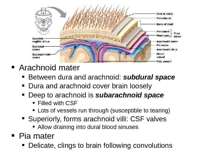
Arachnoid mater Between dura and arachnoid: subdural space Dura and arachnoid cover brain loosely Deep to arachnoid is subarachnoid space Filled with CSF Lots of vessels run through (susceptible to tearing) Superiorly, forms arachnoid villi: CSF valves Allow draining into dural blood sinuses Pia mater Delicate, clings to brain following convolutions
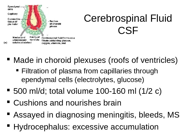
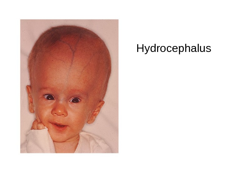
Cerebrospinal Fluid CSF Made in choroid plexuses (roofs of ventricles) Filtration of plasma from capillaries through ependymal cells (electrolytes, glucose) 500 ml/d; total volume 100 -160 ml (1/2 c) Cushions and nourishes brain Assayed in diagnosing meningitis, bleeds, MS Hydrocephalus: excessive accumulation
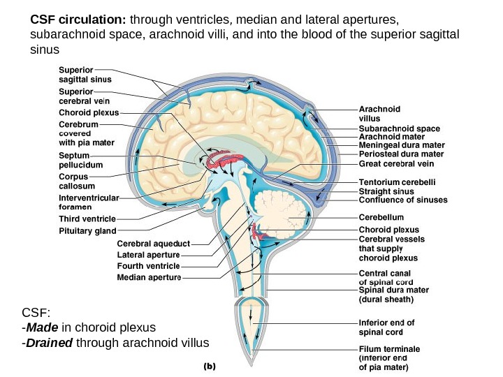
CSF circulation: through ventricles, median and lateral apertures, subarachnoid space, arachnoid villi, and into the blood of the superior sagittal sinus CSF: — Made in choroid plexus — Drained through arachnoid villus
Hydrocephalus
Blood-Brain Barrier Tight junctions between endothelial cells of brain capillaries, instead of the usual permeability Highly selective transport mechanisms Allows nutrients, O 2, CO 2 Not a barrier against uncharged and lipid soluble molecules; allows alcohol, nicotine, and some drugs including anesthetics
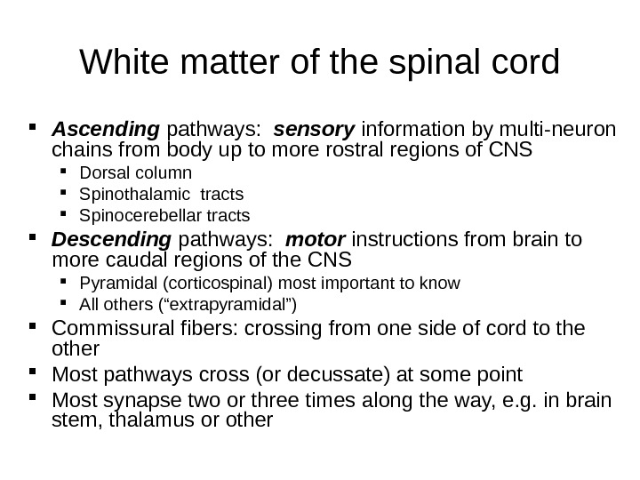
White matter of the spinal cord Ascending pathways: sensory information by multi-neuron chains from body up to more rostral regions of CNS Dorsal column Spinothalamic tracts Spinocerebellar tracts Descending pathways: motor instructions from brain to more caudal regions of the CNS Pyramidal (corticospinal) most important to know All others (“extrapyramidal”) Commissural fibers: crossing from one side of cord to the other Most pathways cross (or decussate) at some point Most synapse two or three times along the way, e. g. in brain stem, thalamus or other
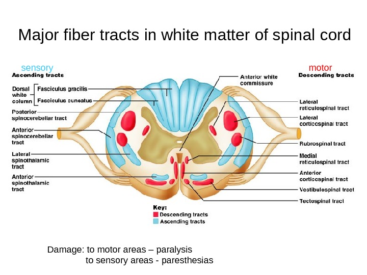
Major fiber tracts in white matter of spinal cord Damage: to motor areas – paralysis to sensory areas — paresthesiassensory motor
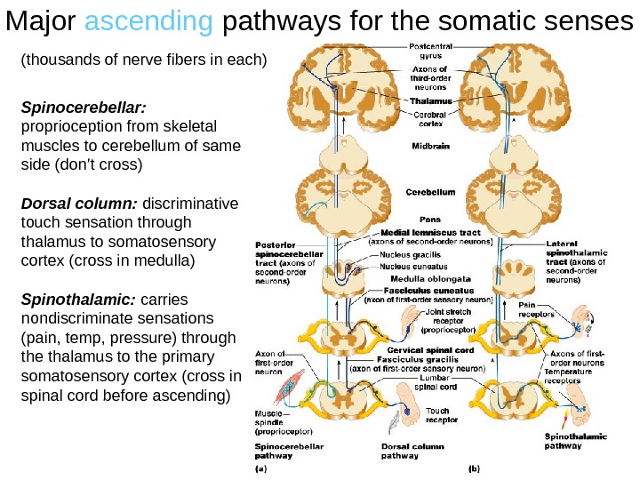
Major ascending pathways for the somatic senses Spinocerebellar: proprioception from skeletal muscles to cerebellum of same side (don’t cross) Dorsal column: discriminative touch sensation through thalamus to somatosensory cortex (cross in medulla) Spinothalamic: carries nondiscriminate sensations (pain, temp, pressure) through the thalamus to the primary somatosensory cortex (cross in spinal cord before ascending) (thousands of nerve fibers in each)
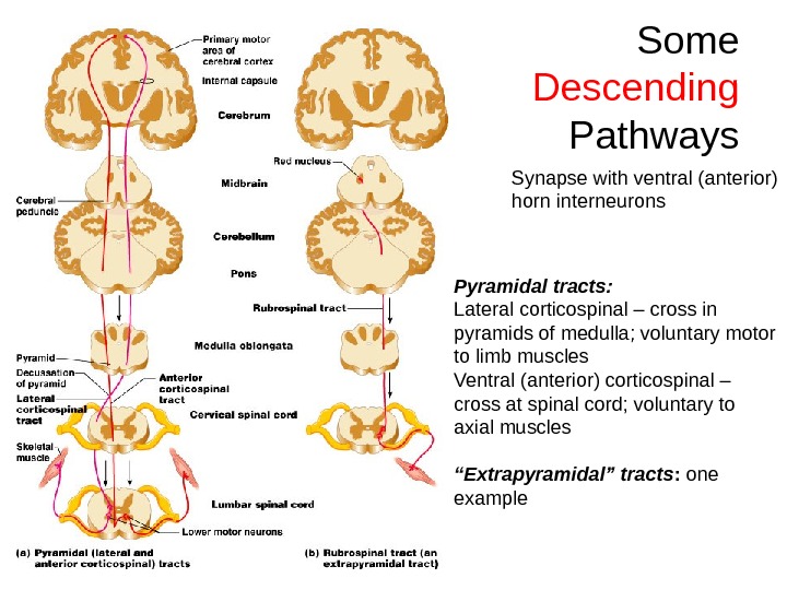
Some Descending Pathways Pyramidal tracts: Lateral corticospinal – cross in pyramids of medulla; voluntary motor to limb muscles Ventral (anterior) corticospinal – cross at spinal cord; voluntary to axial muscles “ Extrapyramidal” tracts : one example Synapse with ventral (anterior) horn interneurons
Check out: Medical gross anatomy atlas images (good teaching pics): http: //anatomy. med. umich. edu/atlas_index. ht ml (can access from Paul Wissman’s site also: -anatomy and physiology -brain and spinal cord -brain pics at U. Mich) Really good site for photos of human brain dissections: http: //library. med. utah. edu/Web. Path/HISTHTML/N EURANAT/NEURANCA. html
Hints & additional pics Unless your prints of the slides are very large and clear, look at the pictures from the book on your computer screen or in the book itself so you can read all the labels Anything in bold, italicized or repeated should be learned Remembering the terminology from the quiz will help you figure things out Anterior horn cells = ventral motor neurons Forget funiculi; know dorsal column (spinal cord)
Know the names of the ventricles and which ones connect to which, in order You don’t need to know the #s of the Brodman areas You do need to know where are the: primary somatosensory, primary motor, broca’s speech, visual cortex, the lobes of the brain, main sulci and fissures, precentral and postcentral gyri and which go with which of motor and sensory, etc For the most part, the medical info is FYI
From this site, which also has text explanations: http: //www. emc. maricopa. edu/facul ty/farabee/BIOBK/Bio. Book. NERV. ht ml
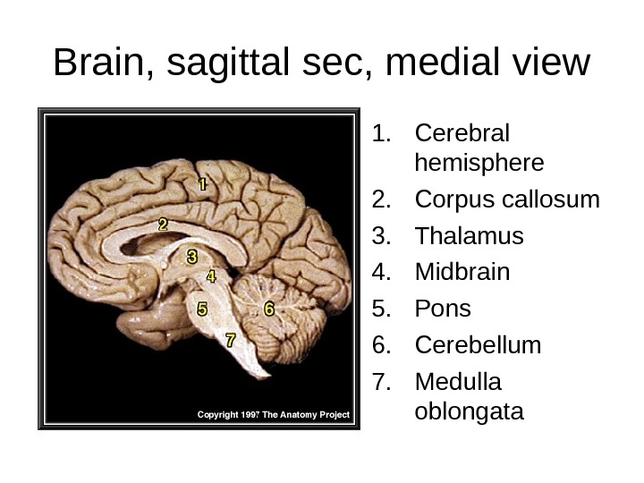
Brain, sagittal sec, medial view 1. Cerebral hemisphere 2. Corpus callosum 3. Thalamus 4. Midbrain 5. Pons 6. Cerebellum 7. Medulla oblongata
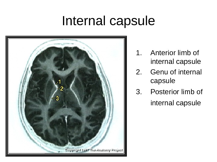
Internal capsule 1. Anterior limb of internal capsule 2. Genu of internal capsule 3. Posterior limb of internal capsule
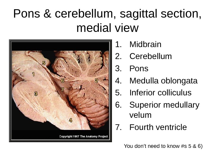
Pons & cerebellum, sagittal section, medial view 1. Midbrain 2. Cerebellum 3. Pons 4. Medulla oblongata 5. Inferior colliculus 6. Superior medullary velum 7. Fourth ventricle You don’t need to know #s 5 & 6)
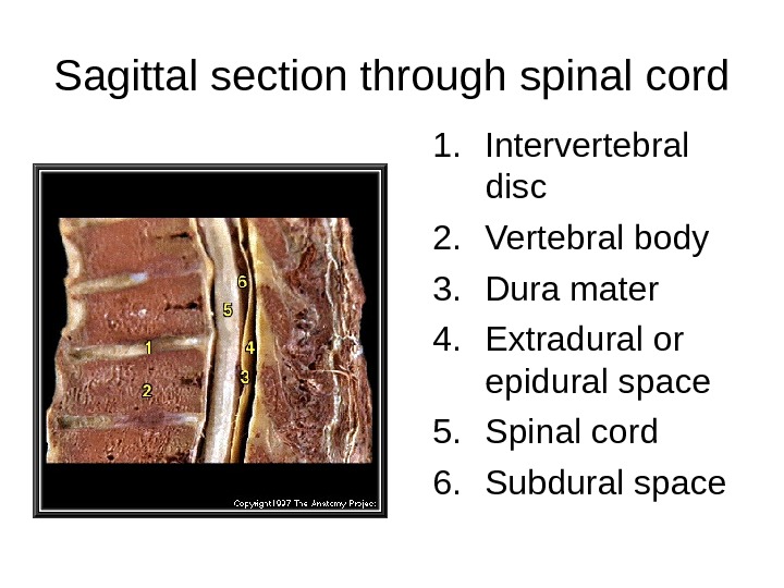
Sagittal section through spinal cord 1. Intervertebral disc 2. Vertebral body 3. Dura mater 4. Extradural or epidural space 5. Spinal cord 6. Subdural space