
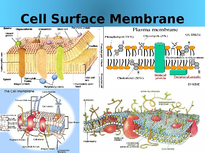
Cell Surface Membrane 03/19/16 Pork Chop Willie cell


l3.cell_membrane_final_2013.ppt
- Размер: 9.6 Mегабайта
- Количество слайдов: 57
Описание презентации Cell Surface Membrane 03/19/16 Pork Chop Willie cell по слайдам
Cell Surface Membrane 03/19/16 Pork Chop Willie cell membrane structure
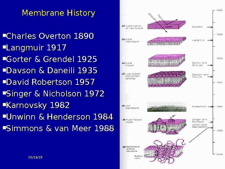
Membrane History Charles Overton 1890 Langmuir 1917 Gorter & Grendel 1925 Davson & Daneili 1935 David Robertson 1957 Singer & Nicholson 1972 Karnovsky 1982 Unwinn & Henderson 1984 Simmons & van Meer 1988 03/19/
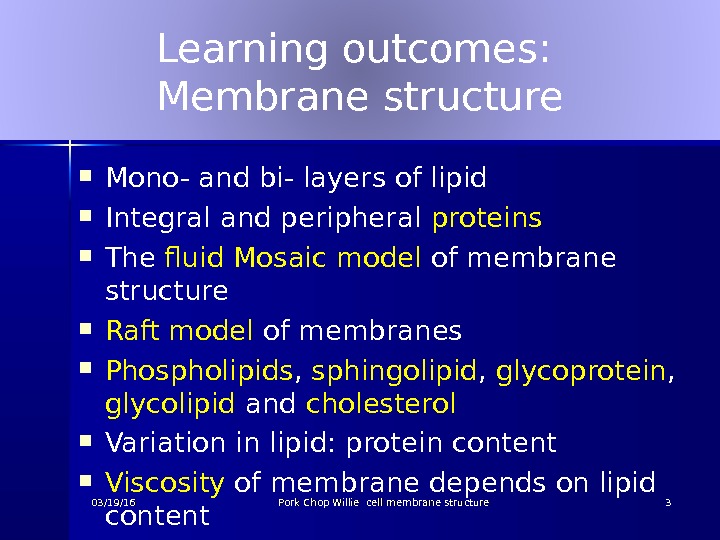
Learning outcomes: Membrane structure Mono- and bi- layers of lipid Integral and peripheral proteins The fluid Mosaic model of membrane structure Raft model of membranes Phospholipids , sphingolipid , glycoprotein , glycolipid and cholesterol Variation in lipid: protein content Viscosity of membrane depends on lipid content 03/19/16 Pork Chop Willie cell membrane structure
Learning outcomes: Role of membrane Boundary layer but also an active part of the biochemical functioning of the cell Passage of hydrophili c c and hydrophobic material across the membrane Pores 03/19/16 Pork Chop Willie cell membrane structure
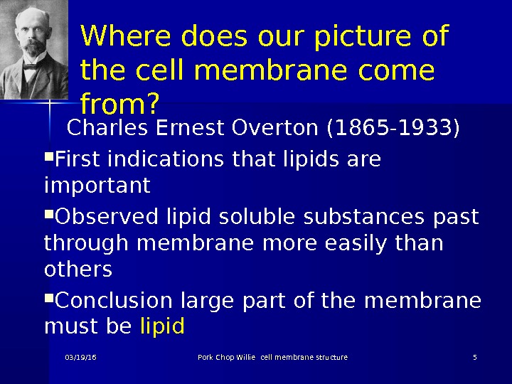
Where does our picture of the cell membrane come from? Charles Ernest Overton (1865 -1933) First indications that lipids are important Observed lipid soluble substances past through membrane more easily than others Conclusion large part of the membrane must be lipid 03/19/16 Pork Chop Willie cell membrane structure
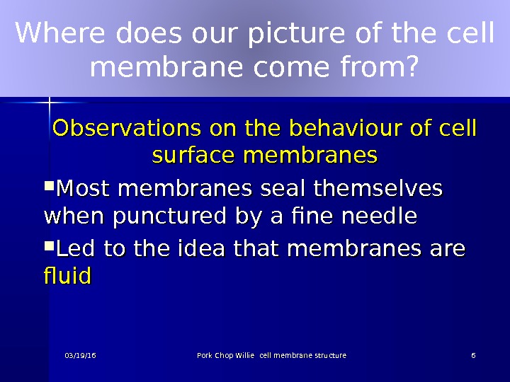
Where does our picture of the cell membrane come from? Observations on the behaviour of cell surface membranes Most membranes seal themselves when punctured by a fine needle Led to the idea that membranes are fluid 03/19/16 Pork Chop Willie cell membrane structure
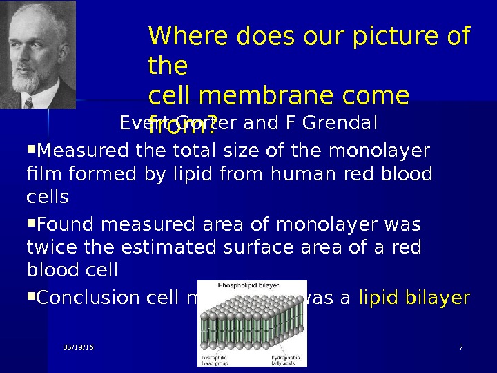
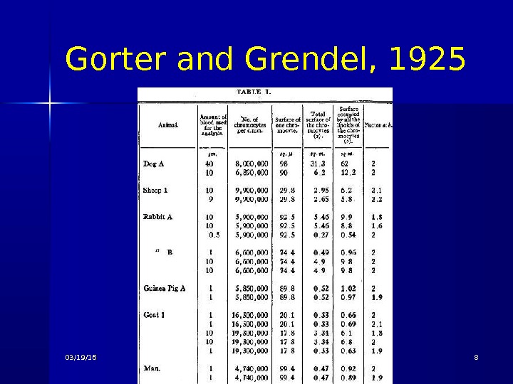
Where does our picture of the cell membrane come from? Evert Gorter and F Grendal Measured the total size of the monolayer film formed by lipid from human red blood cells Found measured area of monolayer was twice the estimated surface area of a red blood cell Conclusion cell membrane was a lipid bilayer 03/19/
Gorter and Grendel, 1925 03/19/16 Pork Chop Willie cell membrane structure
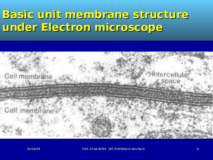
Basic unit membrane structure under Electron microscope 03/19/16 Pork Chop Willie cell membrane structure
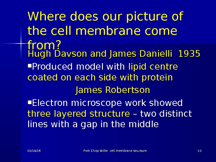
Where does our picture of the cell membrane come from? Hugh Davson and James Danielli 1935 Produced model with lipid centre coated on each side with protein James Robertson Electron microscope work showed three layered structure – two distinct lines with a gap in the middle 03/19/16 Pork Chop Willie cell membrane structure
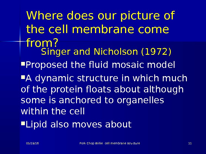
Where does our picture of the cell membrane come from? Singer and Nicholson (1972) Proposed the fluid mosaic model A dynamic structure in which much of the protein floats about although some is anchored to organelles within the cell Lipid also moves about 03/19/16 Pork Chop Willie cell membrane structure
03/19/16 Pork Chop Willie cell membrane structure
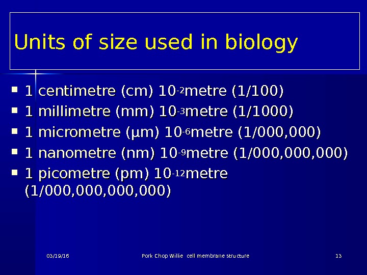
Units of size used in biology 1 1 centimetre (cm) 10 -2 -2 metre (1/100) 1 1 millimetre (mm) 10 -3 -3 metre (1/1000) 1 1 micrometre ( ( µµ m) 10 -6 -6 metre (1/000, 000) 1 1 nanometre (nm) 10 -9 -9 metre (1/000, 000) 1 1 picometre (pm) 10 -12 -12 metre (1/000, 000, 000) 03/19/16 Pork Chop Willie cell membrane structure
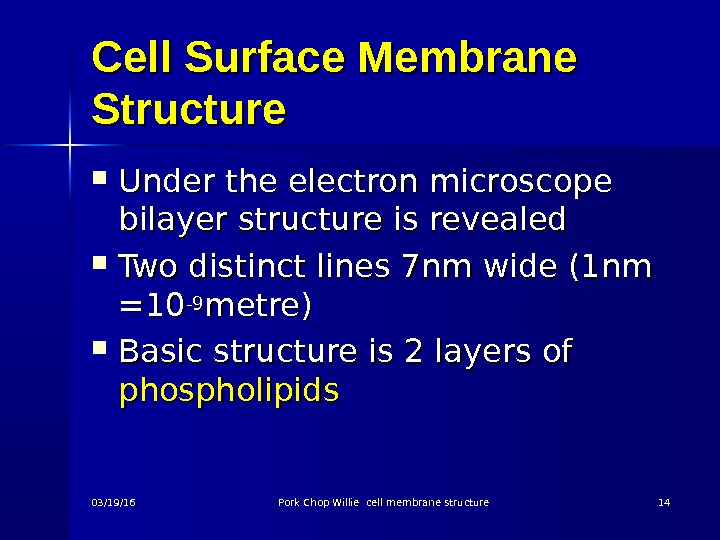
Cell Surface Membrane Structure Under the electron microscope bilayer structure is revealed Two distinct lines 7 nm wide (1 nm =10=10 -9 -9 metre) Basic structure is 2 layers of phospholipids 03/19/16 Pork Chop Willie cell membrane structure
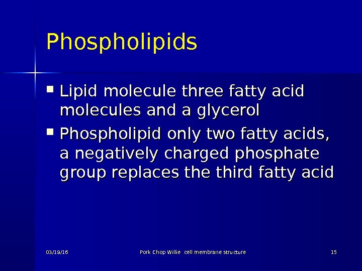
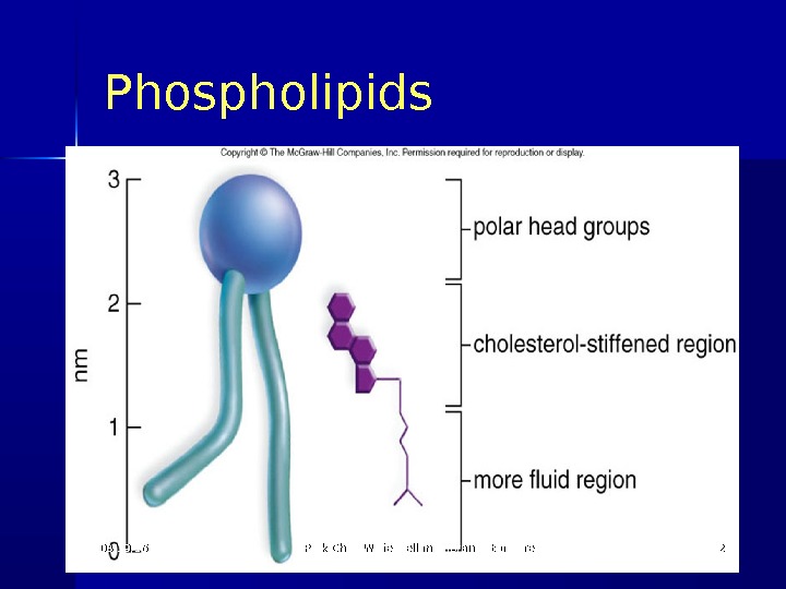
Phospholipids Lipid molecule three fatty acid molecules and a glycerol Phospholipid only two fatty acids, a negatively charged phosphate group replaces the third fatty acid 03/19/16 Pork Chop Willie cell membrane structure
Lipid molecule 03/19/16 Pork Chop Willie cell membrane structure
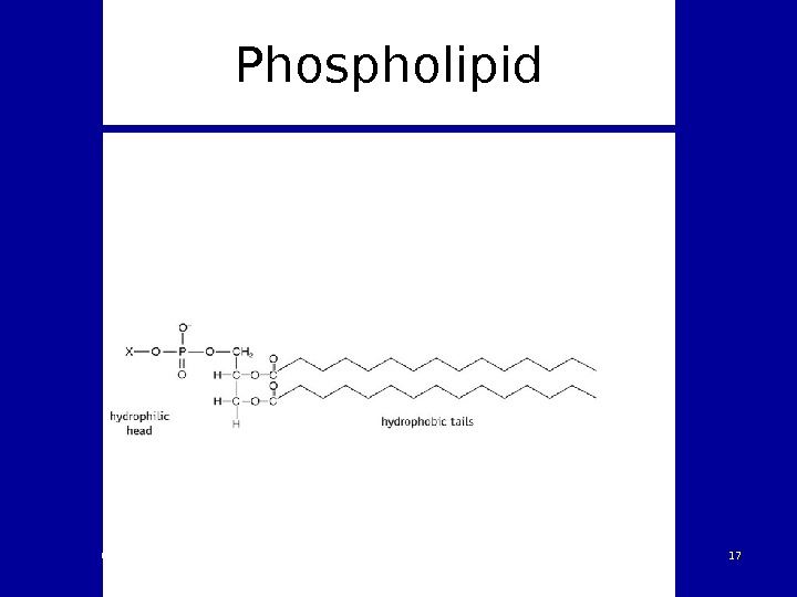
Phospholipid 03/19/16 Pork Chop Willie cell membrane structure
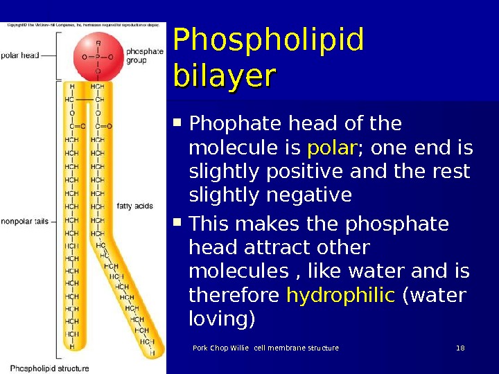
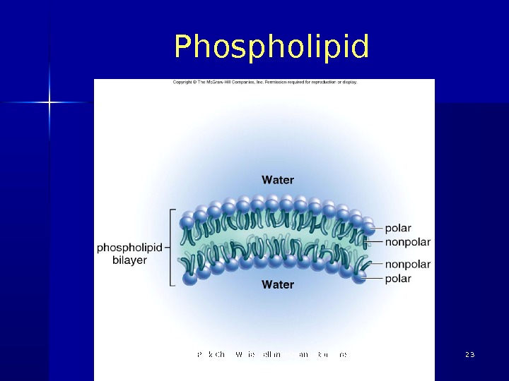
Phospholipid bilayer Phophate head of the molecule is polar ; one end is slightly positive and the rest slightly negative This makes the phosphate head attract other molecules , like water and is therefore hydrophilic (water loving) 03/19/16 Pork Chop Willie cell membrane structure
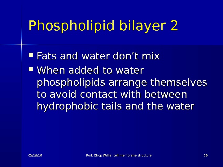
Phospholipid bilayer 2 Fats and water don’t mix When added to water phospholipids arrange themselves to avoid contact with between hydrophobic tails and the water 03/19/16 Pork Chop Willie cell membrane structure
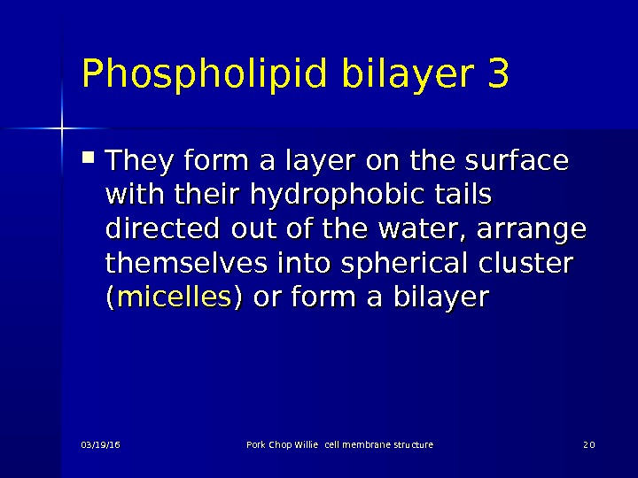
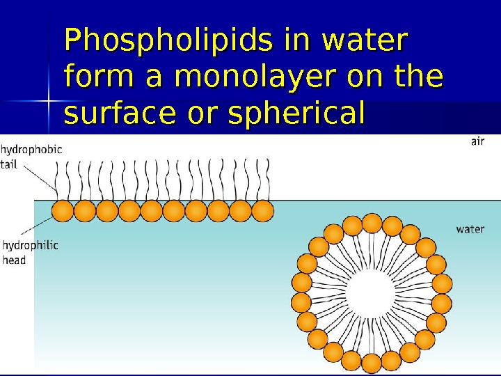
Phospholipid bilayer 3 They form a layer on the surface with their hydrophobic tails directed out of the water, arrange themselves into spherical cluster (( micelles ) or form a bilayer 03/19/16 Pork Chop Willie cell membrane structure
Phospholipids in water form a monolayer on the surface or spherical micelles 03/19/16 Pork Chop Willie cell membrane structure
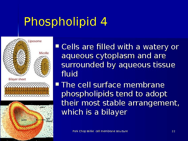
Phospholipid 4 Cells are filled with a watery or aqueous cytoplasm and are surrounded by aqueous tissue fluid The cell surface membrane phospholipids tend to adopt their most stable arrangement, which is a bilayer 03/19/16 Pork Chop Willie cell membrane structure
Phospholipid Pork Chop Willie cell membrane structure
Phospholipid 5 This arrangement avoids the hydrophobic fatty acid tails having any contact with water on either side of the membrane but ensures that the hydrophilic phosphate heads are in contact with the water. 03/19/16 Pork Chop Willie cell membrane structure
Phospholipids 03/19/16 Pork Chop Willie cell membrane structure
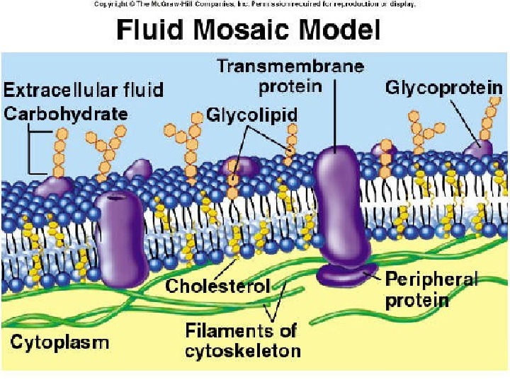
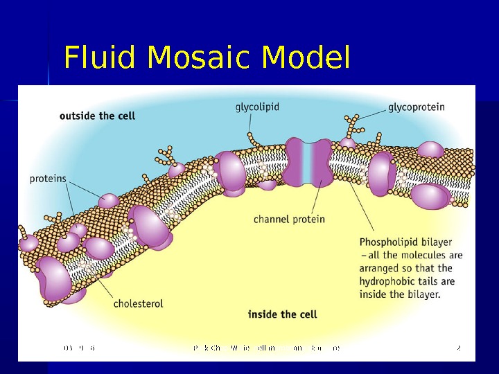
Fluid-Mosaic Model 1 The cell surface membrane is not just a phospholipid bilayer It also contains proteins , cholesterol , glycoproteins (protein molecule with polysaccharide attached) and glycolipid (lipid molecule with polysaccharide attached) 03/19/16 Pork Chop Willie cell membrane structure
Fluid-Mosaic Model 2 Some of the proteins span the layer Other proteins are found only within the inner layer or only within the outer layer Membrane proteins have hydrophobic areas and these are positioned within the membrane bilayer 03/19/16 Pork Chop Willie cell membrane structure
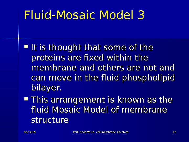
Fluid-Mosaic Model 3 It is thought that some of the proteins are fixed within the membrane and others are not and can move in the fluid phospholipid bilayer. This arrangement is known as the fluid Mosaic Model of membrane structure 03/19/16 Pork Chop Willie cell membrane structure
Fluid Mosaic Model 03/19/16 Pork Chop Willie cell membrane structure
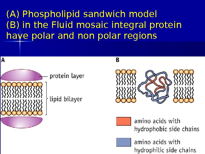
Evidence for the model 1 The most widely accepted model until the early 1970 s was a three layer protein-lipid layer sandwich based on electron micrographs (diagram A) However this model does not allow the hydrophillic head to come into contact with water 03/19/16 Pork Chop Willie cell membrane structure
(A) Phospholipid sandwich model (B) in the Fluid mosaic integral protein have polar and non polar regions 03/19/16 Pork Chop Willie cell membrane structure
Evidence for the model 2 Experiments showed that there were two types of protein- those that could be dissociated easily by increasing the ionic strength of the surrounding solution and those that could only be removed with detergent This evidence indicated some proteins were loosely attached and some are fully embedded 03/19/16 Pork Chop Willie cell membrane structure
Evidence for the model 3 Several integral proteins were shown to have regions at their ends that had polar hydrophilic amino acids, with the middle portion composed mainly of non polar hydrophobic amino acids (diagram B) 03/19/16 Pork Chop Willie cell membrane structure
(A) Phospholipid sandwich model (B) in the Fluid mosaic integral protein have polar and non polar regions 03/19/16 Pork Chop Willie cell membrane structure
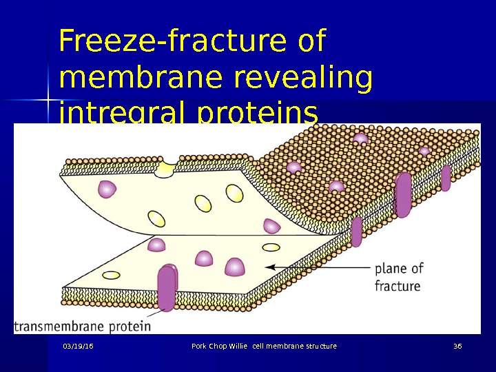
Evidence for the model 4 Additional evidence for integral proteins came from freeze-fracture electron microscope studies Freeze-fracture sections were fractured along their weak point between lipid layers Scanning Electron microscopy gave a a three dimensional image 03/19/16 Pork Chop Willie cell membrane structure
Freeze-fracture of membrane revealing intregral proteins 03/19/16 Pork Chop Willie cell membrane structure
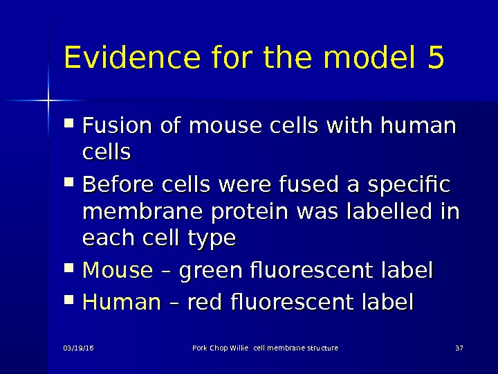
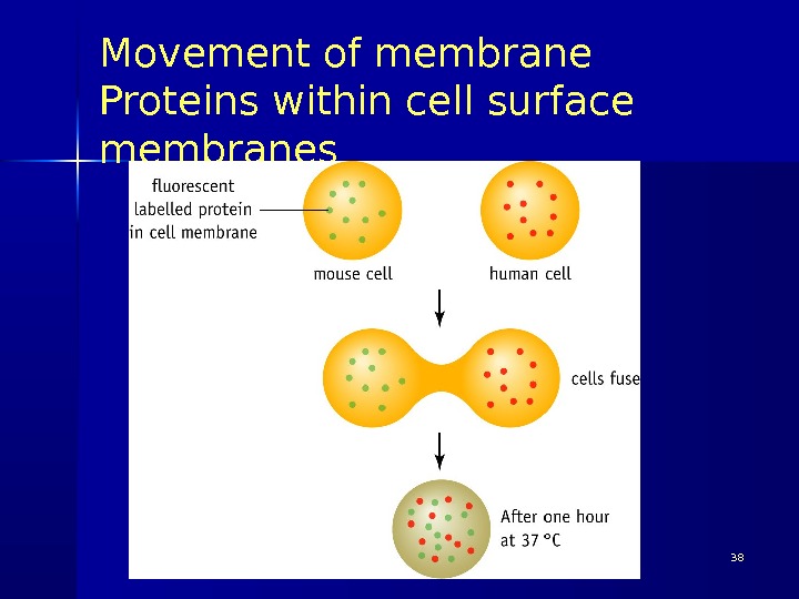
Evidence for the model 5 Fusion of mouse cells with human cells Before cells were fused a specific membrane protein was labelled in each cell type Mouse – green fluorescent label Human – red fluorescent label 03/19/16 Pork Chop Willie cell membrane structure
Movement of membrane Proteins within cell surface membranes Pork Chop Willie cell membrane structure
Membrane Protein Diversity
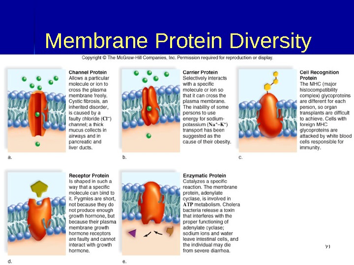
Functions of Membrane Proteins Channel Proteins : : Tubular Allow passage of molecules through membrane Carrier Proteins : : Combine with substance to be transported Assist passage of molecules through membrane Cell Recognition Proteins : : Provides unique chemical ID for cells Help body recognize foreign substances Receptor Proteins : : Binds with messenger molecule Causes cell to respond to message Enzymatic Proteins: Carry out metabolic reactions directly 03/19/16 Pork Chop Willie cell membrane structure
More unsaturated phospholipids – more fluid The more phospholipids containing unsaturated fatty acids the more fluid the membrane The ‘kinks’ in the hydrocarbon tails of the unsaturated tails prevents them from packing closely together, so more movement is possible 03/19/16 Pork Chop Willie cell membrane structure
Cholesterol reduces the fluidity of the membrane by preventing movement of the phospholipids 03/19/16 Pork Chop Willie cell membrane structure
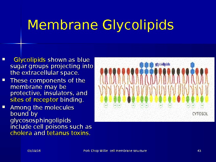
Membrane Glycolipids shown as blue sugar groups projecting into the extracellular space. These components of the membrane may be protective, insulators, and sites of receptor binding. Among the molecules bound by glycososphingolipids include cell poisons such as cholera and tetanus toxins. . 03/19/16 Pork Chop Willie cell membrane structure
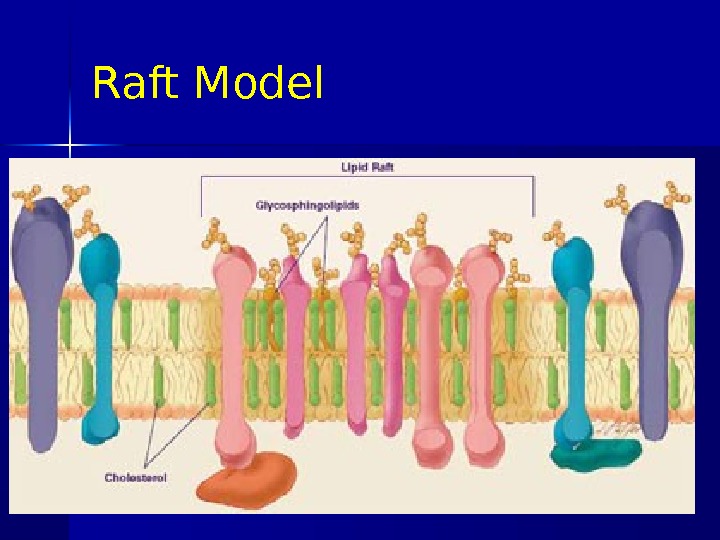
Sphingolipid Structural lipid of which the parent structure is sphingosine rather than glycerol. Synthesised in the Golgi complex Form the lipid rafts 03/19/16 Pork Chop Willie cell membrane structure
Raft Model 03/19/16 Pork Chop Willie cell membrane structure
Raft Model Lipid rafts are possible island like structure present in cellular membranes. They are enriched in cholesterol and sphingolipids. Cellular membranes with lipid rafts have a higher concentration of glycosphingolipids and cholesterol than do non-raft parts of membrane. 03/19/16 Pork Chop Willie cell membrane structure
Raft Model The existence of lipid rafts in cell membrane has not yet been approved completely by all scientists, but many think they serve as communication hubs by recruiting proteins that need to come together in order to transmit a signal. 03/19/16 Pork Chop Willie cell membrane structure
03/19/16 Pork Chop Willie cell membrane structure 4848 Q 1 According to the fluid-mosaic model for the plasma membrane, there is a ______ bilayer in which proteins are scattered throughout the membrane. The _____ (water loving) polar heads of the phospholipids face the intracellular and extracellular fluid. The _______ (water hating) nonpolar tails of the phospholipid molecules face each other.
03/19/16 Pork Chop Willie cell membrane structure 4949 A 1 According to the fluid-mosaic model for the plasma membrane, there is a phospholipid bilayer in which proteins are scattered throughout the membrane. The hydrophilic (water loving) polar heads of the phospholipids face the intracellular and extracellular fluid. The hydrophobic (water hating) nonpolar tails of the phospholipid molecules face each other.
Q 2 Q 2 Phospholipids have their hydrophilic polar heads facing the __________ and _______ fluid. The hydrophobic nonpolar tails face each other. The other two types of lipids present in the plasma membrane are the _______ and ______________. 03/19/16 Pork Chop Willie cell membrane structure
A 2 Phospholipids have their hydrophilic polar heads facing the intracellular and extracellular fluid. The hydrophobic nonpolar tails face each other. The other two types of lipids present in the plasma membrane are the glyco lipids and cholesterol. 03/19/16 Pork Chop Willie cell membrane structure
Q 3 The proteins found in the plasma membrane may be _____ proteins, which are found within the membrane, or ______ proteins, which occur either on the cytoplasmic side or the outer surface side of the membrane. 03/19/16 Pork Chop Willie cell membrane structure
A 3 The proteins found in the plasma membrane may be integral proteins, which are found within the membrane, or peripheral proteins, which occur either on the cytoplasmic side or the outer surface side of the membrane. 03/19/16 Pork Chop Willie cell membrane structure
Q 4 Q 4 State two roles of cholesterol in the membrane (2 marks) 03/19/16 Pork Chop Willie cell membrane structure
A 4 State two roles of cholesterol in the membrane (2 marks) Regulates membrane fluidity; Mechanical stability; Reduces leakage of polar ions by diffusion; 03/19/16 Pork Chop Willie cell membrane structure
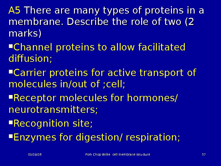
Q 5 Q 5 There are many types of proteins in a membrane. Describe the role of two (2 marks) 03/19/16 Pork Chop Willie cell membrane structure
A 5 There are many types of proteins in a membrane. Describe the role of two (2 marks) Channel proteins to allow facilitated diffusion; Carrier proteins for active transport of molecules in/out of ; cell; Receptor molecules for hormones/ neurotransmitters; Recognition site; Enzymes for digestion/ respiration; 03/19/16 Pork Chop Willie cell membrane structure