Blood Red el.ppt
- Количество слайдов: 49


Blood Physiology

Состав тела и жидкие среды организма Вода в теле человека содержится в разных компартментах: (Fig. 2. 1): ● Внутриклеточная (ICF) ● Внеклеточная (ECF): состоит из: ● 75% интерстициальная (ISF): ● 25% плазма крови (система кровооорбращения.
")
Трансцеллюлярная жидкость (незначительный % веса тела)

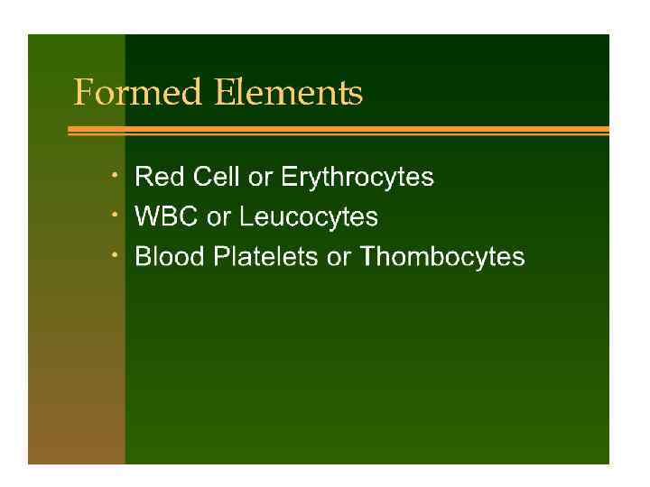
Состав крови
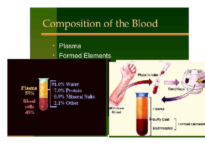

Determination of the haematocrit from a centrifuged blood sample.
")
Blood volume This can be derived from the haematocrit (fraction of total blood volume) and the plasma volume: Total blood volume (TBV) = Plasma volume 1− haematocrit

BLOOD VOLUME: PHYSIOLOGICAL VARIATIONS • AGE • SEX • TEMPERATURE • BODY WEIGHT • BODY SURFACE AREA • PREGNANCY • EXERCISE • POSTURE • HYPOXIA • EMOTIONS

BLOOD VOLUME & AGE AT BIRTH BLOOD VOLUME in Liters 0. 3 6 Months 0. 5 1 year 2 yrs 6 months 4 years 7 years 10 years ADULTS 0. 7 1. 0 1. 3 1. 7 2. 5(Girls); 3. 2 (Boys) 5 (Men) 4. 5 (Women)

BLOOD VOLUME: PHYSIOLOGICAL VARIATIONS 1. SEX: – Males have more blood volume than females. 2. TEMPERATURE: – Acute exposure to cold causes reduction in blood volume due to Plasma water loss to tissues.

BLOOD VOLUME: PHYSIOLOGICAL VARIATIONS 3. BODY WEIGHT: – It is usually 7% of the Body Weight. 4. BODY SURFACE AREA (BSA ): – 2. 8 L/ M 2 of BSA

BLOOD VOLUME: PHYSIOLOGICAL VARIATIONS 5. PREGNANCY: – Increases by 20 – 30% due to mass of fetus. 6. EXERCISE: – Vigorous exercise causes an increase. 7. POSTURE: – Changing from lying down to erect.

BLOOD VOLUME: PHYSIOLOGICAL VARIATIONS 8. HYPOXIA: – Seen in High altitudes. – Erythrocytes – So Blood volume. 9. EMOTIONS: – Excitement causes an increase in the Blood volume.
 4. DEHYDRATION: – Diarrhoea – Cholera – Gastroenteritis – Burns")
HYPOVOLEMIA: CAUSES (contd) 4. DEHYDRATION: – Diarrhoea – Cholera – Gastroenteritis – Burns
 5. ANEMIA: – Decreased RBC volume – Plasma may increase. 6.")
HYPOVOLEMIA: CAUSES (contd) 5. ANEMIA: – Decreased RBC volume – Plasma may increase. 6. OBESITY: – Blood volume per body weight decreases though Blood volume per BSA may be normal. 7. HYPOTHYROIDISM (MYXEDEMA): – Decrease in Blood volume. 8. ACUTE COLD: Decreases blood volume.

 or deuterium (2")
Total body water Radioactive water – tritium (3 H 2 O) or deuterium (2 H 2 O) – is used by the dilutional principle. Normal total body water values are: ● 63% in males = 45 L of 70 kg body weight. ● 52% in females = 36 L of 70 kg body weight owing to greater proportion of fat; fat cells have a lower water content than muscle.

Water Daily water intake can be from two sources: ● Ingestion of fluids as liquids and as water in food: 30 -35 мл/кг массы тела (2000 m. L). ● Oxidative metabolism of food: 400 m. L.

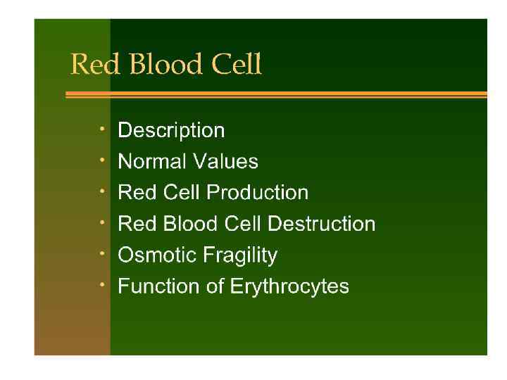
ЭРИТРОЦИТЫ
 can be measured in a variety of ways, depending on")
Red cell volume (RCV) can be measured in a variety of ways, depending on what information is known: 1. RCV = TBV − plasma volume 2. RCV = Haematocrit x plasma volume 1 - haematocrit 3. Direct dilutional method: radioactive chromium (51 Cr) is used to label or tag red blood cells. The fraction of red blood cells tagged is measured.

non-nucleated

HEMOLYSIS 3. HEMOLYSIS: – Mismatched transfusion – Snake bite – Black water fever – Hemorrhagic Plagues/Dengue – Measles
 2α 2β Hemoglobin A 2 2α")
THE HEMOGLOBINS Hemoglobin Polypeptides Hemoglobin A 1 (Adult) 2α 2β Hemoglobin A 2 2α 2δ Hemoglobin F (Fetal) 2α 2γ
. ● Females")
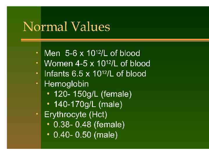
Haemoglobin • Men have 150 g Hb/L of blood (15 g/d. L). ● Females have 130 g Hb/L of blood (13 g/d. L). In an adult man with 15 g of Hb/d. L blood, the O 2 -carrying capacity will be: 15 × 1. 34 = 20. 1 m. L O 2/d. L blood Schematic diagram of haemoglobin.

Components of haemoglobin

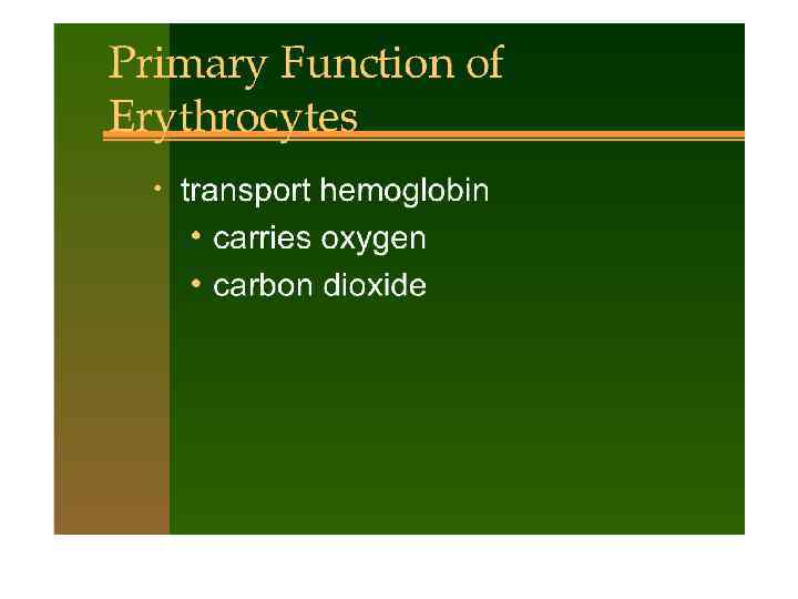
Other forms of haemoglobin Myoglobin This haem protein is found within muscle. Fetal haemoglobin (Hb. F- γ chains replace the β chains of Hb. A) The fetal circulation has a lower PO 2 (30 mm. Hg after deoxygenation) Carboxyhaemoglobin (carboxy. Hb) is formed when carbon monoxide (CO) binds to Hb. The affinity of CO for Hb is significantly higher (250 times greater) than that of O 2

RED BLOOD CELLS • IRON METABOLISM – Total body iron: 4 -5 gms • 65% hemoglobin • 4 % myoglobin • 1 % heme compounds (cytochrome, peroxidase, catalase) • 0. 1 % combined with protein transferrin • 15 -30% storage pool as ferritin (RES, liver)

RED BLOOD CELLS • Daily, a normal adult, destroys 15 ml of senescent • • red cells and produces an equal quantity of new red cells. For the daily production of erythrocytes, about 15 mg of iron is required. Almost all of the iron needed is drawn from stores, which was recycled from the destruction of old red cells. The vast majority of the iron is retained in the body as it is moved from one compartment to another. Very little is iron is obtained from the diet.

RED BLOOD CELLS • Transport and Storage of Iron – Iron absorbed from the intestine combines with apotransferrin to form transferrin – Transferrin carries the iron to the cells – In the cell cytoplasm, iron combines with apoferritin to form ferritin (storage iron) • When in excess, stored as insoluble hemosiderin – When needed, the ferritin storage pool is transported as transferrin • Transferrin binds strongly to the cell membranes of the erythroblasts • Ingested by the erythroblasts and iron is directly delivered to the mitochondria (heme synthesis)

RED BLOOD CELLS • • Recycling of the iron by the body is not a tight system. A normal healthy person loses around 1 to 2 mg of iron everyday. – Menstruation – GIT (sloughed epithelium) – Exfoliated skin
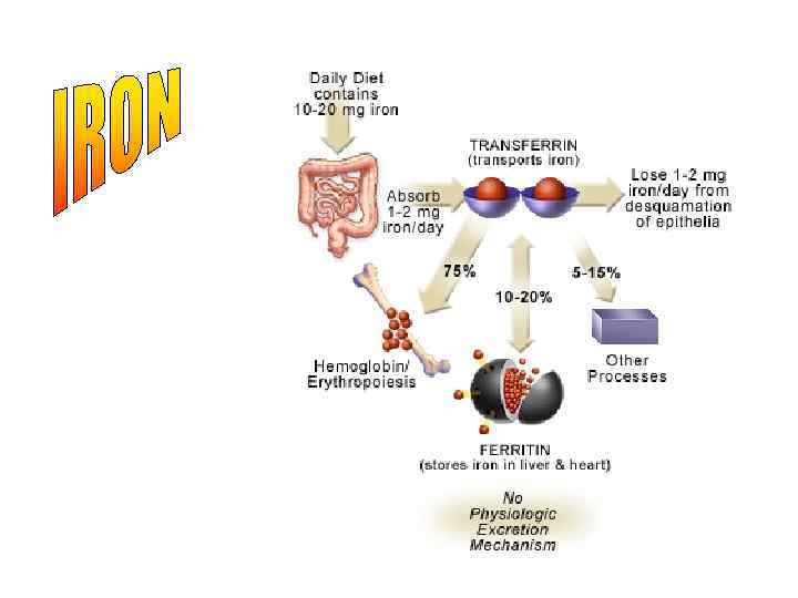

RED BLOOD CELLS • ABSORPTION OF IRON FROM THE GIT – Liver secretes apotransferrin to the bile – In the duodenum, the apotransferrin binds with free iron and other iron compounds (hemoglobin and myoglobin) to form transferrin – Transferrin binds to the receptors in the membranes of the intestinal epithelial cells – By pinocytosis, the transferrin is absorbed into the epithelial cells and released into the blood capillaries
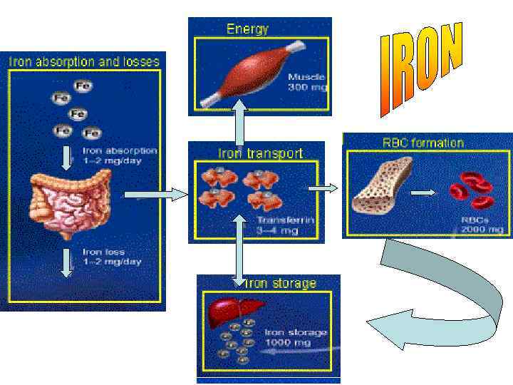

RED BLOOD CELLS • IRON REGULATION – Through altering the rate of absorption – When all the apoferritin in the storage areas are filled up with iron, the rate of GIT absorption slows down – Increase in the absorption occurs with iron depletion
Blood Red el.ppt