
Anatomy & Physiology of Heart Geu-Ru Hong, MD,


1._heart_anatomy_and_physiology.ppt
- Размер: 4.9 Mегабайта
- Количество слайдов: 44
Описание презентации Anatomy & Physiology of Heart Geu-Ru Hong, MD, по слайдам
Anatomy & Physiology of Heart Geu-Ru Hong, MD, Ph. D Associate Professor Director of Echocardiography Yonsei University, Seoul, Korea
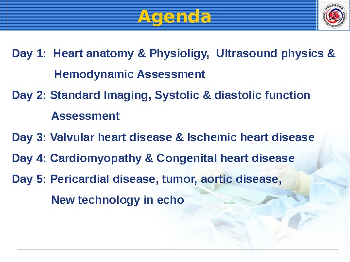
Agenda Day 1: Heart anatomy & Physioligy, Ultrasound physics & Hemodynamic Assessment Day 2: Standard Imaging, Systolic & diastolic function Assessment Day 3: Valvular heart disease & Ischemic heart disease Day 4: Cardiomyopathy & Congenital heart disease Day 5: Pericardial disease, tumor, aortic disease, New technology in echo
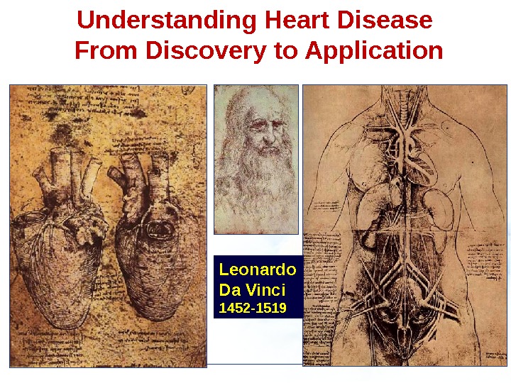
Leonardo Da Vinci 1452 -1519 Understanding Heart Disease From Discovery to Application
Edler – cardiologist at the Dept. of Cardiology at the University of Lund. Hertz — graduate in Physics. Borrowed an Ultrasonic Reflectroscope from a Shipyard in Malmo used for testing Metals. May 1953 — Detected moving echoes by the Ultrasound Reflectroscope.
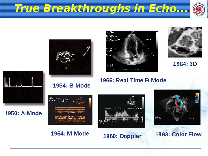
1966: Real-Time B-Mode. True Breakthroughs in Echo. . . 1950: A-Mode 1954: B-Mode 1964: M-Mode 1984: 3 D 1980: Doppler 1983: Color Flow
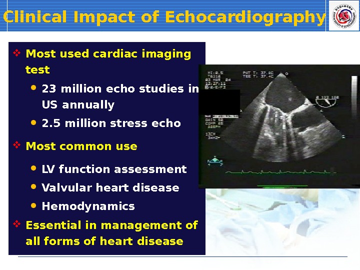
Clinical Impact of Echocardiography Most used cardiac imaging test 23 million echo studies in US annually 2. 5 million stress echo Most common use LV function assessment Valvular heart disease Hemodynamics Essential in management of all forms of heart disease
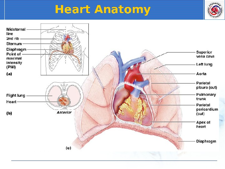
Heart Anatomy
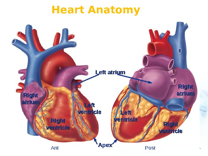
Heart Anatomy Left ventricle Right ventricle Left atrium Right atriumatrium Right ventricle. Left ventricle Apex Ant Post
Heart Anatomy Aorta Superior vena cava Inferior vena cava Ant Post
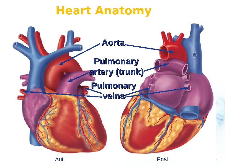
Heart Anatomy Aorta Pulmonary artery (trunk) Pulmonary veins Ant Post
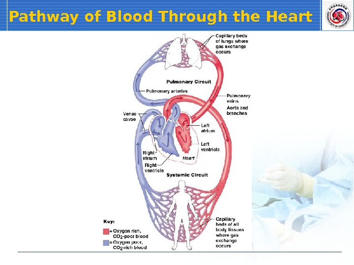
Pathway of Blood Through the Heart
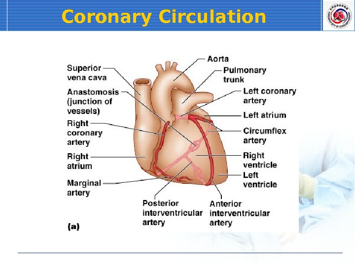
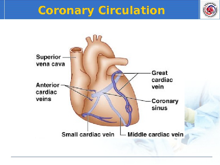
Heart Anatomy Aorta Coronary arteries Cardiac veins Coronary sinus Ant Post
Coronary Circulation
Coronary Circulation
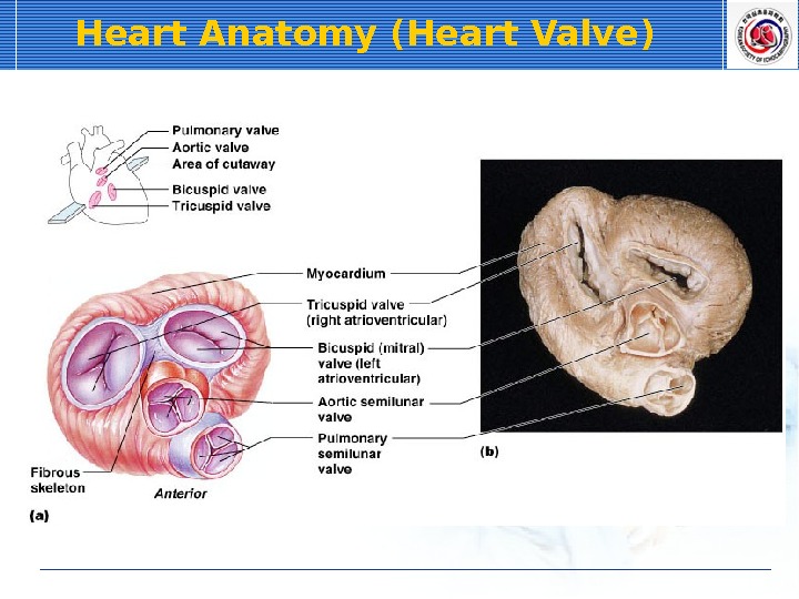
Heart Anatomy (Heart Valve)
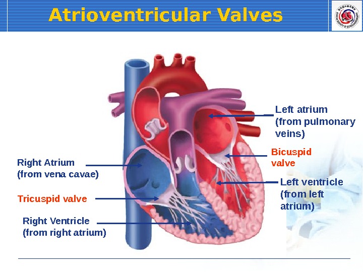
Atrioventricular Valves Right Atrium (from vena cavae) Right Ventricle (from right atrium) Left atrium (from pulmonary veins) Left ventricle (from left atrium)Tricuspid valve Bicuspid valve
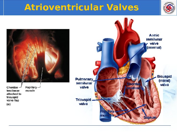
Atrioventricular Valves Chordae tendieae Tricuspid valve Bicuspid (mitral) valve. Pulmonary semilunar valve Papillary muscles Septum Aortic semilunar valve (internal)
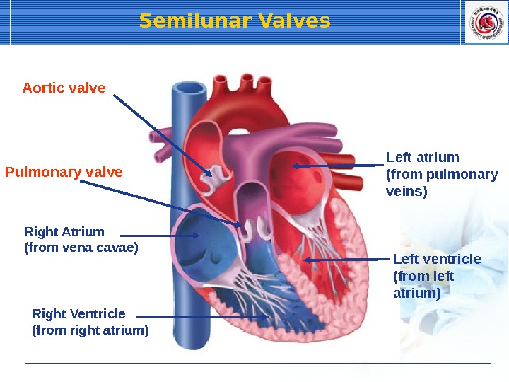
Right Atrium (from vena cavae) Right Ventricle (from right atrium) Left atrium (from pulmonary veins) Left ventricle (from left atrium)Aortic valve Pulmonary valve Semilunar Valves
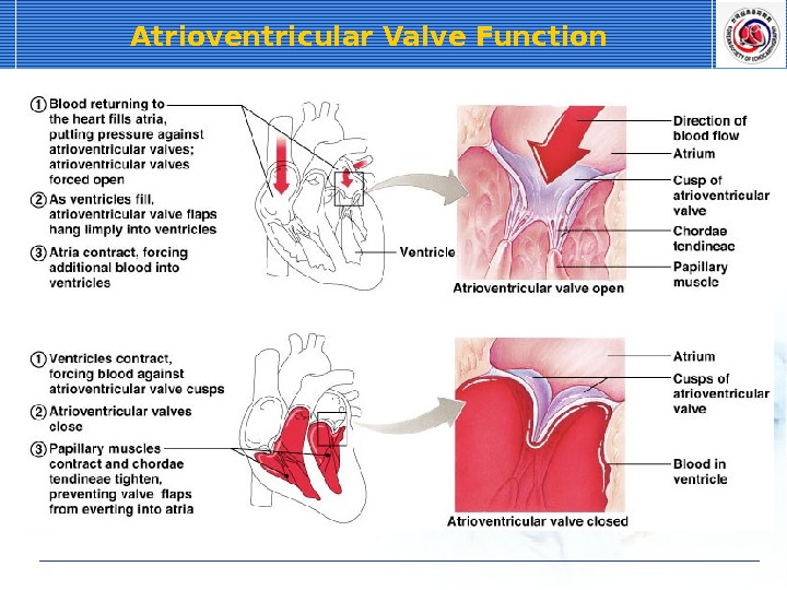
Atrioventricular Valve Function
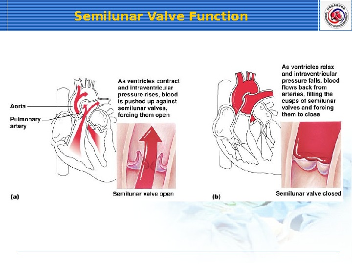
Semilunar Valve Function
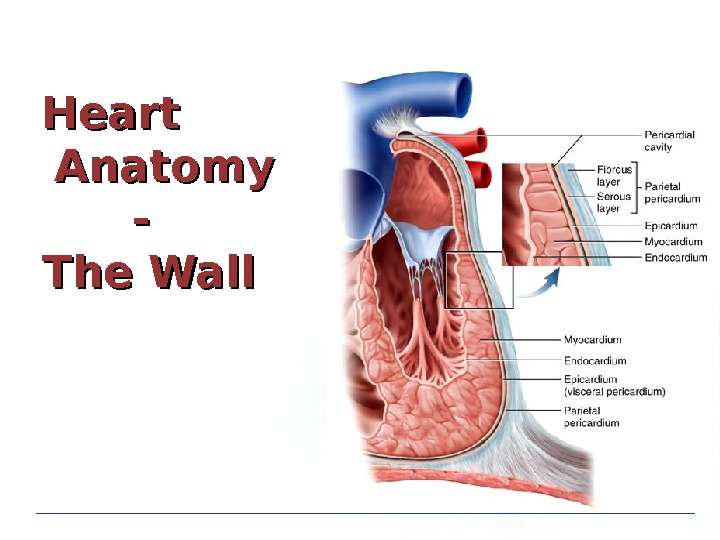
Heart Anatomy — The Wall
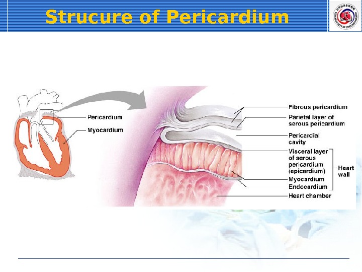
Strucure of Pericardium
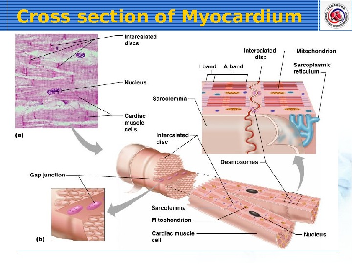
Cross section of Myocardium
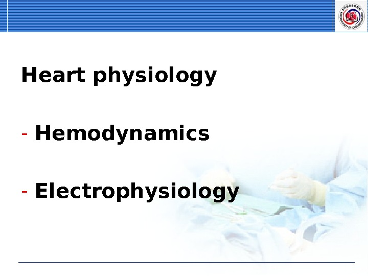
Heart physiology — Hemodynamics — Electrophysiology
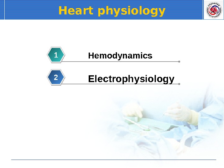
Heart physiology Hemodynamics 1 Electrophysiology
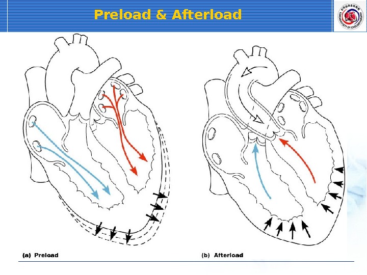
Preload & Afterload
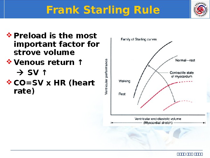
Frank Starling Rule Preload is the most important factor for strove volume Venous return ↑ SV ↑ CO=SV x HR (heart rate) 심심심심
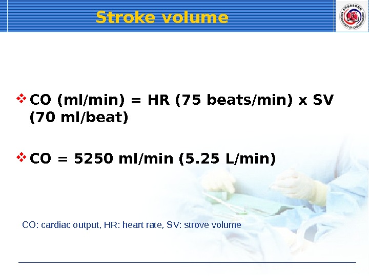
Stroke volume CO (ml/min) = HR (75 beats/min) x SV (70 ml/beat) CO = 5250 ml/min (5. 25 L/min) CO: cardiac output, HR: heart rate, SV: strove volume
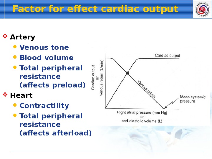
Factor for effect cardiac output Artery Venous tone Blood volume Total peripheral resistance (affects preload) Heart Contractility Total peripheral resistance (affects afterload)
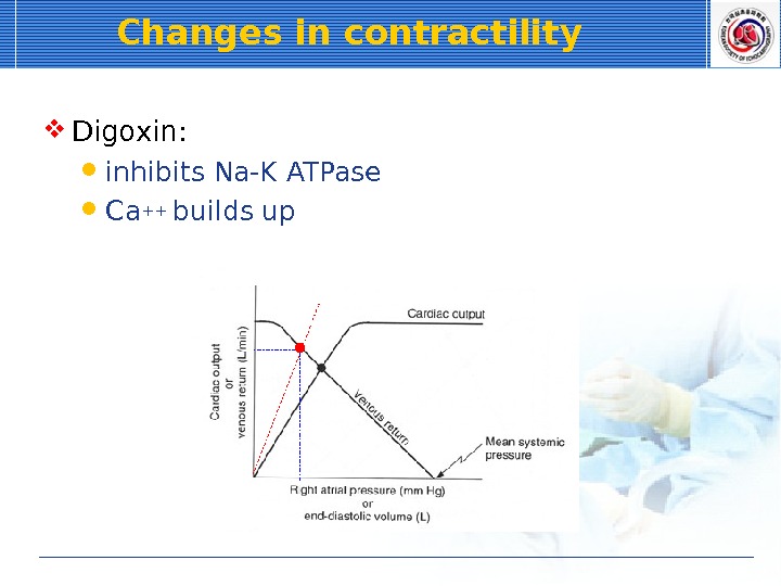
Changes in contractility Digoxin: inhibits Na-K ATPase Ca++ builds up
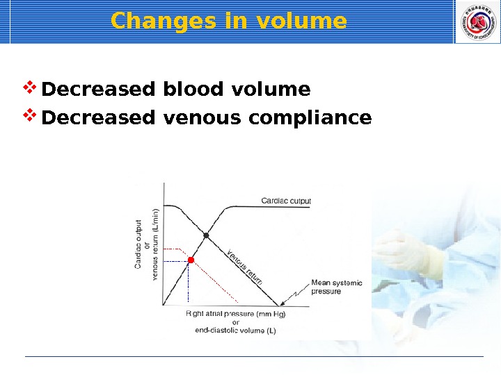
Changes in volume Decreased blood volume Decreased venous compliance
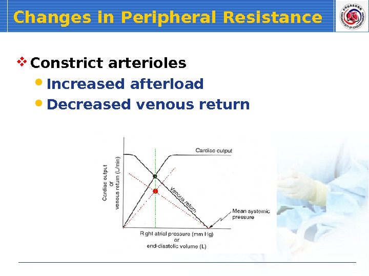
Changes in Peripheral Resistance Constrict arterioles Increased afterload Decreased venous return
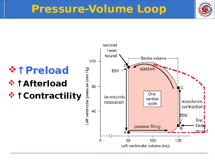
Pressure-Volume Loop Factors Preload Afterload Contractility Normal
Pressure-Volume Loop ↑ Preload ↑ Afterload ↑ Contractility
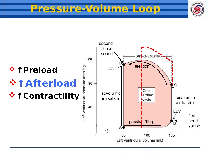
Pressure-Volume Loop ↑ Preload ↑ Afterload ↑ Contractility
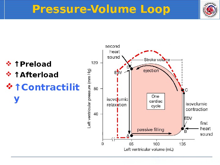
Pressure-Volume Loop ↑ Preload ↑ Afterload ↑ Contractilit y
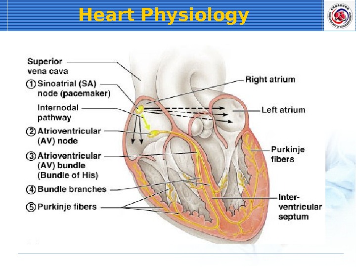
Heart Physiology
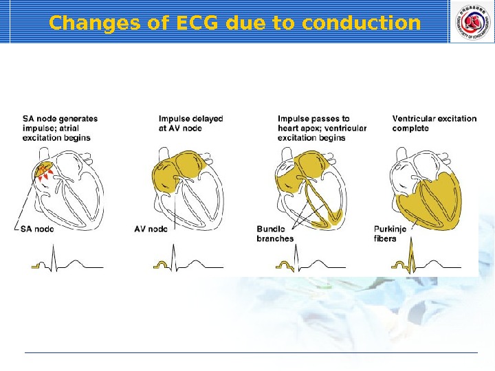
Changes of ECG due to conduction
Cardiac Cycle Cardiac cycle Systole – contraction of heart muscle Diastole – relaxation of heart muscle
Phases of the Cardiac Cycle Atria relax Rising ventricular pressure results in closing of AV valves Isovolumetric contraction phase Ventricular ejection phase opens semilunar valves
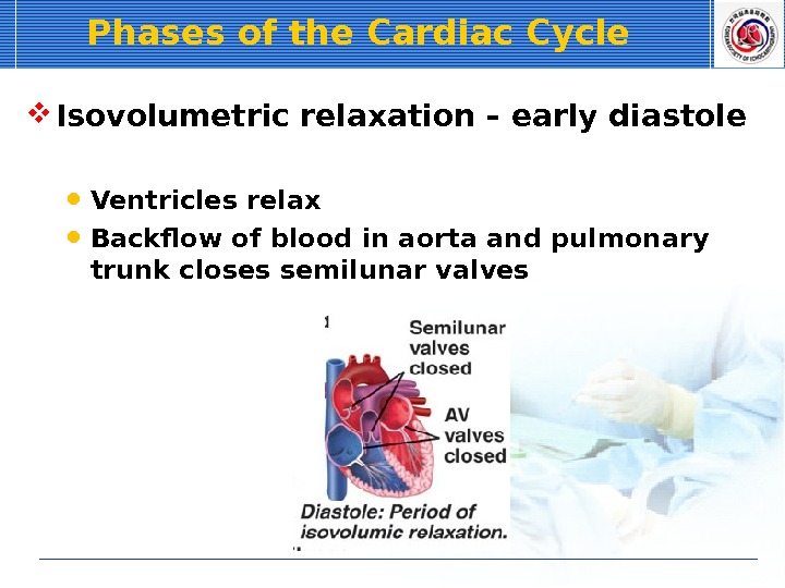
Phases of the Cardiac Cycle Isovolumetric relaxation – early diastole Ventricles relax Backflow of blood in aorta and pulmonary trunk closes semilunar valves
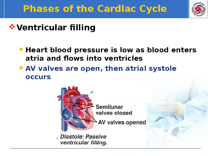
Phases of the Cardiac Cycle Ventricular filling Heart blood pressure is low as blood enters atria and flows into ventricles AV valves are open, then atrial systole occurs
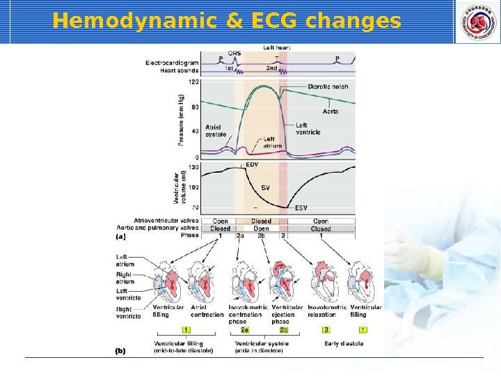
Hemodynamic & ECG changes