ahmad ata 1 Anatomy & physiology of cells

38660-cell_.ppt
- Количество слайдов: 53
 ahmad ata 1 Anatomy & physiology of cells Chapter 3
 ahmad ata 2 Objectives 1. label the components, name a term that describes the cell. 2. Distinguish between passive and active transport processes. 3. Define the terms diffusion, osmosis, filtration and facilitated diffusion, and give an example of each. 4. Define the terms active transport, endocytosis, and exocytosis. 5. List a function(s) for each cellular component and/or organelle. 6. Describe the structure of each cellular organelle.
 ahmad ata 3 INTRODUCTION The cell is the basic unit of structure and function in living things. Cells vary in their shape size, and arrangements but all cells have similar components, each with a particular function. Some of the 100 trillion of cells make up human body. All human cell are microscopic in size, shape and function. The diameter range from 7.5 micrometer (RBC) to 150 mm (ovum).
 ahmad ata 4 Introduction Cell is defined as the fundamental living unit of any organism. Cell is important to produce energy for metabolism (all chemical reactions within a cell) Cell can mutate (change genetically) as a result of accidental changes in its genetic material (DNA). Cytology: the study of the structure and functions of cells.
 ahmad ata 5
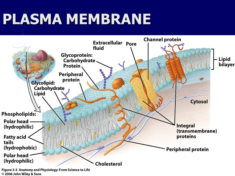
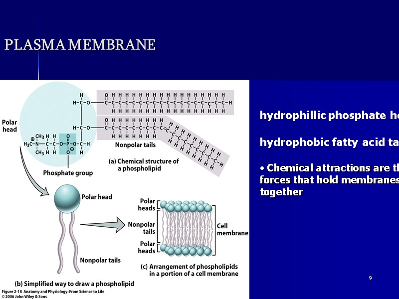
 ahmad ata 6 Cell structure 1) THE CELL (PLASMA) MEMBRANE The cell membrane is a thin, dynamic membrane that encloses the cell and controls what enters and leaves the cell. Fluid Mosaic Model composed of a double layer (bilayer) of phospholipid molecules with many protein molecules dispersed within it;
 ahmad ata 7 Fluid Mosaic Model a. The surfaces of the membrane are "hydrophilic" due to the polar phosphate heads; b. The internal portion of the membrane is "hydrophobic" due to the non-polar fatty acid tails; c. The membrane proteins also have both hydrophilic and hydrophobic
 ahmad ata 8 PLASMA MEMBRANE
 ahmad ata 9 PLASMA MEMBRANE hydrophillic phosphate head hydrophobic fatty acid tails Chemical attractions are the forces that hold membranes together
 ahmad ata 10 Function of plasma membrane Serves as boundary of the cell. Serve as markers that identify the cells. Play significant role in transportation. Cell recognition proteins-allow cell to recognize other cells.
 ahmad ata 11 Membrane proteins Some membrane proteins have carbohydrates attached to them, forming glycoproteins that act as identification markers Some membrane proteins are receptors that react to specific chemicals, sometimes permitting a process called signal transduction
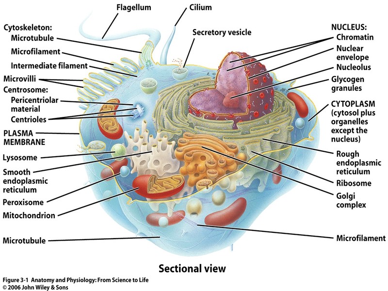
 ahmad ata 12 Cytoplasm Is a gel-like matrix of water, enzymes, nutrients, wastes, and gases and contains cell structures (organelles). Fluid around the organelles called cytosol. Most of the cells metabolic reactions occur in the cytoplasm.
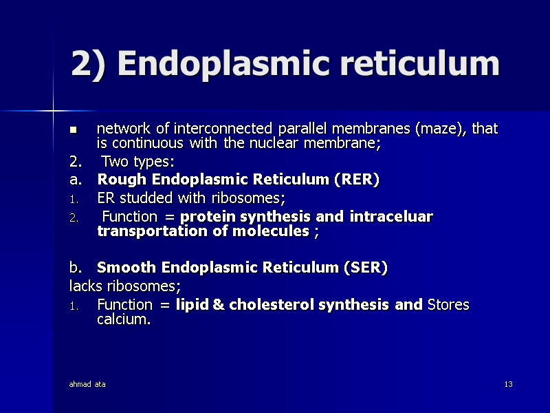
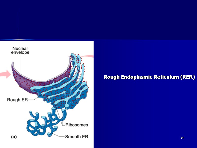
 ahmad ata 13 2) Endoplasmic reticulum network of interconnected parallel membranes (maze), that is continuous with the nuclear membrane; 2. Two types: a. Rough Endoplasmic Reticulum (RER) ER studded with ribosomes; Function = protein synthesis and intraceluar transportation of molecules ; b. Smooth Endoplasmic Reticulum (SER) lacks ribosomes; Function = lipid & cholesterol synthesis and Stores calcium.
 ahmad ata 14 Rough Endoplasmic Reticulum (RER)
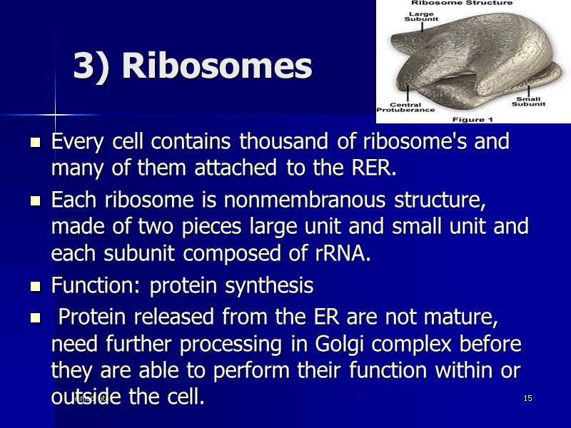
 ahmad ata 15 3) Ribosomes Every cell contains thousand of ribosome's and many of them attached to the RER. Each ribosome is nonmembranous structure, made of two pieces large unit and small unit and each subunit composed of rRNA. Function: protein synthesis Protein released from the ER are not mature, need further processing in Golgi complex before they are able to perform their function within or outside the cell.
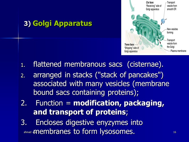
 ahmad ata 16 3) Golgi Apparatus flattened membranous sacs (cisternae). arranged in stacks ("stack of pancakes") associated with many vesicles (membrane bound sacs containing proteins); 2. Function = modification, packaging, and transport of proteins; 3. Encloses digestive enyzymes into membranes to form lysosomes.
 ahmad ata 17 4) Lysosomes 1. spherical membranous sacs containing digestive enzymes; 2. "suicide sacs" which destroy anything the cell no longer wants or needs. 3. Autolysis is the process by which worn cell parts are digested by autophagy.
 ahmad ata 18 Peroxisomes: 1. membranous sacs containing oxidase enzymes; 2. Function = detoxification of harmful or toxic substances (i.e. alcohol, formaldehyde, oxygen free radicals); H2O2 (peroxide) ----> water
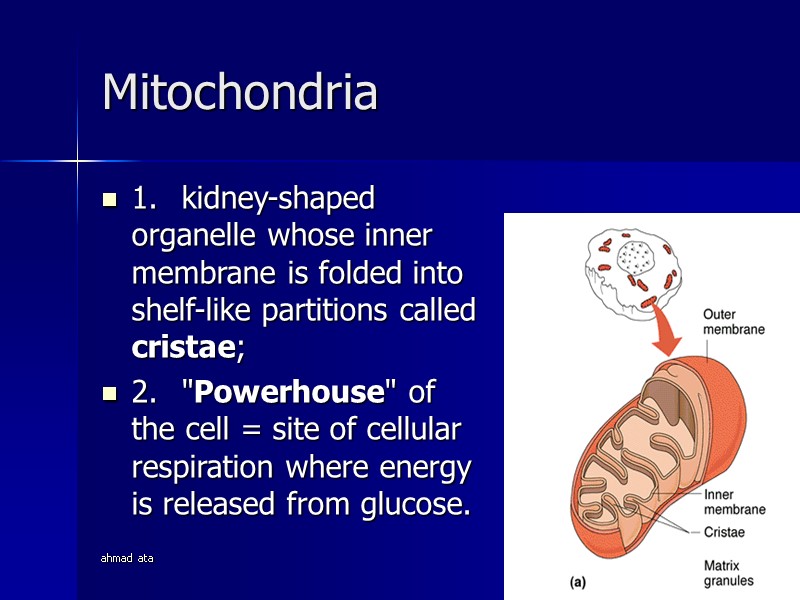
 ahmad ata 19 Mitochondria 1. kidney-shaped organelle whose inner membrane is folded into shelf-like partitions called cristae; 2. "Powerhouse" of the cell = site of cellular respiration where energy is released from glucose.
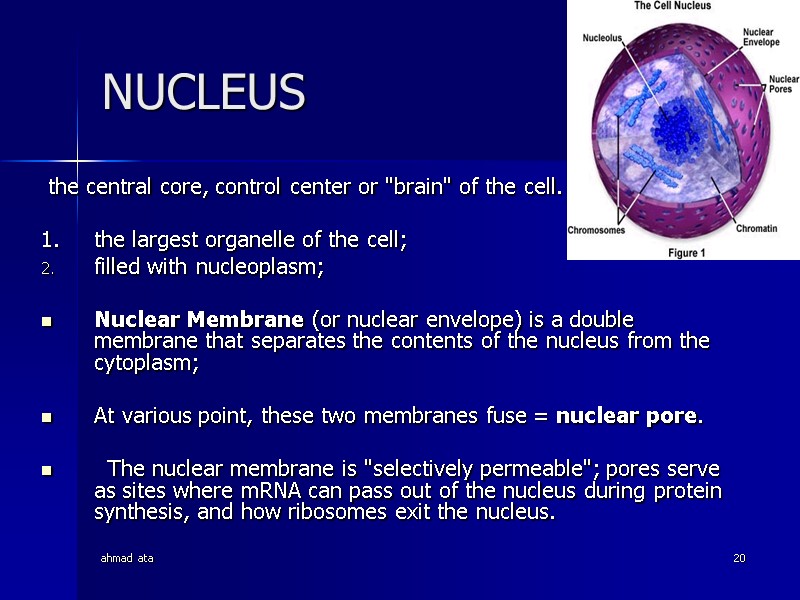
 ahmad ata 20 NUCLEUS the central core, control center or "brain" of the cell. 1. the largest organelle of the cell; filled with nucleoplasm; Nuclear Membrane (or nuclear envelope) is a double membrane that separates the contents of the nucleus from the cytoplasm; At various point, these two membranes fuse = nuclear pore. The nuclear membrane is "selectively permeable"; pores serve as sites where mRNA can pass out of the nucleus during protein synthesis, and how ribosomes exit the nucleus.
 ahmad ata 21 Nucleoli Nucleolus (s) = a spherical body within the nucleus; composed of RNA and proteins; Function = synthesis of ribosomes.
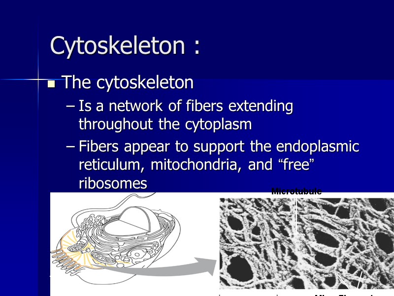
 ahmad ata 22 Cytoskeleton : The cytoskeleton Is a network of fibers extending throughout the cytoplasm Fibers appear to support the endoplasmic reticulum, mitochondria, and “free” ribosomes
 ahmad ata 23 The cytoskeleton Gives mechanical support to the cell Is involved in cell motility, which utilizes motor proteins Rodlike pieces that provide support and allow movement and mechanisms that can move the cell or its parts
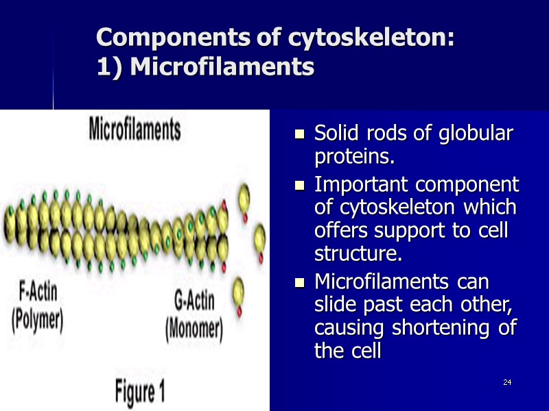
 ahmad ata 24 Components of cytoskeleton: 1) Microfilaments Solid rods of globular proteins. Important component of cytoskeleton which offers support to cell structure. Microfilaments can slide past each other, causing shortening of the cell
 ahmad ata 25 Components of cytoskeleton: 2) Intermediate filaments Intermediate filaments are twisted protein strands slightly thicker than microfilaments; they form much of the supporting framework in many types of cells
 ahmad ata 26 Components of cytoskeleton: 3) Microtubules Microtubules Shape the cell Guide movement of organelles (their function is to move things around in the cell) Help separate the chromosome copies in dividing cells
 ahmad ata 27 Components of cytoskeleton: 4) Microtubules Centrosomes and Centrioles The centrosome An area of the cytoplasm near the nucleus that coordinates the building and breaking of microtubules in the cell Its considered to be a “microtubule-organizing center” Plays an important role during cell division Contains a pair of centrioles
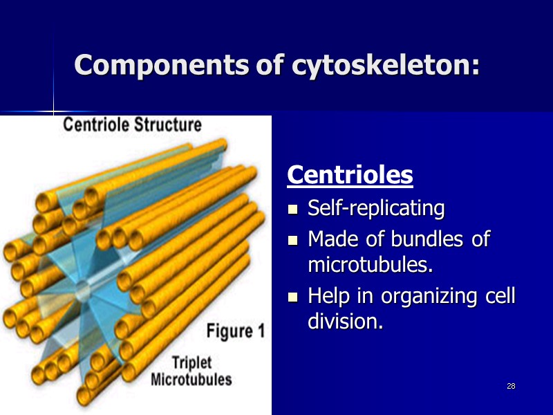
 ahmad ata 28 Components of cytoskeleton: Centrioles Self-replicating Made of bundles of microtubules. Help in organizing cell division.
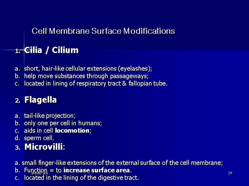
 ahmad ata 29 Cell Membrane Surface Modifications Cilia / Cilium a. short, hair-like cellular extensions (eyelashes); b. help move substances through passageways; c. located in lining of respiratory tract & fallopian tube. Flagella a. tail-like projection; b. only one per cell in humans; c. aids in cell locomotion; d. sperm cell. Microvilli: a. small finger-like extensions of the external surface of the cell membrane; b. Function = to increase surface area. c. located in the lining of the digestive tract.
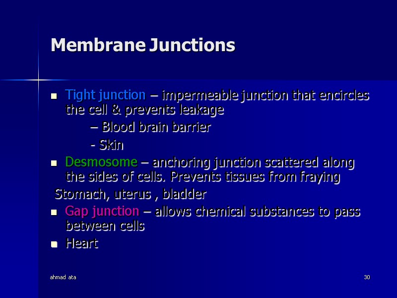
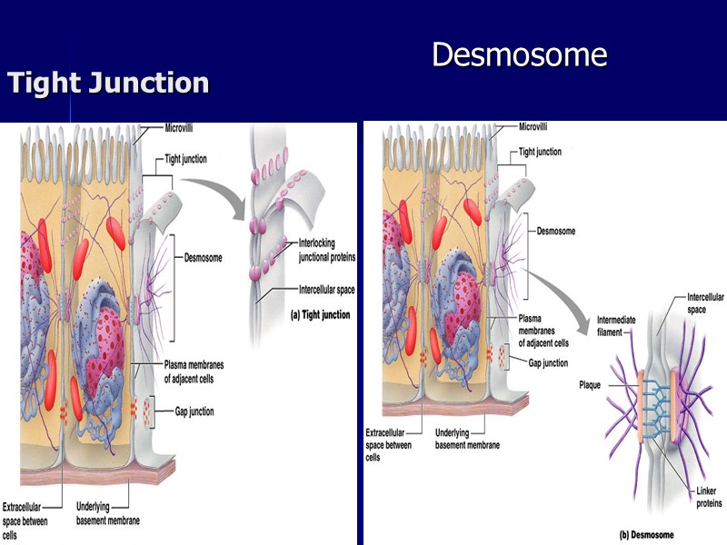
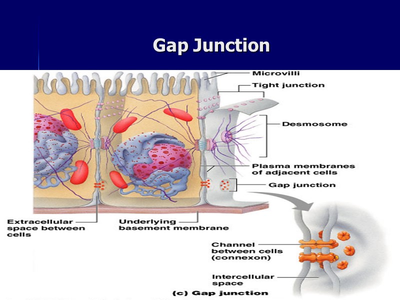
 ahmad ata 30 Membrane Junctions Tight junction – impermeable junction that encircles the cell & prevents leakage – Blood brain barrier - Skin Desmosome – anchoring junction scattered along the sides of cells. Prevents tissues from fraying Stomach, uterus , bladder Gap junction – allows chemical substances to pass between cells Heart
 ahmad ata 31 Tight Junction Desmosome
 ahmad ata 32 Gap Junction
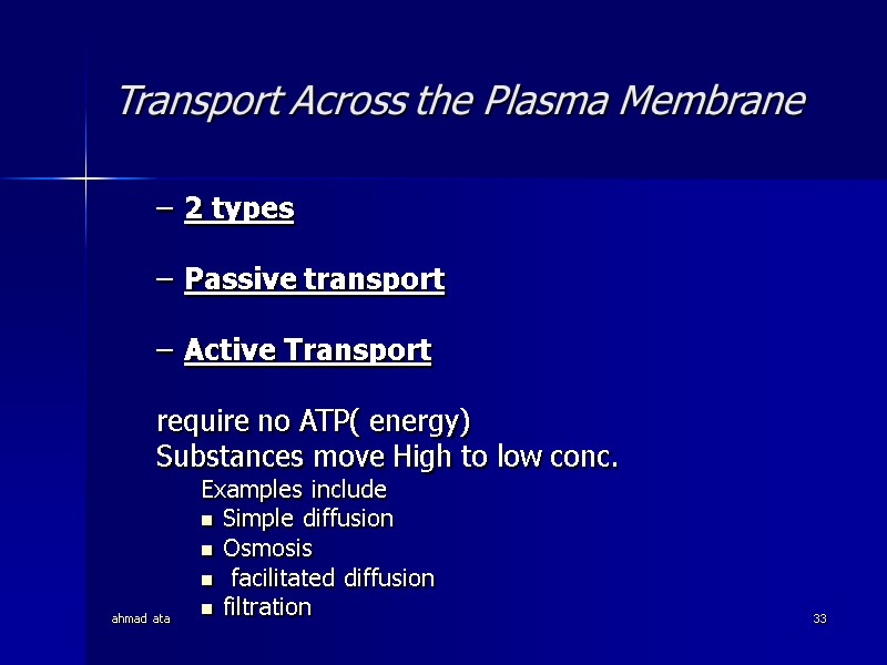
 ahmad ata 33 Transport Across the Plasma Membrane 2 types Passive transport Active Transport require no ATP( energy) Substances move High to low conc. Examples include Simple diffusion Osmosis facilitated diffusion filtration
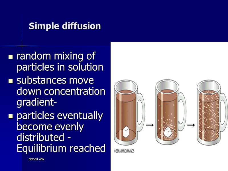
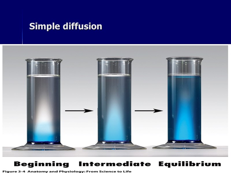
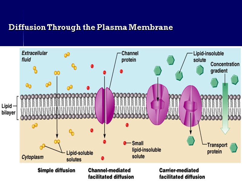
 ahmad ata 34 Simple diffusion random mixing of particles in solution substances move down concentration gradient- particles eventually become evenly distributed - Equilibrium reached
 ahmad ata 35 Simple diffusion
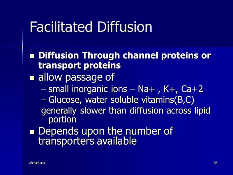
 ahmad ata 36 Facilitated Diffusion Diffusion Through channel proteins or transport proteins allow passage of small inorganic ions – Na+ , K+, Ca+2 Glucose, water soluble vitamins(B,C) generally slower than diffusion across lipid portion Depends upon the number of transporters available
 ahmad ata 37 Diffusion Through the Plasma Membrane
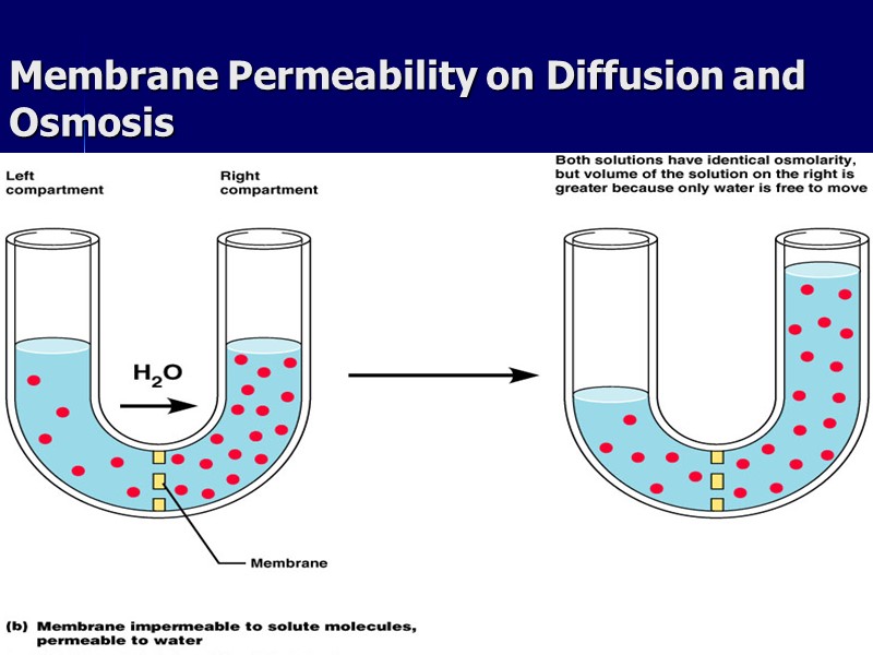
 ahmad ata 38 Osmosis passive process diffusion of water across a selectively permeable membrane from Hi. Conc. of WATER ( low solute) to lower concentration of WATER( Hi. solute)
 ahmad ata 39 Membrane Permeability on Diffusion and Osmosis
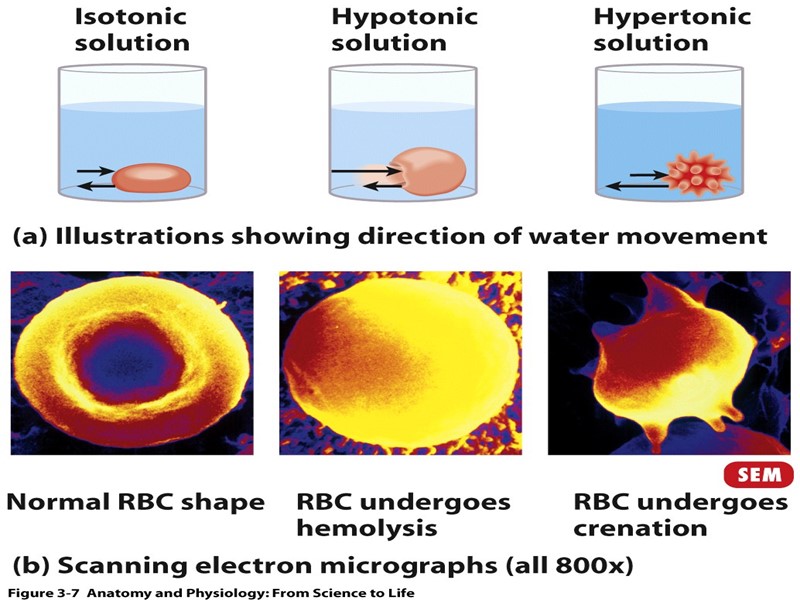
 ahmad ata 40 Tonicity Describes how a solution affects cell volume hypertonic solution with more solutes Blood cells shrink and crenate hypotonic solution with less solutes Blood cells swell up and hemolyse isotonic both solutions have similar concentrations of solutes. Cell size is unchanged
 ahmad ata 41
 ahmad ata 42 Active transport - movement of a substance from a lower concentration to a higher concentration using a carrier and energy Endocytosis - brings substances into the cells
 ahmad ata 43 solutes moving against concentration gradient-Uses carrier proteins can be driven by ATP use or via energy stored in ionic concentration Types : Primary active transport Secondary active transport Endocytosis Exocytosis Tanscytosis
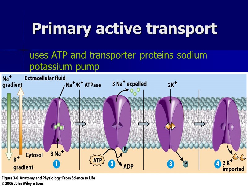
 ahmad ata 44 Primary active transport uses ATP and transporter proteins sodium potassium pump
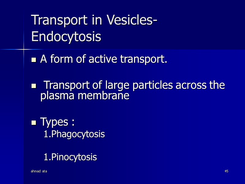
 ahmad ata 45 Transport in Vesicles- Endocytosis A form of active transport. Transport of large particles across the plasma membrane Types : Phagocytosis Pinocytosis
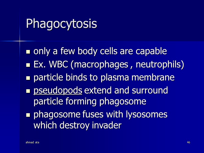
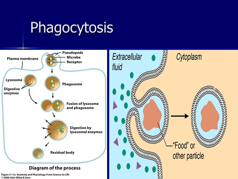
 ahmad ata 46 Phagocytosis only a few body cells are capable Ex. WBC (macrophages , neutrophils) particle binds to plasma membrane pseudopods extend and surround particle forming phagosome phagosome fuses with lysosomes which destroy invader
 ahmad ata 47 Phagocytosis
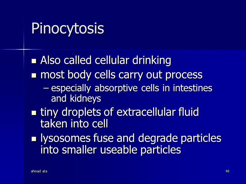
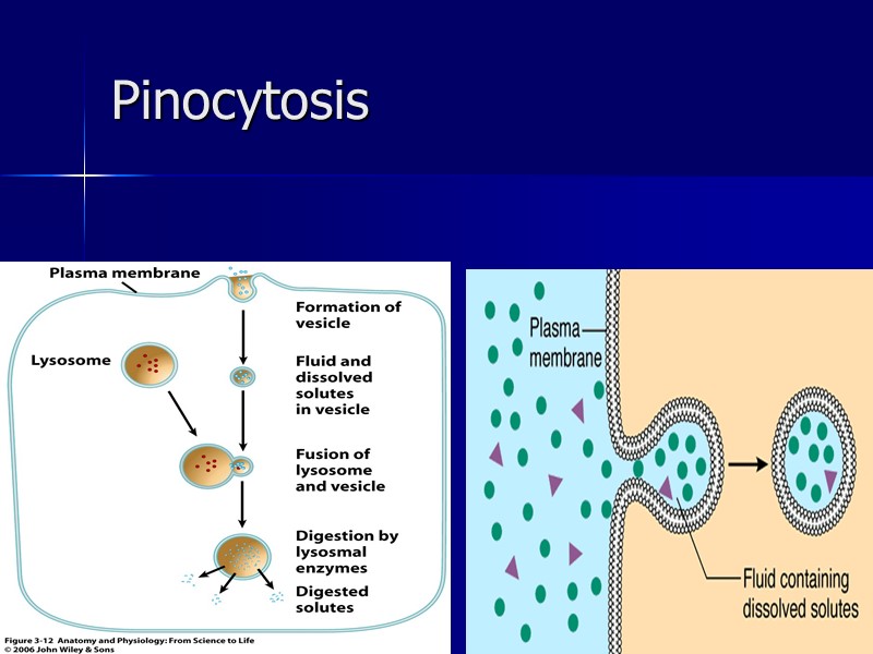
 ahmad ata 48 Pinocytosis Also called cellular drinking most body cells carry out process especially absorptive cells in intestines and kidneys tiny droplets of extracellular fluid taken into cell lysosomes fuse and degrade particles into smaller useable particles
 ahmad ata 49 Pinocytosis
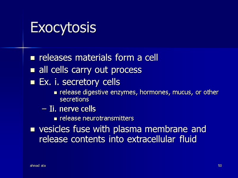
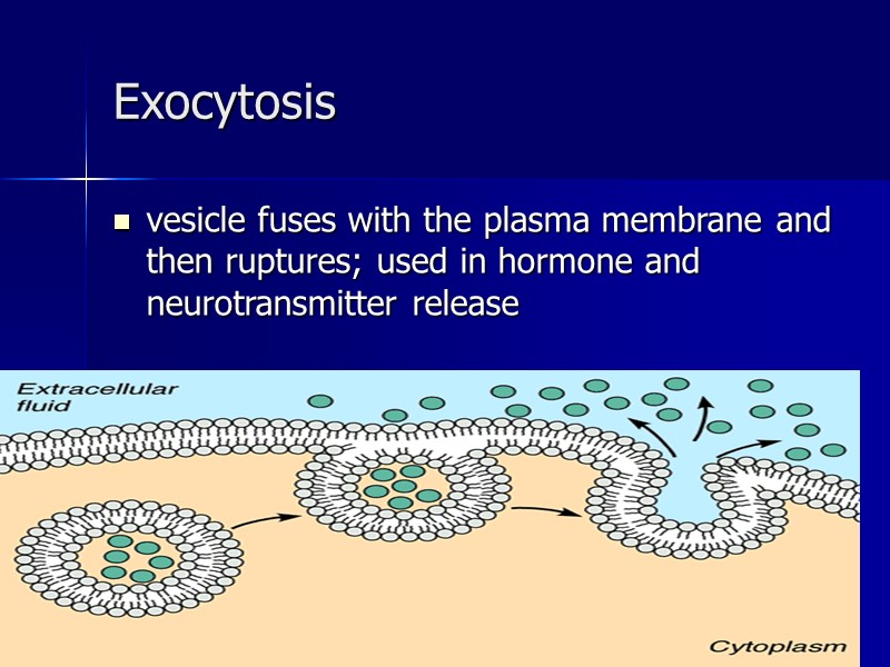
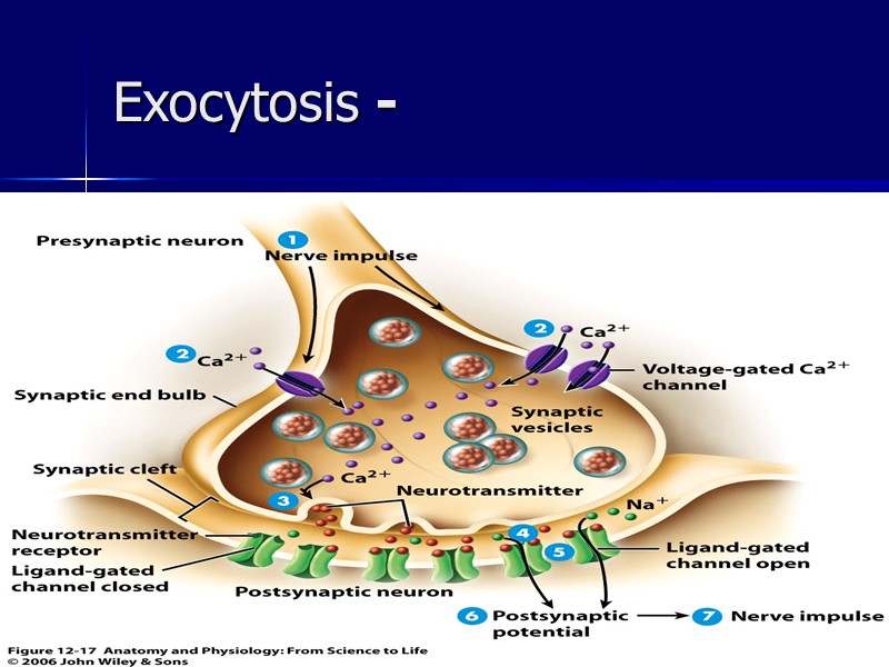
 ahmad ata 50 Exocytosis releases materials form a cell all cells carry out process Ex. i. secretory cells release digestive enzymes, hormones, mucus, or other secretions Ii. nerve cells release neurotransmitters vesicles fuse with plasma membrane and release contents into extracellular fluid
 ahmad ata 51 Exocytosis vesicle fuses with the plasma membrane and then ruptures; used in hormone and neurotransmitter release
 ahmad ata 52 Exocytosis -
 ahmad ata 53 The End
