
1 Neoplasms of the Nose and Paranasal Sinus


- Размер: 228.5 Кб
- Количество слайдов: 39
Описание презентации 1 Neoplasms of the Nose and Paranasal Sinus по слайдам
1 Neoplasms of the Nose and Paranasal Sinus University of Texas Medical Branch Steven T. Wright, M. D. Anna M. Pou, M. D. May 19,
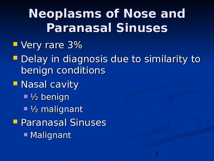
2 Neoplasms of Nose and Paranasal Sinuses Very rare 3% Delay in diagnosis due to similarity to benign conditions Nasal cavity ½ benign ½ malignant Paranasal Sinuses Malignant
3 Neoplasms of Nose and Paranasal Sinuses Multimodality treatment Orbital Preservation Minimally invasive surgical techniques
4 Epidemiology Predominately of older males Exposure: Wood, nickel-refining processes Industrial fumes, leather tanning Cigarette and Alcohol consumption No significant association has been shown
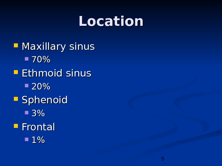
5 Location Maxillary sinus 70%70% Ethmoid sinus 20%20% Sphenoid 3%3% Frontal 1%1%
6 Presentation Oral symptoms: 25 -35% Pain, trismus, alveolar ridge fullness, erosion Nasal findings: 50% Obstruction, epistaxis, rhinorrhea Ocular findings: 25% Epiphora, diplopia, proptosis Facial signs Paresthesias, asymmetry
7 Radiography CTCT Bony erosion Limitations with periorbita involvement MRIMRI 94 -98% correlation with surgical findings Inflammation/retained secretions: low T 1, high T 2 Hypercellular malignancy: low/intermediate on both Enhancement with Gadolinium
8 Benign Lesions Papillomas Osteomas Fibrous Dysplasia Neurogenic tumors
9 Papilloma Vestibular papillomas Schneiderian papillomas derived from schneiderian mucosa (squamous) Fungiform: 50%, nasal septum Cylindrical: 3%, lateral wall/sinuses Inverted: 47%, lateral wall
10 Inverted Papilloma 4% of sinonasal tumors Site of Origin: lateral nasal wall Unilateral Malignant degeneration in 2 -13% (avg 10%)
11 Inverted Papilloma Resection Initially via transnasal resection: 50 -80% recurrence Medial Maxillectomy via lateral rhinotomy: Gold Standard 10 -20% Endoscopic medial maxillectomy: Key concepts: Identify the origin of the papilloma Bony removal of this region Recurrent lesions: Via medial maxillectomy vs. Endoscopic resection 22%22%
12 Osteomas Benign slow growing tumors of mature bone Location: Frontal, ethmoids, maxillary sinuses When obstructing mucosal flow can lead to mucocele formation Treatment is local excision
13 Fibrous dysplasia Dysplastic transformation of normal bone with collagen, fibroblasts, and osteoid material Monostotic vs Polyostotic Surgical excision for obstructing lesions Malignant transformation to rhabdomyosarcoma has been seen with radiation
14 Neurogenic tumors 4% are found within the paranasal sinuses Schwannomas Neurofibromas Treatment via surgical resection Neurogenic Sarcomas are very aggressive and require surgical excision with post op chemo/XRT for residual disease. When associated with Von Recklinghausen’s syndrome: more aggressive (30% 5 yr survival).
15 Malignant lesions Squamous cell carcinoma Adenoid cystic carcinoma Mucoepidermoid carcinoma Adenocarcinoma Hemangiopericytoma Melanoma Olfactory neuroblastoma Osteogenic sarcoma, fibrosarcoma, chondrosarcoma, rhabdomyosarcoma Lymphoma Metastatic tumors Sinonasal undifferentiated carcinoma
16 Squamous cell carcinoma Most common tumor (80%) Location: Maxillary sinus (70%) Nasal cavity (20%) 90% have local invasion by presentation Lymphatic drainage: First echelon: retropharyngeal nodes Second echelon: subdigastric nodes
17 Treatment 88% present in advanced stages (T 3/T 4) Surgical resection with postoperative radiation Complex 3 -D anatomy makes margins difficult
18 Adenoid Cystic Carcinoma 33 rdrd most common site is the nose/paranasal sinuses Perineural spread Anterograde and retrograde Despite aggressive surgical resection and radiotherapy, most grow insidiously. Neck metastasis is rare and usually a sign of local failure Postoperative XRT is very important
19 Mucoepidermoid Carcinoma Extremely rare Widespread local invasion makes resection difficult, therefore radiation is often indicated
20 Adenocarcinoma 22 ndnd most common malignant tumor in the maxillary and ethmoid sinuses Present most often in the superior portions Strong association with occupational exposures High grade: solid growth pattern with poorly defined margins. 30% present with metastasis Low grade: uniform and glandular with less incidence of perineural invasion/metastasis.
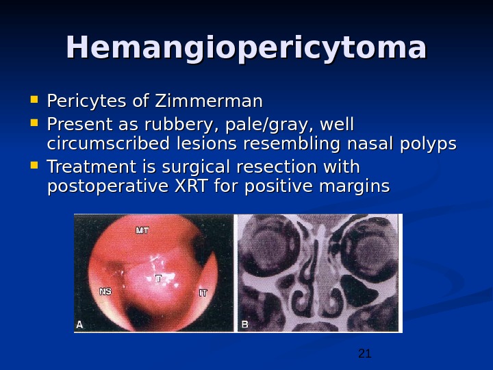
21 Hemangiopericytoma Pericytes of Zimmerman Present as rubbery, pale/gray, well circumscribed lesions resembling nasal polyps Treatment is surgical resection with postoperative XRT for positive margins
22 Melanoma 0. 5 — 1. 5% of melanoma originates from the nasal cavity and paranasal sinus. Anterior Septum: most common site Treatment is wide local excision with/without postoperative radiation therapy END not recommended AFIP: Poor prognosis 5 yr: 11% 20 yr: 0. 5%
23 Olfactory Neuroblastoma Esthesioneuroblastoma Originate from stem cells of neural crest origin that differentiate into olfactory sensory cells. Kadish Classification A: confined to nasal cavity B: involving the paranasal cavity C: extending beyond these limits
24 Olfactory Neuroblastoma Esthesioneuroblastoma UCLA Staging system T 1: Tumor involving nasal cavity and/or paranasal sinus, excluding the sphenoid and superior most ethmoids T 2: Tumor involving the nasal cavity and/or paranasal sinus including sphenoid/cribriform plate T 3: Tumor extending into the orbit or anterior cranial fossa T 4: Tumor involving the brain
25 Olfactory Neuroblastoma Esthesioneuroblastoma Aggressive behavior Local failure: 50 -75% Metastatic disease develops in 20 -30% Treatment: En bloc surgical resection with postoperative XRT
26 Sarcomas Osteogenic Sarcoma Most common primary malignancy of bone. Mandible > Maxilla Sunray radiographic appearance Fibrosarcoma Chondrosarcoma
27 Rhabdomyosarcoma Most common paranasal sinus malignancy in children Non-orbital, parameningeal Triple therapy is often necessary Aggressive chemo/XRT has improved survival from 51% to 81% in patients with cranial nerve deficits/skull/intracranial involvement. Adults, Surgical resection with postoperative XRT for positive margins.
28 Lymphoma Non-Hodgkins type Treatment is by radiation, with or without chemotherapy Survival drops to 10% for recurrent lesions
29 Sinonasal Undifferentiated Carcinoma Aggressive locally destructive lesion Dependent on pathological differentiation from melanoma, lymphoma, and olfactory neuroblastoma Preoperative chemotherapy and radiation may offer improved survival
30 Metastatic Tumors Renal cell carcinoma is the most common Palliative treatment only
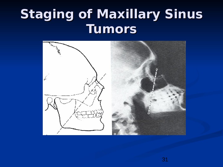
31 Staging of Maxillary Sinus Tumors
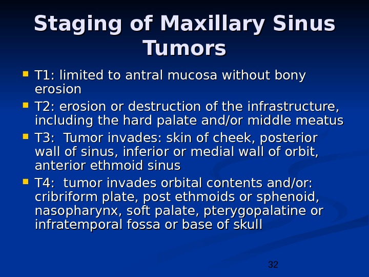
32 Staging of Maxillary Sinus Tumors T 1: limited to antral mucosa without bony erosion T 2: erosion or destruction of the infrastructure, including the hard palate and/or middle meatus T 3: Tumor invades: skin of cheek, posterior wall of sinus, inferior or medial wall of orbit, anterior ethmoid sinus T 4: tumor invades orbital contents and/or: cribriform plate, post ethmoids or sphenoid, nasopharynx, soft palate, pterygopalatine or infratemporal fossa or base of skull
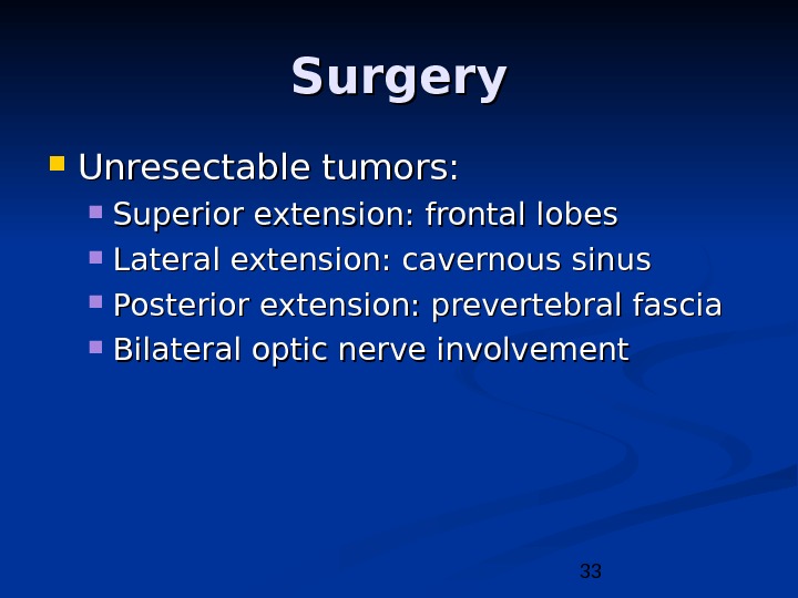
33 Surgery Unresectable tumors: Superior extension: frontal lobes Lateral extension: cavernous sinus Posterior extension: prevertebral fascia Bilateral optic nerve involvement
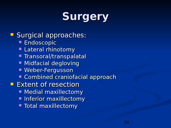
34 Surgery Surgical approaches: Endoscopic Lateral rhinotomy Transoral/transpalatal Midfacial degloving Weber-Fergusson Combined craniofacial approach Extent of resection Medial maxillectomy Inferior maxillectomy Total maxillectomy
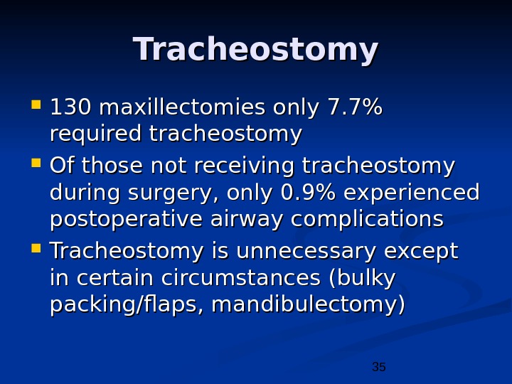
35 Tracheostomy 130 maxillectomies only 7. 7% required tracheostomy Of those not receiving tracheostomy during surgery, only 0. 9% experienced postoperative airway complications Tracheostomy is unnecessary except in certain circumstances (bulky packing/flaps, mandibulectomy)
36 Treatment of the Orbit Before 1970’s orbital exenteration was included in the radical resection Preoperative radiation reduced tumor load and allowed for orbital preservation with clear surgical margins Currently, the debate is centered on what “degree” of orbital invasion is allowed.
37 Current indications for orbital exenteration Involvement of the orbital apex Involvement of the extraocular muscles Involvement of the bulbar conjunctiva or sclera Lid involvement beyond a reasonable hope for reconstruction Non-resectable full thickness invasion through the periorbita into the retrobulbar fat
38 Conclusions Neoplasms of the nose and paranasal sinus are very rare and require a high index of suspicion for diagnosis Most lesions present in advanced states and require multimodality therapy
39 Bibliography Bhattacharyya N. Cancer of the Nasal Cavity: Survival and Factors Influencing Prognosis. Archives of Oto-HNS. Vol 128(9). September 2002. Pp 1079 -1083. Bradley P, Jones N, Robertson I. Diagnosis and Management of Esthesioneuroblastoma. Current Opinion in Oto-HNS. Vol 11(2). April 2003. Pp 112 -118. Carrau R, Segas J, Nuss D, et al. Squamous Cell Carcinoma of the Sinonasal Tract Invading the Orbit. Laryngoscope. Vol 109 (2, part 1). February 1999. Pp 230 -235. Devaiah A, Larsen C, Tawfik O, et al. Esthesioneuroblastoma: Endoscopic Nasal and Anterior Craniotomy Resection. Laryngoscope. Vol 113(12). December 2003. Pp 2086 -2090. Han J, Smith T, Loehrl T, et al. An Evolution in the Management of Sinonasal Inverting Papilloma. Laryngoscope. Vol 111(8). August 2001. Pp 1395 -1400. Imola M, Schramm V. Orbital Preservation in Surgical Management of Sinonasal Malignancy. Laryngoscope. Vol 112(8). August 2002. Pp 1357 -1365. Katzenmeyer K, Pou A. Neoplasms of the Nose and Paranasal Sinus. Dr. Quinn’s Online Textbook of Otolaryngology. June 7, 2000. Kraft M, Simmen D, Kaufmann T, et al. Laryngoscope. Vol 113(9). September 2003. Pp 1541 -1547. Mc. Cary S, Levine P, Cantrell R. Preservation of the eye in the Treatment of Sinonasal Malignant Neoplasms with Orbital Involvement: A Confirmation of the Original Treatise. Archives of Oto-HNS. Vol 122(6). June 1996. Pp 657 -659. Myers E, Suen J. Cancer of the Head and Neck, 3 rd Edition: Neoplasms of the Nose and Paranasal Sinuses. W. B. Saunders Company. 1996. Myers L, Nussenbaum B, Bradford C, et al. Paranasal Sinus Malignancies: An 18 -Year Single Institution Experience. Laryngoscope. Vol 112(11). November 2002. Pp 1964 -1969.